ISSN Online: 2164-3199 ISSN Print: 2164-3180
DOI: 10.4236/ojbd.2018.84007 Oct. 12, 2018 61 Open Journal of Blood Diseases
Surrogate Role of CD85k on Monocytic Lineage
Involved Leukemogenesis Biology and
Clinical Aspect
Hasnaa A. Abo-Elwafa
1*, Shereen P. Aziz
1, Heba A. Ahmed
1, Elsayed Mostafa Ali
2, Doaa S. Elsaied
11Clinical Pathology Department, Sohag Faculty of Medicine, Sohag University, Sohag, Egypt 2Clinical Oncology Department, Sohag Faculty of Medicine, Sohag University, Sohag, Egypt
Abstract
Background: Unique receptor involved in leukemogenesis is CD85k; an im-muneglobulin receptor for immune tolerance, CD36 is glycoprotein mediates cellular adhesion and metastatic spread, CD14, CD15 considered common monocytic markers. Aims: to investigate CD85k with monocytic lineage in-volved leukemia (MLIL) markers in leukemia pathogenesis and clinical presen-tation. Patients and Methods: 47 patients (32 diagnosed acute myeloid leuke-mia (AML); 15 non-malignant hematological disease as a control), were in-cluded, aged from 2 to 80 years, all subjected to peripheral blood (P.Bl) and bone marrow (B.M) examination, immunophenotyping (IPT) using FASC Canto four color flow cytometer (FCM) Becton Dickenson (BD) USA, for CD13, CD33, MPO, HLA-DR, CD34, CD38, CD117, CD14, CD15 and CD36 the Mo Abs supplied by B.D Bioscience, and anti CD85k Mo Abs by Aveda de Coimbra Flamenco, reference No. 1399990130. Results: Frequency of CD85k is 19/32 (59.37%) of AML; 14/14 (M4/M5) 100% positive CD85k, insignificant correlations of CD85k to sex, lymphadenopathy or organomegaly, platelets count and P.Bl blast (P > 0.05), significant to age < 35 years, WBCs > 50,000 × 109/l, Hb < 7 g/dl, BM blasts, CD34 and HLA-DR CD33, CD13, CD38 (P < 0.05), insignificant correlations to CD36, CD14, CD15 and CD117 (P > 0.05). Conclusion: Although CD85k is MLIL associated marker, it is not correlated with other MLIL markers with frequency 100% in MLIL and 59.37% in AML, age predisposition is <35 years with no sex variation, significant correlation to progenitor and myeloid markers, it’s a crucial role in leukemogenesis biology, not in clinical presentations, considered good follow up predictor MLIL marker.
Keywords
CD85k, Monocytic Lineage Leukemia How to cite this paper: Abo-Elwafa, H.A.,
Aziz, S.P., Ahmed, H.A., Ali, E.M. and El-saied, D.S. (2018) Surrogate Role of CD85k on Monocytic Lineage Involved Leukemo-genesis Biology and Clinical Aspect. Open Journal of Blood Diseases, 8, 61-73. https://doi.org/10.4236/ojbd.2018.84007
Received: April 7, 2017 Accepted: October 9, 2018 Published: October 12, 2018
Copyright © 2018 by authors and Scientific Research Publishing Inc. This work is licensed under the Creative Commons Attribution International License (CC BY 4.0).
DOI: 10.4236/ojbd.2018.84007 62 Open Journal of Blood Diseases
1. Introduction
The unique synergistic action of membrane bound proteins in leukemogenesis in monocytic lineage involved (MLIL) leukemia include immune escape by im-munoglobulin inhibitory receptor CD85k and cellular adhesion and metastatic spread by glycoprotein CD36 [1] [2]. CD36 mediating adhesion process to en-dothelial cells and in promoting tumor spreading and organ infiltrations [3]. Monocytoid dendritic cells with high ILT3 levels suppress T cell activation and are tolerogenic, ILT3 levels are higher in patients being treated with type I IFN, supporting the concept that IFN-induced ILT3 expression is immunosuppres-sive [1] [2] [4]. The cytoplasmic tail of ILT3 contains immune tyrosine-based inhibition receptor recruits and activates tyrosine phosphatases. Ligation of CD85k on dendritic cells blocks the activation and downstream signaling [5] [6] [7]. CD85k expressed by, monocytes, dendritic cells and endothelial cells. It is encoded in chromosome 19 [8]. CD85k has a crucial role in tolerogenic activity of antigen presenting cells and tumor escape [9]. Also it promotes conversion alloreactive CD4 to regulatory T cell (Terg). It inhibits T cell proliferation and induces CD8 differentiation. Crosslinking of CD85k to monocyte receptor de-crease activation [10]. In AML CD85k is sensitive marker with CD36 for mono-cytic differentiation [3] [11]. In MLIL CD85k co-expressed with CD34 and CD117 progenitor cells so it has a role in leukemogenesis [10]. AML with mo-nocytic differentiation has a high risk of extra medullary disease, high leukocytic count and coagulation defect also genetic and cytogenetic abnormality [8]. The early clinical findings of AML are often vague and nonspecific [12]. Splenic en-largement and generalized lymphadenopathy are rare in AML [13]. Some pa-tients may experience swelling of the gums because of infiltration of leukemic cells [14]. AML has several subtypes; treatment and prognosis differ between them, several markers can predict which drug may work best [15].
CD85k mainly has prognostic value in leukemia so it should be incorporated into the initial diagnosis work-up and leukemia monitoring [9]. CD85k is an important target for anticancer therapy [10]. Lack of CD85k expression leads to leukemia remission increase survival rate in animal model, also block leukemia development in transplantation [16].
2. Subjects and Methods
2.1. Ethical Approval
The present study was revised by the Scientific Ethical Committee of Sohag University Hospital; a written informed consent was taken from all patients groups. It was in accordance with the ethical standards of the institutional and/or national research committee and with the 1964 Helsinki declaration.
2.2. Patients Selection
DOI: 10.4236/ojbd.2018.84007 63 Open Journal of Blood Diseases from October 2014 to October 2016, they were aged from 2 to 80 years old with mean age (31.77 ± 19.49), 19 males and 13 females and 15 subjects of non-malignant hematological disease (ITP), 8 males, 7 females; their ages ranged from 3 to 60 years with mean of (15.47 ± 15.46) of the same ethnic group as a control.
2.3. Inclusion Criteria
Newly diagnosed AML especially MLIL. All were subjected to:
Through history and clinical examination with stress on the presence and ex-tent of leukemia involvement of liver, spleen, lymphadenopathy and gum hyper-trophy.
2.4. Laboratory Investigations
2.4.1. Sampling3 ml of venous blood were collected from each one, dispensed into (K-EDTA) B.D tube used for P.Bl hemogram. BM examinations (aspirate/biopsy) were done for all groups, diagnosis of AML based on morphological features of P.Bl and B.M smears, cytochemical tests and IPT data. Blood count using Cell-Dyne-Ruby, automated cell counter, ABBOTT diagnostic (USA), with micro-scopic examination of stained P.Bl. and BMA smears for differential leucocytes count, blast cells percentage, morphological features and cytochemical stains.
2.4.2. IPT of Blast Cells
FCM FASC Canto four colors B.D; USA was used and the MoAbs supplied by B.D Bioscience, USA. The panel of fluorescein isothiocyanate (FITC), phycoe-rythrin (PE) and Peridinin chlorophyll (PerCP) conjugated MoAbs were used for each sample. Common progenitor marker, CD34, HLA-DR, CD38, CD117 Myeloid markers CD13, CD33, MPO, monocytic marker CD14, CD15, CD36, CD65, CD68. Lymphoid markers: B cell markers CD19, CD22, CD10, T cell markers CD2, CD3, CD5, CD7, FITC labeled MoAbs for detection of CD36 (B.D Bioscience, Cat. No.656151 USA). PE labeled Mo Abs for CD85k provided by Aveda de Coimbra Flamenco, Reference No. 1399990130.
2.5. Procedure of Surface Membrane Markers
Expression on blast cells:Reagents
DOI: 10.4236/ojbd.2018.84007 64 Open Journal of Blood Diseases
2.6. Procedure
Blood was diluted with (PBS) so that WBCs count was adjusted between 5 and 10 × 109/l. For each sample, sets of tubes were labeled for all the MoAbs to be used, including 1 tube for the appropriate negative isotypic control.
50 µL of diluted samples were delivered in each tube. 5 µL of each MoAbs as well as of the isotypic negative control. The tubes were vortexed and incubated in the dark at room temperature for 15 minutes. The tubes were centrifuged at 500 rpm for 5 minutes and the supernatant was discarded. Lysing solution (1.5 mL) was added to each tube. The tubes were vortexed and incubated for 5 - 10 minutes in the dark at room temperature. 2 ml PBS was added and the tubes were vortexed. The tubes were centrifuged at 500 rpm for 5 minutes and the su-pernatant was discarded. Cells were suspended in 500 µL PBS to be ready for acquiring data by the FCM.
The expression of blast cells for CD85k was determined as a percentage from the gated blast cells population. Cells were considered positive for a certain marker when ≥20% of cells expressed it, except for CD34, cytoplasmic MPO and CD85K where its expression by 10% of cells was sufficient to confer positivity.
2.7. Statistical Analysis
The collected data were tabulated and analyzed using statistical package of social science (SPSS) version 17 software. Suitable statistical techniques were computed ANOVA test, Student’s t test, Mann Whitney test and Kruskal Wallis for non-parametric values, correlation coefficient were used as tests of significance. Qualitative data were described in the form of number and percentage. Quantit-ative data were described in the form of mean ± standard deviation (SD), range and median.
2.8. Results
The present study was carried on 47 patients thirty two diagnosed as AML in-cluding fourteen MLIL (M4/M5 cases), their age ranged from 2 to 80 years, with median age 29 years old (mean is 31.77 ± 19.49), they were nineteen males/thirteen females, and fifteen cases of non-malignant hematological disease (ITP); their age ranged from 3 to 60 with median age 15 years old (mean 15.47 ± 15.46); seven males/eight females as a control.
DOI: 10.4236/ojbd.2018.84007 65 Open Journal of Blood Diseases
Table 1. Demographic data with clinical features and hematological variables in the studied patients.
Patients
Variables AML group CD85k + Ve AML subgroup (MLIL) Control p-value
Patients No 32 14 15 NS
Age (years) Median (range)
<35 >35
19 (2 - 80) 14 (12)
18 (7)
20(2 - 74) 10
4
15 (3 - 60) NS 0.06 S 0.03 NS 0.08
Sex
M/F 19/13 (10/9) 6/8 8/7 NS 0.5
Hepatomegaly Normal size 18 (12) 14 (7) 12 2
Normal NS 0.3
Splenomegaly Normal size 20 (15) 12 (4) 12 2
Normal NS 0.4
Lymphadenopathy No lymphadenopathy 14 (10) 18 (9) 10 2
Normal NS 0.2
WBCs × 109/l
Range <50 >50
1.7 - 245 21 11
23 - 245 9 5
3.2 - 12.8
S 0.02
Hb g/dl Range
<7 >7
3.7 - 12.6 19 13
4.5 - 8 10
4
11 - 13.5
S 0.023
Platelets × 109/l
Range <100 >100
12 - 456 24
8
14 - 38 14
0
243 - 427
S 0.02
P.Bl. blasts %
Range 11 - 91 14 - 69 0.0 NS 0.06
B.M blasts %
Range 24 - 93 23 - 78 0.0 NS 0.5
CD85k + Ve % Range Mean ± SD
(0.63 - 87.6) 26.92 ± 25.29
(19) (23 - 87.6) 14
0.0
HS 0.001
DOI: 10.4236/ojbd.2018.84007 66 Open Journal of Blood Diseases 50% positive expression of CD85k (7 cases). Splenomegaly was observed in 20/32 ML patients 62.5%, from which 15 patients were positive for CD85k (75%) and five were negative; and the other twelve patients with normal size spleen showed 30% positive for CD85k (4 cases). Lymphadenopathy was observed in 14/32 AML patients (43.75%); nine patients from them showed positive CD85k 9/14 (64.28%), and 10/18 patients without lymphadenopathy showed positive CD85k (55.55%). All these data were insignificant to CD85k expression (p > 0.05). The positive rate expression of both CD 85k and other MLIL markers were zero within the control group. As regard the hematological variables, also in ta-ble-1; significant correlations of CD85k was found when WBCs count was more than 50 × 109/l (p < 0.05), Hb value was less than 7 g/dl (p < 0.05), also signifi-cant correlation to the percentage of BM blasts. While platelet counts, the P.Bl blasts showed insignificant correlations (p > 0.05). We found that; the number of AML cases positive for progenitor markers were seventeen patients were positive for CD34, thirty were positive for CD38, 6 cases were positive for CD117 and twenty four patients were positive for HLA-DR. While the number of positive cases for the rest MLIL markers were 6 patients were positive for CD14, 8 pa-tients were positive for CD15 and only 3 cases were positive forCD36. Myeloid markers positivity showed that; CD13 was positive in twenty eight cases and CD33 was positive in thirty patients.
Table 2 represents the correlation of CD85k to the progenitors, myeloid and MLIL FCM markers. We notice that, there was insignificant correlation between CD85K and CD16, CD117, CD36, 235a.There was no correlation between CD85K and CD45, CD14 and CD15. There was positive significant correlation between CD85K and HLA-DR, CD34 and CD38, also CD13 and CD33 showed significant correlation. Details of FCM analysis were illustrated in Figure 1, Figure 2.
Comparison between MLIL and other types of AML as regards IPT were pre-sented in Table 3, we found that the mean expression of CD45, CD13, CD33 was increased in MLIL than other AML group, with significant increase in CD45 (p = 0.002). On the other hand, the mean expression of MPO, CD38, CD117, CD34 was increased in other AML group than MLIL with significant p value in CD 34, CD38, CD117 (P0.01, 0.005, 0.011 respectively). While the mean expression of HLA-DR, CD14, CD15 showed significant increase in MLIL group than other AML group (p < 0.001) for all markers. The mean expression of CD36 and CD61 were higher in other AML group than MLIL group (P 0.027 and 0.01 respective-ly). High significant increase in the mean expression of 235a in other AML group than MLIL group (p < 0.001). Finally the mean expression of CD85k is highly significant increase in MLIL group than other AML group (43.23 ± 18.46 to 14.23 ± 22.07); p < 0.001.
3. Discussion
DOI: 10.4236/ojbd.2018.84007 67 Open Journal of Blood Diseases
Table 2. Spearmans’ correlation of CD85k to clinical and hematological variables in AML
patients.
FCM markers Spearman’s correlation p value
Age 0.33 0.06 (NS)
Sex 0.137 0.09 (NS)
WBCs
<50 × 109/l 0.111 0.148 (NS)
>50 × 109/l 0.35 0.048 (S)
Hb < 7 g/dl 0.346 0.034 (S)
Hb > 7 g/dl 0.23 0.22 (NS)
Platelets 0.06 0.73 (NS)
P.Bl blasts 0.32 0.07 (NS)
B.M blasts 0.42 0.02 (S)
Progenitors
CD34 0.494 0.03 (S)
HLA-DR 0.544 0.004 (S)
CD38 0.471 0.035 (S)
CD117 0.0435 0.734 (NS)
Myeloid
CD13 0.399 0.011 (S)
CD33 0.457 0.003 (S)
Monocytic
CD14 0.267 0.26 (NS)
CD15 −0.363 0.12 (NS)
CD36 0.064 0.651 (NS)
DOI: 10.4236/ojbd.2018.84007 68 Open Journal of Blood Diseases
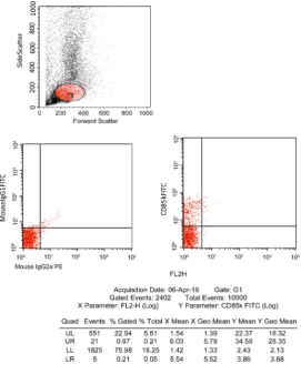
[image:8.595.233.516.429.711.2]Figure 1. FCM analysis of CD85k in AML M4 patient.
DOI: 10.4236/ojbd.2018.84007 69 Open Journal of Blood Diseases
Table 3. Comparison betweenMLIL and other types of AML as regards IPT.
FCM Markers
Expression % (No. = 14) MLIL Other AML (No. = 18) P value
CD45
Mean ± SD 74.70 ± 17.63 89.744 ± 7.0932 0.002 (S)
CD33
Mean ± SD 79.9 ± 12.93 63.40 ± 30.57 0.06 (NS)
CD13
Mean ± SD 58.31 ± 21.41 52.69 ± 28.0343 0.539 (NS)
MPO
Mean ± SD 2.302 ± 2.27 6.737 ± 21.56 0.451 (NS)
CD38
Mean ± SD 36.88 ± 36.34 67.32 ± 26.79 0.01 (S)
CD34
Mean ± SD 0.760 ± 0.460 26.15 ± 31.58 0.005 (S)
CD 117
Mean ± SD 1.98 ± 1.843 25.24 ± 31.951 0.011(S)
HLADR 69.321 ± 23.642 37.989 ± 31.379 <0.001 (HS)
CD15
Mean ± SD 31.731 ± 26.567 4.972 ± 3.808 <0.001 (HS)
CD14
Mean ± SD 38.421 ± 39.79 5.47 ± 4.43 <0.001 HS
CD 36
Mean ± SD 0.9800 ± 1.004 15.69 ± 23.616 0.027 (S)
235a
Mean ± SD 0.7143 ± 0.3697 17.798 ± 16.831 <0.001 (HS)
CD 61
Mean ± SD 2.881 ± 1.749 8.308 ± 7.216 0.01 (S)
CD85K
Mean ± SD 43.235 ± 19.465 14.232 ± 22.070 <0.001 (HS)
DOI: 10.4236/ojbd.2018.84007 70 Open Journal of Blood Diseases differs in leukemia development, CD85k has a role in clonality association and immune escape, other results were correlated with clinical features, peripheral blood lymphocytosis, platelets count and hemoglobin level [21]. Also our results was consistent with Dobrowolska et al. (2013); who reported that there were no significant differences between CD85k positive or negative groups for the fol-lowing parameters: age, sex, WBC, hemoglobin, and platelet count. In their work, they revealed that; the inhibitory receptor CD85k was sensitive and spe-cific marker for MLIL diagnosis and follow up [22]. In this study there is no correlation between CD85K and P.Bl variables as WBCs less than 50,000 and Hb more than 7 g/dl or platelet count in AML group and control group, while Cag-netta et al. (2014), in their study showed that the expression of CD 85K was re-lated to age, WBC counts, and Platelet counts in P.Bl respectively (p < 0.05), CD85k was poorly associated with of hemoglobin level [23]. Our study found that there was positive significant correlation between CD85K and each of HLA-DR, negative significant correlation between CD85K and each of CD16, CD117, CD36, and no correlation with other types of IPT. When we made com-parison between (M4, M5) and other types of AML, we found that there was sig-nificant statistical value of CD45, CD14, CD15, CD34, CD38, CD36, CD16, CD85K, HLADR, 235a. Dobrowolska et al. (2013), found in their research that CD85k was co-expressed by positive CD34 /negative (CD117/ CD14), M4/M5, also by more differentiated (negative CD34, CD117, CD14) leukemic cells. Overall, the co-expression with CD117 was presented 50%, while co-expression with CD34 was seen in 39% of MLIL [22]. According to Zhang (2015) and Petz et al. (2015); whom found that; the co-expression of CD85k and progenitor markers may be interpreted as asynchronous proliferation of leukemic cells [9] [24]. Thus, our findings are in accordance of Hao Cheng et al. (2011) and Cag-netta et al. (2014), whom reported that segregation of myeloid and monocytic precursors occurs at an early step of hematopoietic differentiation [1] [23]. Our results demonstrated that; the expression of ILT3 is absent in the control group, ILT3 is expressed in 19 patients in AML group (59.38%), ILT3 was expressed by 14/14 cases of AML (M4/M5). This is in agreement with Dobrowolska et al. (2013) that show in their study that the inhibitory receptor ILT3 is a highly sen-sitive and specific marker for the diagnosis and monitoring of AML with mono-cytic differentiation, ILT3 was expressed by all cases of AML with monomono-cytic differentiation and in none of the AML cases, which included M1/M2 and M3. The distinction between monocytic AML and other AML types is extremely im-portant particularly in the differential diagnosis of AML with monocytic diffe-rentiation which requires different treatment strategies [22]. It is essential for the generation of regulatory T cells in humans; up regulation of this inhibitory re-ceptor plays a crucial role in graft adaptation and protection against the reci-pient’s immune response as reported by Xunlei et al. (2016) [10] [25].
4. Conclusion
DOI: 10.4236/ojbd.2018.84007 71 Open Journal of Blood Diseases By the use of specific CD85k Mo Abs rendering leukemic cells more susceptible to anti-tumor T cell regulation. In practice it has no role in diagnosis or clinical assessment of the disease, but it’s a main role in prediction after chemotherapy, so it is a good marker for follow up. In patients with ITP (control), CD85k has no biological role in pathophysiology of the disease. Here we find that in leuke-mia, CD85k expression is up regulated the reverse occurred in ITP, down regu-lation of CD85k, the usefulness of these data is that; some experimental mani-pulations in cellular signal transduction of ILT-3 in these diseases by downregu-lation in leukemia or upregudownregu-lation in ITP can alter the fate of the disease. And be useful in therapy.
Limitations of This Study
The small sample size of the patients, most patients were died before complete the follow up.
Conflicts of Interest
The authors declare no conflicts of interest regarding the publication of this pa-per.
References
[1] Cheng, H., Mohammed, F., Nam, G., Chen, Y., Qi, J. and Garner, L.I. (2011) Crystal Structure of Leukocyte Ig-Like Receptor LILRB4 (ILT3/LIR-5/CD85k): A Myeloid Inhibitory Receptor Involved in Immune Tolerance. The Journal of Biological Chemistry, 286, 18013-18025.https://doi.org/10.1074/jbc.M111.221028
[2] Deng, M., Lu, Z., Zheng, J., Wan, X., Chen, X., Hirayasu, K., Sun, H., Lam, Y., Chen, L. and Wang, Q. (2014) A Motif in LILRB2 Critical for Angptl2 Binding and Acti-vation. Blood, 124, 924-935. https://doi.org/10.1182/blood-2014-01-549162
[3] Podrez, E.A., Poliakov, E., Shen, Z., Zhang, R., Deng, Y., Sun, M., Finton, P.J., Shan, L., Gugiu, B., Fox, P.L., Hoff, H.F., Salomon, R.G. and Hazen, S.L. (2012) Identifica-tion of a Novel Family of Oxidized Phospholipids That Serve as Ligands for the Macrophage Scavenger Receptor CD36. Journal of Biological Chemistry, 277, Ar-ticle ID: 3850316.
[4] Zhang, Z., Hatano, H., Shaw, J., Olde Nordkamp, M., Jiang, G., Li, D. and Kollnberger, S. (2015) The Leukocyte Immunoglobulin-Like Receptor Family Member LILRB5 Binds to HLA-Class I Heavy Chains. PLoS One, 10, Article ID: e0129063. https://doi.org/10.1371/journal.pone.0129063
[5] Meyers, J., Yu, Y., Kaye, J.A. and Davis, K.L. (2013) Medicare Fee-For-Service Enrollees with Primary Acute Myeloid Leukemia: An Analysis of Treatment Pat-terns, Survival, and Healthcare Resource Utilization and Costs. Applied Health Economics and Health Policy, 11, 275-286.
https://doi.org/10.1007/s40258-013-0032-2
[6] Matutes, E., Morilla, R. and Morilla, A.M. (2012) Immunophenotyping Immuno-logical Markers in Acute Leukemia. In: Dacie and Lewis Practical Hematology, 11th Edition, Churchill Livingstone Elsevier, London, 362-364.
Interfe-DOI: 10.4236/ojbd.2018.84007 72 Open Journal of Blood Diseases
ron Beta-1b. Multiple Sclerosis, 16, 30-38.
https://doi.org/10.1177/1352458509352794
[8] Han, L., Qiu, P., Zeng, Z., Cortes, J. and Steven, M. (2015) Single-Cell Mass Cyto-metry Reveals Intracellular Survival/Proliferative Signaling in FLT3-ITD-Mutated AML Stem/Progenitor Cells. Cytometry Part A, 87, 346-356.
https://doi.org/10.1002/cyto.a.22628
[9] Zhang, P., Yu, S., Li, H., Liu, C., Li, J., Lin, W., Gao, A., Wang, L., Gao, W. and Sun, Y. (2015) ILT4 Drives B7-H3 Expression via PI3K/AKT/m TOR Signaling and ILT4/B7-H3 Co-Expression Correlates with Poor Prognosis in Non-Small Cell Lung Cancer. Science China Life Sciences, 58, 1216
https://doi.org/10.1016/j.febslet.2015.06.037
[10] Kang, X., Kim, J., Deng, M., John, S., Chen, H., Wu, G., Phan, H. and Zhang, C. (2016) Inhibitory Leukocyte Immunoglobulin like Receptors Immune Checkpoint Protein and Tumor Sustaining Factors. Cell Cycle, 15, 25-40.
https://doi.org/10.1080/15384101.2015.1121324
[11] Miller, K.B. and Pihan, G. (2013) Clinical Manifestations of Acute Myeloid Leuke-mia. In: Hoffman, R., Benz, E.J., Shattil, S.J., Furie, B., Cohen, H.J., Silberstein, L.E., McGlave, P. and Heslop, H.E., Eds., Hematology Basic Principles and Practice, 5th Edition, Chapter 11, Elsevier, Amsterdam, 933-950.
[12] Yonal, I., Hindilerden, F., Coskun, R., Dogan, O.L. and Nalcaci, M. (2011) A Leu-kemic Leukemia Cutis, the First Manifestation of Acute Myeloid Leukemia. Case Reports in Oncology, 4, 547-554.https://doi.org/10.1159/000334745
[13] Suciu-Foca, N., Vlad, G., Chang, C.-C., Liu, Z. and Colovai, A.L. (2013) Diagnosis and Treatment of Cancer Expressing ILT3 or ILT3 Ligand. Patentscope, WO/2013/033734.
[14] Greer, J.P., Baer, M.B. and Kinney, M.C. (2012) Acute Myeloid Leukemia in Adults. In: Greer, J.P., Foerster, J., Rodgers, G.M., Paraskevas, F., Glader, B., Arder, D.A. and Means, R.T., Eds., Wintrobe’s Clinical Hematology, 12th Edition, Chapter 79, Lippincott Williams and Wilkins, Philadelphia, 1843-1888.
[15] Lu, H.K., Rentero, C., Raftery, M.J., Raftery, M.J., Borges, L., Bryant, K. and Tedla, N. (2011) Leukocyte Ig-Like Receptor B4 (LILRB4) Is A Potent Inhibitor of FcgammaRI-Mediated Monocyte Activation via Dephosphorylation of Multiple Kinases. The Journal of Biological Chemistry, 284, 34839-34848.
https://doi.org/10.1074/jbc.M109.035683
[16] Kang, X., Lu, Z., Cui, C., Deng, M., Fan, Y., Dong, B., Han, X., Xie, F., Tyner, J.W. and Coligan, J.E. (2015) The ITIM Containing Receptors LAIR1 Is Essential for Acute Myeloid Leukemia Development. Nature Cell Biology, 17, 665-677.
https://doi.org/10.1038/ncb3158
[17] Sharma, P. and Allison, J.P. (2015) Immune Checkpoint Targeting in Cancer Ther-apy: Toward Combination Strategies with Curative Potential. Cell, 161, 205-214. https://doi.org/10.1016/j.cell.2015.03.030
[18] Gupta, A., Pal, A. and Nelson, S.S. (2015) Immunophenotyping in Acute Leukemia: A Clinical Study. International Journal of Scientific Study, 3, 129-136.
[19] Arber, D.A., Orazi, A., Hasserjian, R., Thiele, J., Borowitz, M.J., Le Beau, M.M., Bloomfield, C.D., Cazzola, M. and Vardiman, J.W. (2016) The 2016 Revision to the World Health Organization Classification of Myeloid Neoplasms and Acute Leu-kemia. Blood, 127, 2391-2405.
DOI: 10.4236/ojbd.2018.84007 73 Open Journal of Blood Diseases
Myelodysplastic Syndromes and Acute Myeloid Leukemia Are Highly Predictive for Outcome. Haematologica, 98, 208-216.
https://doi.org/10.3324/haematol.2012.067892
[21] Ghosh, S., Shinde, S.C., Kumaran, G.S., Sapre, R.S., Dhond, S.R. and Badrinath, Y. (2003) Haematologic and Immunophenotypic Profile of Acute Myeloid Leukemia. An Experience of Tata Memorial Hospital. Indian Journal of Cancer, 40, 71-76. [22] Dobrowolska, H., Gill, K.Z., Serban, G., Ivan, E., Lio, Q., Qiao, P., Suciu-Foca, N.,
Savage, D., Alobeid, B., Bhagat, G. and Colovia, A.I. (2013) Expression of Immune Inhibitory Receptor ILT3 in Acute Myeloid Leukemia with Monocytic Differentia-tion. Cytometry Part B: Clinical Cytometry, 84, 21-29.
[23] Cagnetta, A., Adamia, S., Acharya, C., Patrone, F., Miglino, M. and Nencioni, A. (2014) Role of Genotype-Based Approach in the Clinical Management of Adult Acute Myeloid Leukemia with Normal Cytogenetics. Leukemia Research, 38, 649-659.https://doi.org/10.1016/j.leukres.2014.03.006
[24] Betz, B.L. and Hess, J.L. (2015) Acute Myeloid Leukemia Diagnosis in the 21st Century. Archives of Pathology & Laboratory Medicine, 134, 1427-1433.
[25] Boer, J.M., Veer, A.V., Rizopoulos, D., Fiocco, M., Sonneveld, E., Stanulla, M. and Conter, V. (2016) Prognostic Value of Rare IKZF1 Deletion in Childhood B-Cell Precursor Acute Lymphoblastic Leukemia: An International Collaborative Study.