A STUDY OF ACUTE INTESTINAL OBSTRUCTION
Associate Professor, Dept. of General Surgery, Thanjavur Medical
ARTICLE INFO ABSTRACT
Objectives
1.To evaluate the common causes of acute intestinal obstruction in 2.To identify the etio
3.To evaluate the various modes of presentation.
4.To study the various modalities of treatment in this centre.
5.To evaluate the morbidity and mortality of acute intestinal obstruction
Methods Study design Study centre
Tamil Nadu, India
Study
Duration of study:
Total number of cases studied
Inclusion criteria: Exclusion criteria Conclusion
external hernias; strangulation rate is comparatively much lower; adhesive obstruction accounts for most; sigmoid volvulus ranking fourth; plain X
before surgery; early diagnosis and early surgical intervention is the key to reduce mortality
Copyright © 2016, Antony prabakar. This is an open access article distributed under the Creative Commons Att distribution, and reproduction in any medium, provided the original work is properly cited.
INTRODUCTION
Acute intestinal obstruction continues to be a frequent emergency till date. It is one of the gravest emergencies that reveal the talents of surgeon in all aspects. It was said by Berkeley Moynihan’s in 1926 “when called upon to deal with a case of acute intestinal obstruction the surgeon is confronted with of the gravest and most disastrous emergencies. The patient may be and often is a man or woman in the
life in full enjoyment of vigorous health, who, without, is suddenly seized with the most intolerable pain in the abdomen….. “ Physical signs and their importance reach a high pinnacle of importance in the diagnosis. Frequently an urgent and all important decision has to be reached by their aid alone. It is one of the emergencies where as quickly as possible we act, the result will be remarkable. This fact indicated by Sir Heneage Ogilvie that “in the acute abdominal emergencies the difference between the best and worst surgery is infinitely less
*Corresponding author: Dr. Antony prabakar
Associate Professor, Dept. of General Surgery, Thanjavur Medical College, Thanjavur, Tamilnadu, India.
ISSN: 0975-833X
Article History:
Received xxxxxxxxxx, 2016 Received in revised form xxxxxxxxxxxxxxxxx, 2016 Accepted xxxxxxxxxx, 2016
Published online xxxxxxx,2016
Citation: Dr. Antony Prabakar,2016. “A study of acute intestinal obstruction
Article History:
Received 20th February, 2016
Received in revised form
23rd March, 2016
Accepted 19th April, 2016
Published online 10th May,2016
Key words: Intestinal obstruction, Strangulation, Volvulus, Adhesion, Small bowel, Large bowel.
RESEARCH ARTICLE
A STUDY OF ACUTE INTESTINAL OBSTRUCTION
*Dr. Antony Prabakar
Associate Professor, Dept. of General Surgery, Thanjavur Medical College, Thanjavur, Tamilnadu, India
ABSTRACT
Objectives
To evaluate the common causes of acute intestinal obstruction in To identify the etio-pathogenesis
To evaluate the various modes of presentation.
To study the various modalities of treatment in this centre.
To evaluate the morbidity and mortality of acute intestinal obstruction
Methods
Study design: Retrospective
Study centre: Department Of General Surgery, Thanjavur Medical College Hospital, Thanjavur, Nadu, India
Study period: May 2007 – November 2009
Duration of study: 30 months Total number of cases studied :229
Inclusion criteria: both men and women > 12 years of age
Exclusion criteria: pediatric patients are not included
Conclusion: Majority of acute intestinal obstruction is contributed by small bowel; major cause being external hernias; strangulation rate is comparatively much lower; adhesive obstruction accounts for most; sigmoid volvulus ranking fourth; plain X-ray abdomen is a valuable and minimal
before surgery; early diagnosis and early surgical intervention is the key to reduce mortality
is an open access article distributed under the Creative Commons Attribution License, which distribution, and reproduction in any medium, provided the original work is properly cited.
Acute intestinal obstruction continues to be a frequent emergency till date. It is one of the gravest emergencies that aspects. It was said by Berkeley Moynihan’s in 1926 “when called upon to deal with a case of acute intestinal obstruction the surgeon is confronted with of the gravest and most disastrous emergencies. The patient may be and often is a man or woman in the prime of life in full enjoyment of vigorous health, who, without, is suddenly seized with the most intolerable pain in the Physical signs and their importance reach a high pinnacle of importance in the diagnosis. Frequently an important decision has to be reached by their aid alone. It is one of the emergencies where as quickly as possible we act, the result will be remarkable. This fact indicated by Sir Heneage Ogilvie that “in the acute abdominal emergencies the tween the best and worst surgery is infinitely less
Associate Professor, Dept. of General Surgery, Thanjavur Medical
than that between early and late surgery, and
of all is the sacrifice of time” more apt in the present context.
Classification and etiology of acute intestinal obstruction
Intestinal obstruction is divided into two main types
In mechanical obstruction, the intestinal contents
from passing along the bowels by acute obstruction of the lumen of the gut. In Neurogenic obstruction, the peristalsis ceases and no true propulsive waves occur and so the intestinal contents do not traverse the bowel.
Mechanical obstruction is further classified: According to site of obstruction it is International Journal of Current Research
Vol. 8, Issue, 05, pp.30986-30993, May, 2016
INTERNATIONAL
A study of acute intestinal obstruction”, International Journal of Current Research
ge, Thanjavur, Tamilnadu, India
To evaluate the common causes of acute intestinal obstruction in this region.
To evaluate the morbidity and mortality of acute intestinal obstruction
Thanjavur Medical College Hospital, Thanjavur,
ntestinal obstruction is contributed by small bowel; major cause being external hernias; strangulation rate is comparatively much lower; adhesive obstruction accounts for ray abdomen is a valuable and minimal investigation before surgery; early diagnosis and early surgical intervention is the key to reduce mortality
ribution License, which permits unrestricted use,
than that between early and late surgery, and greatest sacrifice of all is the sacrifice of time” more apt in the present context.
Classification and etiology of acute intestinal obstruction
Intestinal obstruction is divided into two main types
In mechanical obstruction, the intestinal contents are prevented from passing along the bowels by acute obstruction of the In Neurogenic obstruction, the peristalsis ceases and no true propulsive waves occur and so the intestinal contents do not traverse the bowel.
is further classified: According to site of obstruction it is classified into
INTERNATIONAL JOURNAL OF CURRENT RESEARCH
Small bowel obstruction
Large bowel obstruction
According to etiology: this is the most useful type of
classification which reveals the underlying cause for obstruction
Causes in the lumengall stone, food bolus, fecal impaction, barium, bezoars, worms, etc.
Causes in the wallcongenital atresia, bowel neoplasms, inflammatory strictures, etc.
Causes outside the wall strangulated internal hernia, external hernia, Obstruction due to adhesions, volvulus, ntussusceptions
The etiological factors for intestinal obstruction are diverse and show variation from country to country, decade to decade.
Age incidence
As per world literature, intestinal obstruction may occur at any age. Its incidence rises in middle age and reaches a plateau in those over 50. It’s comparatively rare in children and in young adults.
Common causes of intestinal obstruction at each age group
Sex incidence: Intestinal obstruction is roughly equal in male
and female
Site: About 70 percent incidence of intestinal obstructions
occurs in the small bowel and about 30 % in the large bowel in the western world.
Pathophysiology
Though, simple mechanical obstruction, strangulated obstruction and ileus have much in common, there are important differences in pathophysiology and management. Also, colonic obstruction differs in some aspects from small bowel obstruction.
A). Simple mechanical obstruction of the small intestine
The principal physiological derangements of the mechanically obstructed intestine with intact blood supply are
Accumulation of fluid and gas above the level of bstruction
Altered bowel motility
Fluid and electrolyte disturbances
The bowel immediately above the obstruction is the most affected initially. The ileum above the obstruction ceases to
absorb sodium and water. So these substances get accumulated in the intestinal lumen and as time passes, the rates of their secretion increase. Potassium, normally secreted by ileum, was secreted at an even greater rate after gut had been obstructed. In early stages there is accumulation of water, K+, Na+ due to retarded absorption. After 48 hours, water, K+, Na+ get accumulated into the lumen above the obstruction at an increasing rate due to accelerated secretion of those substances. Prostaglandin release in response to bowel distension is thought to be a mechanism by which secretion into obstructed loop is increased. The ileum below the obstruction showed only moderate changes in its absorptive and secretary capacity. The most striking change was a two fold increase in K+ secretion into the lumen. Since the fluid lost into the intestinal lumen was isotonic with the body fluids, the concentration of electrolytes in the serum was not altered until later in obstruction. The fluid is made up by whatever fluids the patient ingest as well as the various digestive juices – 8000 ml per 24 hours
Above pylorus 4000 mlsaliva 1500 ml Gastric2500 ml
Below pylorus 4000 mlbile and pancreatic 1000 ml Succus entericus3000 ml
In obstruction, absorption from the gut is retarded but the exact concentration of excretion of water and electrolytes especially sodium, potassium, chloride, bicarbonate, varies depending on the particular site of intestinal obstruction. The severity of depletion and the speed with which it manifests depends upon the level of obstruction. It is most severe and occurs early in high intestinal obstruction, later in ileal obstruction and is slow to appear in colonic obstruction. The second route of fluid and electrolyte loss is into the wall of the involved bowel, accounting for the boggy edematous appearance of the bowel. Thirdly some of this fluid exudes from the serosal surface of the bowel, resulting in free peritoneal fluid. Fourthly, and most obvious route of fluid and electrolyte loss is by vomiting or nasogastric tube aspiration after treatment is initiated. Above said causes rapidly depletes the extra cellular fluid space, leading progressively to hemo-concentration, hypovoluemia, metabolic acidosis, renal insufficiency, shock and death unless treatment is prompt and resolute
Intestinal gas
This is also responsible for distension of bowel above the obstruction. This consists of swallowed atmospheric air (68%) diffusion from blood into the bowel lumen (22%) and the products of digestion and bacterial activity (10%). The O2 and CO2 (8%) has been absorbed into blood stream, the resultant mixture is made up of nitrogen (90%) and hydrogen sulphide. The enormous increase in the intestinal gas is mainly due to the marked increase in the gut bacteria both anaerobes and aerobic organic organisms.
Bowel motility
from 48 hours to several days. The more distal the point of obstruction, the longer does it remains vigorous. If the obstruction is not relieved, the increasing distension causes peristalsis to become feebler; finally the peristalsis ceases, and the obstructed intestine becomes flaccid and paralyzed. The intestine below the point of obstruction exhibits normal peristalsis and absorption from it continues for 2 to 3 hours following the obstruction, until the residue of its contents has been passed onwards. Then the distal empty intestine becomes immobile, contracted and pale
Strangulated obstruction
Occlusion of blood supply to a segment of bowel in addition to obstruction of the lumen is usually referred to as strangulated obstruction. The first effect of strangulation is to compress the veins, and its involved mesentery to become blue and congested. When the venous return is completely occluded, the colour of the intestine turns from purple to black. There is marked increase in the capillary pressure that results in escape of intravascular fluid and RBCs to the bowel wall, its lumen and the hernial sac or peritoneal cavity. About this time, owing to increased edema at the point of obstruction, the arterial supply is jeopardized. The peritoneal coat loses its glistening appearance, mucous membrane becomes ulcerated and gangrene is imminent. Large amount of blood get sequestered in the strangulated segment which is proportional to the length of the segment. Unlike non-strangulating obstruction, early distension of the proximal intestine is absent. For a time varying from a few minutes to several hours, vigorous peristalsis in the proximal segment but is still unaccompanied by distension. By the time gangrene of the strangulated segment is imminent, retrograde thrombosis proceeds along the related tributaries of the mesenteric vein. Distension occurs, when the venous return is completely obstructed while the arterial supply remains unimpaired. When the wall of the intestine becomes partly devitalized, both bacterial toxins and products of tissue autolysis pass into the peritoneal cavity, there to be absorbed into the circulation. This is followed by the migration of bacteria and peritonitis follows. Delay in the recognition and treatment of intestinal strangulation significantly enhances the mortality.
Bacteriology
The normal upper small intestinal contents are virtually sterile. The distal small gut fluid may yield a scanty growth of fecal flora. The situation is quite different in the presence of obstruction. The bowel above the level of obstruction contain profuse bacterial colonies, predominantly fecal in type(both aerobic and non aerobic) an increase in the anaerobic organisms especially Bacteriodes. Experimental studies demonstrate that Clostridium perfringenes exotoxin contributes to the lethal activity of filter- sterilized strangulation fluids but direct clinical evidence is lacking. The longer the period of obstruction, the higher up the bowel this contamination extended. The major threat to life in intestinal obstruction is the possible absorption of toxins, mainly from Gram Negative organisms in the presence of damaged bowel, particularly when strangulation is present.
Clinical features and diagnosis
Four main complaints
Pain
Insidious or abrupt
Vomiting
Usually occurs after obstruction of bowel. This early vomiting is reflex vomiting. The quiet interval is short in high intestinal obstruction. As low obstruction progresses, the vomit begins to assume feculent character.
Constipation / obstipation
Absolute constipation (obstipation) occurs in complete intestinal obstruction, after the contents of the bowel below the obstruction have been evacuated.
Abdominal Distension
Clinical signs
The presence of an abdominal scar, whether recent or old, always suggests an underlying band or adhesions. In the early stage the vitals are normal. At a late stage, the patient becomes anxious and pale, with a feeble rapid pulse, falling temperature and blood pressure and typical signs of dehydration. Shock may be more marked in strangulated case. Palpation reveals tenderness and rebound tenderness. Guarding may occur. Mass may be detected on palpation such as carcinoma colon, diverticulitis or intussusceptions. Typically in an intestinal obstruction the rectum is ballooned.
Borborygmi: Obstruction is indicated by high pitched
splashing, rushing or tinkling sounds lasting at least a second and having a characteristic gurgling quality.
Diagnostic studies
Labaratory tests
1. Urine - Mild proteinuria or acetonuria 2. Hb% and PCV- Elevated due to hemoconcentration 3. WBC Count - Increased
4. Blood urea - Increased 5. Serum electrolytes- Lowered
Radiological studies
X- ray abdomen erect
Gas shadows
Obstruction of small intestine is revealed by relatively straight loops that generally lie more or less transversely in a step ladder fashion. Jejunum is characterized by its valvulae conniventes giving rise to concertina effect. Ileum is characterless.
Air fluid levels (Harlow et al., 1993)
Table 1. Treatment modalities for external hernia with morbidity and mortality
S. No. Type of hernia Total no.
of cases
Type of obstruction Procedure done
Complications Death
Simple Strangulated Herniorraphy Resection& anastomosis
1 Inguinal 90 78 12 78 12 21 8
2 Femoral 3 1 2 1 2 3 -
3 Para umbilical 7 6 1 6 1 3 -
[image:4.595.38.551.165.292.2]4 Incisional 10 9 1 9 1 5 2
Table 2. Small bowel obstruction other than external hernia procedure done with morbidity and mortality
S. No. Etiology Total no.
of cases
Type of obstruction Procedure done Compli
cations Death
simple Strangulated Conservative adhesiolysis Resection & anastomosis
1 Post surgical 19 18 1 6 12 1 7 -
2 Post inflammatory 6 4 2 - 4 2 3 -
3 Congenital bands 18 17 1 - 17 1 3 1
4 Tb abdomen 15 15 - - 5 10 8 1
5 Small bowel
volvulus
2 1 1 - - 2 1 1
6 Meckels 1 1 - - - 1 1 -
7 Intussusception 2 2 - - - 2 2 -
8 Ileosigmoid knotting 1 1 - - - 1 1 -
9 Miscellaneous 5 2 3 - 1 4 4 -
[image:4.595.59.533.322.410.2]TOTAL 69 61 8 6 39 24 30 3
Table 3. Large bowel neoplasm – treatment modalities with morbidity and mortality
S. No. Site of neoplasm No. of
cases
Modalities of treatment
Complications Death
Staged procedures One stage Hartmann’s
1 Caecum 4 3 1 - 2 1
2 Ascending colon 3 1 2 - 1 1
3 Splenic flexure 1 1 - - 1 -
4 Descending colon 2 1 1 - 1 1
5 Sigmoid colon 2 - 1 1 2 -
6 Recto sigmoid 12 10 - 2 9 -
TOTAL 24 16 5 3 16 3
Table 4. Morbidity and mortality in acute intestinal obstruction
S. No. Type of bowel Total no. of cases Total no. of Percentage %
Complications Death Complications Death
1 Small bowel 179 62 13 34.64 7.26
2 Large bowel 50 26 7 64 14
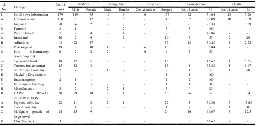
Table 5. Acute intestinal obstruction – etio pathological, sex incidence of 229 cases with morbidity and mortality
S.
No. Etiology
No. of cases
SIMPLE Strangulated Treatment Complication Death
Male Female Male Female Conservative Surgery No. of cases % No. of cases %
I Small bowel obstruction 179 119 35 19 6 6 173 62 34.64 13 7.26
A External hernia 110 81 12 12 5 - 110 32 29.09 10 9.09
i Inguinal 90 76 2 12 - - 90 21 23.33 8 8.89
ii Femoral 3 1 - - 2 - 3 3 100 - -
iii Para umbilical 7 2 4 - 1 - 7 3 42.86 - -
iv Incisional 10 2 6 - 2 - 10 5 50 2 20
B Adhesions 43 22 17 4 - - 37 13 30.23 1 2.32
i Post surgical 19 8 10 1 - 6 13 7 36.84 - -
ii Post inflammatory
(excluding Tb)
6 2 2 2 - 6 6 3 50 - -
iii Congenital band 18 12 5 2 - - 18 3 16.67 1 5.55
C Tuberculous abdomen 15 12 3 - - - 15 8 53.33 1 6.67
D Small bowel volvulus 2 - 1 1 - - 2 1 50 1 50
E Meckel’s Diverticulum 1 - 1 - - - 1 1 100 - -
F Intussusception 2 1 1 - - - 2 2 100 - -
G Ileo sigmoid knotting 1 1 - - - - 1 1 100 - -
H Miscellaneous 5 2 - 2 1 - 5 4 80 - -
II LARGE BOWEL
OBSTRUCTION
50 29 18 2 1 - 50 26 52 7 14
A Sigmoid volvulus 22 11 8 2 1 - 22 8 36.36 3 13.63
B Caecal volvulus 1 1 - - - - 1 - - 1 100
C Malignant growth of
large bowel
24 15 9 - - - 24 16 66.67 3 12.5
D Miscellaneous 3 2 1 - - - 3 2 66.67 - -
[image:4.595.71.510.444.486.2] [image:4.595.43.550.516.767.2]Strangulation
Occlusion of the blood supply to a segment of bowel in addition to obstruction of the lumen is usually referred to as strangulated obstruction
Etiology: secondary to external hernias, intra abdominal
hernias, adhesive band obstruction, volvulus
Clinical features: symptoms begin suddenly. Shock occurs
early in severe strangulation. Colicky pain is present. Fever, elevated WBCs and tachycardia are present. Metabolic acidosis occurs after 12 hours. Rebound tenderness is a distinctive sign.
Mortality is still high in case of strangulated obstruction which can be reduced by
Adopting immediate resuscitative measures
Pre and post operative naso gastric suction
Early surgeries
Resection of dead segment of bowel and adequate lavage of peritoneal cavity with normal saline with or without antibiotics
Small bowel obstruction
In this series the following are major causes of small bowel obstruction (Mark Evers, 2003)
External Hernia (Robert et al.)
A hernia is the protrusion of whole or part of a viscus through an abnormal opening in the wall of its containing cavity.
Etiology: presence of preformed sac; increase in intra
abdominal pressure; weakening of body muscles and tissues
Clinical features: sudden pain occurs initially over the hernia
followed by generalized abdominal pain, paroxysmal in character. Vomiting is forcible and repeated. The hernia is extremely tender, tense and not reducible. In obstructed / strangulated hernia, operative treatment is mandatory.
Adhesions
[image:5.595.314.557.49.190.2]They are the most common cause of intestinal obstruction in western world(Edward et al.)
[image:5.595.314.555.216.351.2]Figure 1. Sigmoid volvulus with distended sigmoid colon
[image:5.595.314.553.376.628.2]Figure 2. Congenital ileal band
Figure 3. Strangulated umbilical hernia
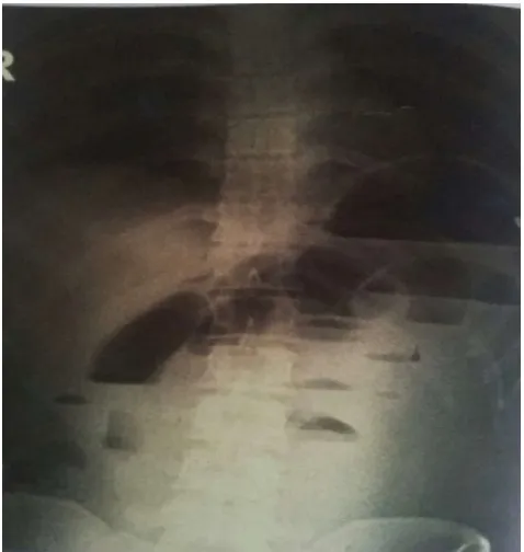
Figure 4. X Rays in acute intestinal obstruction with multiple air fluid levels
Etiology:
1. Ischemic areas -Sites of anastamosis Reperitonealisation of raw areas
2. Foreign bodies -Talc, starch granules, gauze lint, cellulose
[image:5.595.56.271.608.744.2]5. Radiation enteritis 6. Sclerosing peritonitis
Treatment:
1. Non operative treatment with naso-gastric tube drainage combined with iv fluids
2. Adhesiolysis (enterolysis) 3. In cases of recurrent adhesions
a. Noble’s plication operation
b. Child’s – Philip’s transmesentric plication
Prevention
1. Meticulous technique
2. Washing the peritoneal cavity with saline or dextran 3. Avoidance of excessive packing with gauze
4. Covering anastomosis and raw peritoneal surface with greater omentum
5. Leaving raw peritoneal areas unsutured
Obstruction by a band
A band (usually one band only is culpable) is occasionally the cause of acute obstruction.
1. Congenital – obliterated vitello intestinal duct 2. String – thin and fragile, following previous bacterial
peritonitis
3. A portion of greater omentum, adherent to the parietes, constituting an obstructive band
Obstruction by abdominal tuberculosis (Pujari, 2003)
Main source of infection is swallowed sputum
Pathology: commonly the fibrous (plastic) form present as acute intestinal obstruction characterised by production of widespread fibrous adhesion(Das, 1976).
Treatment
1. Anti tuberculous drugs
2. Division of band or adhesiolysis
3. Limited resection of terminal ileum and caecum
4. Stricturoplasty
Volvulus of small intestine(Rebekah et al.)
Meckel’s Diverticulum
It is present in 2% of persons, situated in the anti mesenteric border of small intestine, commonly 2 feet from the ileo caecal valve and it is usually 2 inches long
Intussusception (Iko Bo et al., 1984)
Submucous lipoma is the most frequent benign lesion causing intussusception. It is also due to polyps, papilliferous growth, Meckel’s diverticulum, leiomyoma, etc.
Treatment is invariably surgical
ILEO sigmoid knotting: A loop of ileum wraps around the
base of an elongated sigmoid colon or vice versa. It is a variant of midgut volvulus.
Large bowel obstruction
Sigmoid volvulus (Kelly et al.)
Volvulus is defined as twisting or torsion of a loop of bowel around its related attachments in such a way as to obstruct the lumen of both proximal and distal loop of the segment and a varying degree of impairment of its circulation.
Caecal volvulus (Kelly et al.)
The volvulus is nearly always in a clockwise direction. The first twist obstructs the ascending colon; if a second twist occurs, it obstructs the ileum also.
Large bowel Neoplasms
25% cases of carcinoma from left side of colon present with features of chronic or acute on chronic obstruction because
1. Neoplasms in this situation are of stenosing variety 2. Faecal content is relatively solid
3. Comparatively narrow lumen
Treatment
1. Three staged procedure
2. One stage resection and anastomosis 3. Hartmann’s operation
4. Caecostomy alone
Treatment
1. Naso gastric or gastro intestinal suction drainage (Brolin, 1983)
2. Replacement of fluid and electrolytes 3. Antibiotics
4. Relief of obstruction by surgery a. Exploratory laparotomy
b. Enterostomy, caecostomy, colostomy c. Anastomosis entero enteral, ileo colic d. Resection of bowel
e. Lysis of band or adhesions
f. Enterotomy for gall stones, bezoars
DISCUSSION
External hernia contributes to 61.45% (110 cases) of acute intestinal obstruction. Inguinal hernia contributes to 81.82% (90 cases) of external hernia and 50.2 % of total small bowel obstruction. It ranks FIRST among the causes of acute intestinal obstruction. This is contrast with the world literature, where adhesive obstruction is the principal cause (40 %), and hernia becomes the second cause of obstruction (25%). We have encountered only 3 cases of femoral hernia and 7 cases of par aumbilical hernia and 10 cases of incisional hernia which together contributes only 18.18% of external hernia and 11.17% of total. The high prevalence of inguinal hernia causing obstruction is attributed to
Inadequate knowledge about the disease proper because of low literacy
Reluctance of patient to undergo Elective repair of hernia
High prevalence of Chronic cough (viz. tuberculosis, etc.,)
Even though the prevalence of Inguinal hernia causing obstruction is high, the strangulation rate has come down dramatically to 13.33% (12 cases) due to
Early arrival of patient once obstruction occurs, even though he doesn’t care it before
Early recognition and immediate treatment
In our study, Adhesive obstruction accounts for 18.78% (43 cases) of acute intestinal obstruction ranks second. Among this 44% (19 cases) were due to post-surgical adhesion, 13.95% (6 cases) due to post inflammatory adhesion and 41.86% (18 cases) due to congenital band. Among this strangulation occurred in 9.30% (4 cases) involving the small bowel only. Increased incidence of Cesarean section, hysterectomies and P.I.D accounts for more incidence of adhesive obstruction in females. Still the Abdominal tuberculosis account for 8.38% (15 cases) in total as a cause of small bowel obstruction. Even with advent of potent anti-tuberculous drugs, the reason for failure of improvement of situation is not known to certain. In our study, adhesiolysis could be contemplated in 3 cases because of extensive adhesions. The occurrence of small bowel volvulus was 1.12% (2 cases) in contrast to Agarwal and Misra’s observation who reported 20%. Surprisingly we have come across 2 cases of intussusception and 1 case of Meckel’s diverticulum causing obstruction. One case of small bowel tumour was encountered. Large bowel obstruction, accounted for 21.84% (50 cases) of acute intestinal obstruction. In this series, sigmoid volvulus contributes for large bowel obstruction accounting for 44% (22 cases) and neoplasm of large bowel contributes to 48% (24 cases). The high incidence of Sigmoid volvulus in our place is due to
Diet containing large amount of vegetables, roughages that overloads the colon causing chronic dragging and lengthening of the loop.
Thickened, hypertrophied sigmoid colon
Long freely movable sigmoid loop on a long and freely movable redundant mesosigmoid
The high incidence of neoplasm of large bowel causing obstruction is attributed to
Diet containing fewer amounts of vegetables, fibre.
High fat diet and decreased calcium intake.
The clinical parameters like continuous pain, fever (>37.2`C), tachycardia (>100/min), palpable abdominal mass enable us to detect the presence of strangulation in about 38% of cases, that too mainly in external hernias. Plain x ray abdomen is still useful in diagnosing bowel obstruction. In our study, we were able to get multiple air fluid levels in about 73% cases. We haven’t got positive air fluid level in early stage of obstruction in cases of inguinal hernia. Early recognition by the patient and prompt treatment by surgeon gives good reward and decreases the mortality.
Conclusion
1. Among the causes of acute intestinal obstruction, 78.16% is contributed by Small bowel obstruction and 21.84% by Large bowel obstruction
2. The major cause of acute intestinal obstruction is still External hernia (48.03%) here. Among this, inguinal hernia alone accounts for 81.82% in total.
3. Even though the inguinal hernia causing obstruction is highly prevalent, the strangulation rate comes down dramatically to 13.33% (12 cases).
4. Adhesive obstruction accounts for 18.78% in total, of which the post-surgical adhesion is the major cause
5. Sigmoid volvulus ranks fourth in etiology of acute intestinal obstruction contributing 9.61%, next only to large bowel neoplasms contributing 10.48%
6. Sigmoid volvulus contributes to large bowel obstruction accounting for 44% and neoplasm contributes to 48% 7. Clinical parameters fail to differentiate between simple and
strangulated obstruction exactly
8. Plain x-ray abdomen is a valuable in the diagnosis of the acute obstruction (73%) and hence is considered as minimal investigation before surgery
9. Early surgical intervention and antibiotics has reduced the mortality of the simple bowel obstruction
10.In strangulated obstruction, the mortality rate is still significantly more, due to age, associated diseases and late arrival to hospital
11.Mortality associated with large bowel obstruction is 14% compared to 7.26% with small bowel obstruction
12.Early diagnosis and early surgical intervention is the key to reduce the mortality
REFERENCES
Brolin, R. E. 1983. The role of GI tube decompression in the treatment of Mechanical intestinal obstruction. Am.
Surgeon, 49: 131-137.
Das, P., Shukla, H. S. 1976. Clinical diagnosis of Abdominal Tuberculosis Brit. J. Surg. 63: 941.
Edward, E., Whang, Stanley, W., Ashley and Micheal J. Zinner. small bowel obstruction in Schwartz’s Principles of Surgery, eighth edition, page 1027-1032
same loop of bowel AJR Am. J. Roentgenol., Aug. 161(2):291-5
Iko, Bo, Teal, J. S., Siran, S. M. 1984. Computed tomography of adult intussusception clinical and experimental studies,
AJR 143: 769-772.
Kelly, M. Bullard and David, A. Rothenberger, Volvulus of Sigmoid, Caecal Volvulus, Colonic pseudo-obstruction, ostomies, resections, therapy for rectal and colonic carcinoma in Schwartz’s Principles of Surgery, eighth edition, page 1055-1117
Mark Evers, B. Small intestine obstruction in Sabiston’s Textbook of Surgery, 18th edition, page 1289-1297
Pujari, B.D. 2003. Abdominal Tuberculosis in the ASI Textbook of Surgery, page 501-503
Rebekah, R. White and Danny O.Jacobs, small bowel volvulus in Shackelford’s Surgery of the Alimentary Tract, sixth edition, page 1035-1041
Robert, J. Fitzgibbons, Jr. Charles J. Filipi and Thomas H. Quinn. Hernia accident (incarceration, bowel obstruction, strangulation), groin hernias in women, Schwartz’s Principles of Surgery, eighth edition, page1355-1358, 1390