ROLE OF PLATELET RICH FIBRIN (PRF) IN THE REGENERATION OF OSSEOUS
1
Dr. Nagamalleswar Rao,
4
Dr. Kartheek Gandikota
1,2
Asst.professor, Dept of Oral and Maxillofacial Surgery, KIMS Dental College and Hospital, Amalapuram
3Professor and HOD, Dept of Oral and Maxillofacial Surgery, GITAM Dental College and Hospital, Vishakapatnam
4
Asst., Professor, Dept of Oral
Asst Professor Department of Oral Pathology Kims Dental College Amalapuram
ARTICLE INFO ABSTRACT
Background:
Rich Fibrin (PRF) in osseous defects. A prospective study was performed to determine if periapical osseous defects treated with platelet
graft material. Methods
evaluation of healing was done at 1month, 3 months and 6 months. Histologic evaluation was done at the end of 6 months.
Results
showed resorption of the graft material and by the end of 6 months all the defects showed osseous fill and matured bone formation.
Conclusion defects.
Copyright © 2018, Rao et al. This is an open access distribution, and reproduction in any medium, provided
INTRODUCTION
Osseous regeneration in the periapical defects as a result of trauma orpathology in oral and maxillofacial region has been a challenge. Usually it takes 9-12 months for complete osseous regeneration in the defects after peri apical surgery. Keeping in view of the prolonged time for healing, different bio materials are being tried to accelerate the regenerative process. Though the use of autogenous bone has remained the gold standard in restoring bone defects, there are some limitations like limited quantity available, need for second surgical site, prolonged operative times and donor site morbidity (
2005). To overcome these difficulties alloplastic bone
substitutes have come into use. The most commonly used is synthetic hydroxyapatite due to its chemical composition being similar to that of human bone, its non- toxic nature, its high chemical stability with less inflammation and antigenic reactions. Another important property is that the microstructure can be controlled to promote formation of pores that allow
*Corresponding author: Dr. Poornima Sowjanya,
Asst., professor, Dept of Oral and Maxillofacial Surge Dental College and Hospital, Amalapuram.
ISSN: 0975-833X
Article History:
Received 22nd December, 2017 Received in revised form 19th January, 2018
Accepted 06th February, 2018
Published online 28th March, 2018
Citation: Dr. Nagamalleswar Rao, I., Dr. Poornima Sowjanya, Dr. Satya Bhushan, N.V.V., Dr. Kartheek Gandikota and Dr. Ratnakar Bale “Role of platelet rich fibrin (prf) in the regeneration of osseous defects in the jaws
Key words:
Platelet Growth Factors, Wound Healing, Hydroxyapatite, Platelet rich Fibrin.
RESEARCH ARTICLE
ROLE OF PLATELET RICH FIBRIN (PRF) IN THE REGENERATION OF OSSEOUS
DEFECTS IN THE JAWS
, I.,
2,*Dr. Poornima Sowjanya,
3Dr. Satya Bhushan, N.V.V.
Kartheek Gandikota and Dr. Ratnakar Bale
Asst.professor, Dept of Oral and Maxillofacial Surgery, KIMS Dental College and Hospital, Amalapuram
Professor and HOD, Dept of Oral and Maxillofacial Surgery, GITAM Dental College and Hospital, Vishakapatnam
, Dept of Oral Pathology, KIMS Dental College and Hospital, Amalapuram
Asst Professor Department of Oral Pathology Kims Dental College Amalapuram
ABSTRACT
Background: The healing potential of platelet growth factors has generated interest in using Platelet Rich Fibrin (PRF) in osseous defects. A prospective study was performed to determine if periapical osseous defects treated with platelet-rich fibrin exhibit enhanced healing when along alloplastic bone graft material.
Methods: Periapical defects in ten patients were treated with PRF and hydroxyapatite. Radiological evaluation of healing was done at 1month, 3 months and 6 months. Histologic evaluation was done at the end of 6 months.
Results: Bone Healing was seen in the PRF treated sites. By the end of 3 months all the defects showed resorption of the graft material and by the end of 6 months all the defects showed osseous fill and matured bone formation.
Conclusion: PRF along with bone graft material can be used for rapid rege defects.
access article distributed under the Creative Commons Attribution License, the original work is properly cited.
Osseous regeneration in the periapical defects as a result of trauma orpathology in oral and maxillofacial region has been a 12 months for complete osseous regeneration in the defects after peri apical surgery. Keeping in view of the prolonged time for healing, different bio materials are being tried to accelerate the regenerative process. Though e use of autogenous bone has remained the gold standard in restoring bone defects, there are some limitations like limited quantity available, need for second surgical site, prolonged
(Thorwarth et al.,
vercome these difficulties alloplastic bone substitutes have come into use. The most commonly used is synthetic hydroxyapatite due to its chemical composition being toxic nature, its high inflammation and antigenic reactions. Another important property is that the microstructure can be controlled to promote formation of pores that allow
Dr. Poornima Sowjanya,
Asst., professor, Dept of Oral and Maxillofacial Surgery, KIMS
migration of blood vessels and bone tissues into the material enhancing the osteoconductive property and early resorption of the substitute thereby fastening bone regeneration Shankar, 2011). The next step in osseous regeneration is tissue engineering that is achieved by introduction of platelet concentrates, which utilize the inherent property of platelets to release various growth factors thereby accelerating the healing process. One such recent innovation in dentistry is the preparation and use of platelet rich plasma (PRP), which is a concentration of platelets and growth factors found in platelets. PRP has enjoyed a great increase in popularity in the Oral and Maxillofacial Surgical practice. It is very well documented that application of PRP enhances healing in various procedures
(Shobha et al., 2011). A new platelet concentrate was
introduced in France in the year 1978 by Choukroun called Platelet- rich Fibrin (PRF) which has the ad
way of activation, prolonged release of growth factors, slow polymerization and simplified production protocols
al., 2006). This has encouraged us to radiographicallyevaluate
the efficacy of the platelet rich fibrin along with hydroxyapatite in the healing of periapical defects.
International Journal of Current Research
Vol. 10, Issue, 03, pp.66622-66627, March, 2018
Dr. Nagamalleswar Rao, I., Dr. Poornima Sowjanya, Dr. Satya Bhushan, N.V.V., Dr. Kartheek Gandikota and Dr. Ratnakar Bale Role of platelet rich fibrin (prf) in the regeneration of osseous defects in the jaws”, International Journal of Current Research
Available online at http://www.journalcra.com
z
ROLE OF PLATELET RICH FIBRIN (PRF) IN THE REGENERATION OF OSSEOUS
Dr. Satya Bhushan, N.V.V.,
Asst.professor, Dept of Oral and Maxillofacial Surgery, KIMS Dental College and Hospital, Amalapuram
Professor and HOD, Dept of Oral and Maxillofacial Surgery, GITAM Dental College and Hospital, Vishakapatnam
KIMS Dental College and Hospital, Amalapuram
Asst Professor Department of Oral Pathology Kims Dental College Amalapuram
The healing potential of platelet growth factors has generated interest in using Platelet-Rich Fibrin (PRF) in osseous defects. A prospective study was performed to determine if periapical
healing when along alloplastic bone
Periapical defects in ten patients were treated with PRF and hydroxyapatite. Radiological evaluation of healing was done at 1month, 3 months and 6 months. Histologic evaluation was done at
eated sites. By the end of 3 months all the defects showed resorption of the graft material and by the end of 6 months all the defects showed osseous fill
PRF along with bone graft material can be used for rapid regeneration of osseous
License, which permits unrestricted use,
migration of blood vessels and bone tissues into the material enhancing the osteoconductive property and early resorption of the substitute thereby fastening bone regeneration (Ravi The next step in osseous regeneration is tissue engineering that is achieved by introduction of platelet concentrates, which utilize the inherent property of platelets to release various growth factors thereby accelerating the healing ent innovation in dentistry is the preparation and use of platelet rich plasma (PRP), which is a concentration of platelets and growth factors found in platelets. PRP has enjoyed a great increase in popularity in the Oral and practice. It is very well documented that application of PRP enhances healing in various procedures A new platelet concentrate was introduced in France in the year 1978 by Choukroun called rich Fibrin (PRF) which has the advantage of natural way of activation, prolonged release of growth factors, slow
polymerization and simplified production protocols (Dohan et
This has encouraged us to radiographicallyevaluate the efficacy of the platelet rich fibrin along with hydroxyapatite in the healing of periapical defects.
INTERNATIONAL JOURNAL OF CURRENT RESEARCH
MATERIALS AND METHODS
This study was conducted in the division of Oral and Maxillofacial Surgery, GITAM dental college and hospital. 10 patients who reported to the outpatient clinics of the Department of Oral and Maxillofacial Surgery presenting with the Periapical defects were selected for the study after Ethical committee approval.
Inclusion criteria
Patients above 15 years of age were selected for the study.
Exclusion criteria
Patients suffering from any systemic disorders
Patients with blood dyscrasiasis.
Patients on antiplatelet/ anticoagulant therapy.
Patients with a history of recent myocardial infarction
Pregnant women.
A detailed case history of the patient, informed consent was obtained from the patients regarding the surgery as well as the use of platelet rich fibrin and hydroxyapatite. In the study, all the lesions were diagnosed as radicular cysts except for one case which was diagnosed as dentigerous cyst. The offending teeth were saved conservatively by performing root canal therapy among 7 patients with radicular cysts while in remaining 3 patients offending tooth was extracted due to poor prognosis. The surgical procedure included enucleation of the cystic lining and placement of platelet rich fibrin along with hydroxyapatite material.
Preparation of Platelet Rich Fibrin
The PRF preparation protocol is very simple and armamentarium required is same as that of PRP. Around 5 ml of whole venous blood is collected in each of the two sterile vacutainer tubes of 6 ml capacity without anticoagulant. The vacutainer tubes are then placed in a centrifugal machine at 3000 revolutions per minute (rpm) for 10 minutes, after which it settles into three layers: red lower fraction containing red blood cells, upper straw coloured cellular plasma and the middle fraction containing the fibrin clot.
The upper straw coloured layer is then removed and middle fraction is collected which is the PRF. The mechanism which is followed here is that, fibrinogen which is initially concentrated in the high part of the tube, combines with the circulating thrombin due to centrifugation, to form fibrin. A fibrin clot is then obtained in the middle of the tube, just between the red corpuscles at the bottom and acellular plasma at top. Platelets are trapped massively in the fibrin meshes. The success of this technique entirely depends on the speed of blood collection and transfer to the centrifuge. By driving out the fluids trapped in the fibrin matrix, we can obtain resistant autologous fibrin membranes.
Surgical procedure
Under aseptic conditions local anaesthesia is administered (2%
lidocaine with 1:80000 adrenaline), full thickness
mucoperiosteal flap is raised from the buccal or palatal side, and the defect is exposed. Cystic lining was removed and wound debridement done. Once hemostasis is acheived, PRF is adapted on to the walls and base of the defect and the remaining cavity is filled with hydroxyapatite bone graft. Overfilling and under filling is avoided, as it precludes proper flap closure, there by retarding healing and possibly resulting in loss of graft material.
The wound is closed primarily with interrupted sutures using 3-0 black silk. Postoperative antibiotics and analgesics are prescribed. The patients are recalled on the next day of surgery for check-up and after 7 days for suture removal. Follow up was done at the end of 1, 3 and 6 months. Intra oral periapical radiographs were taken and assessed for bone regeneration based on blending of the bone margins and the presence of the trabecular bone formation.
RESULTS
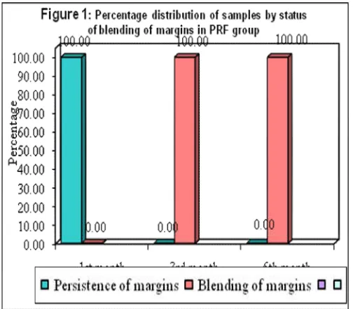
Ten patients were treated with PRF. All the patients were followed for 6 months. Intra oral periapical radiographs were obtained at 1 month, 3 months and 6 months to evaluate the blending of margins and the presence of trabecular bone formation. All the variables were tabulated and analyzed using Mann Whitney U test at 5% level of significance. results are shown in table-1,Fig 1,2. The percentage distribution of status of Blending of margins and Presence of trabecular bone in PRF group are presented in the following Figures.
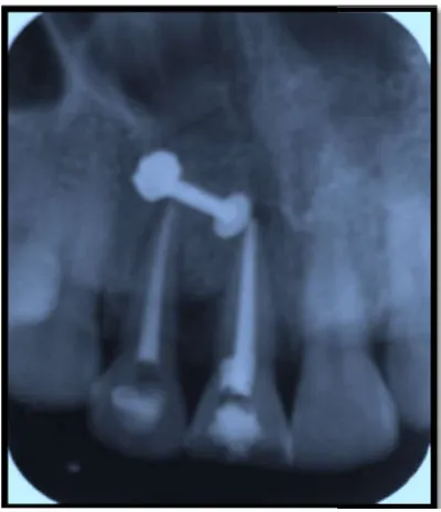
[image:2.595.309.561.556.777.2]Placement of prf, hydroxyapatite in periapical defect
Table 1. Distribution of samples by status of blending margins and Presence of trabecular bone
1st month % 3rd month % 6th month %
Blending of margins Persistence of margins 30 100.0 6 20.0 0 0.0
Blending of margins 0 0.0 24 80.0 30 100.0
Presence of trabecular bone Absence of trabecular bone 30 100.0 15 50.0 0 0.0
Presence of trabecular bone 0 0.0 15 50.0 30 100.0
Fig. 3. pre o.p IOPA showing radiolucency i.r.t 11,12
Fig. 4. Showing osseous defect i.r.t 11,12
[image:3.595.327.536.525.767.2]66624 International Journal of Current Resear
[image:3.595.42.284.545.767.2]Fig. 3. pre o.p IOPA showing radiolucency i.r.t 11,12
Fig. 4. Showing osseous defect i.r.t 11,12
Fig. 5. PRF prepared
Fig 6. Showing placement of PRF+Hydroxyapatite
Fig 7. immediate post o.p IOPA showing graft in place International Journal of Current Research, Vol. 10, Issue, 03, pp.66622-66627, March
Fig. 5. PRF prepared
placement of PRF+Hydroxyapatite
Fig. 8. IOPA of 3rd month follow up
Fig 9. IOPA of 6th month follow up
DISCUSSION
Bone is a specialized form of connective tissue that provides support and protection of vital structures. Even though bone has a good regenerative capacity compared to other tissues, this quality is impaired in cases of decreased healing capacity of the host owing to various systemic or local causes. In such cases the use of bone grafts is indicated to promote healing and
bone regeneration (Reddy et al., 2010). Autogenous bone is
still regarded as a gold standard due to its osteogenic potential. However, this graft has limitations such as limited quantity, secondary surgical site and additional morbidity. To overcome these limitations, bone substitute materials came into existence and are widely used. Recent research in this field suggested that better osteoconductivity would be achieved if synthetic materials could resemble bone in composition and morphology
(Thorwarth et al., 2005).
66625 Dr. Nagamalleswar Rao, Role
month follow up
month follow up
Bone is a specialized form of connective tissue that provides protection of vital structures. Even though bone has a good regenerative capacity compared to other tissues, this quality is impaired in cases of decreased healing capacity of the host owing to various systemic or local causes. In such e grafts is indicated to promote healing and Autogenous bone is still regarded as a gold standard due to its osteogenic potential. However, this graft has limitations such as limited quantity, and additional morbidity. To overcome these limitations, bone substitute materials came into existence and are widely used. Recent research in this field suggested that better osteoconductivity would be achieved if synthetic in composition and morphology
One such material is the synthetic hydroxyapatite, whose chemical composition is similar to human bone. It is nontoxic, has high chemical stability, and causes less inflammatory and antigenic reaction. Another important property is that its microstructure can be controlled to promote the formation of pores that can allow the migration of blood vessels and bone
tissues into the material.2This provoked us to use synthetic
hydroxyapatite (sybograf) of 20
material. We observed less inflammatory reaction, good resorption of the graft material by the end of 3
reduction in the size of the lesion as observed by Showkat
mamun et al (2009). We found it biocompatible with
osteoconductive properties as experienced by Ravi Shankar
al (2011), Mao-Shaun Hung et al
in their studies. Tissue engineering has become a major area of research in bone regeneration of periapical defects in the maxillofacial region.The process of bone regeneration in periapical defects has been greatly enhanced by the identification of growth factors present in platelets. Platelet concentrates prepared from patient’s own blood is something that is new and the strategy i
bone regeneration by enhancing the effect of growth factors
contained in platelets (Reddy, 2010
concentrates, platelet rich plasma (PRP) and platelet rich fibrin (PRF) were introduced into use in Oral and Ma
Surgery for enhancing bone regeneration. Growth factors like platelet derived growth factor (PDGF
growth factor β (TGFβ), insulin like growth factor (IGF), epidermal growth factor are released from platelet α granules. Among them TGF β and PDGF AB are the typical two with the highest amounts, promoting the healing of soft tissues and bone through stimulation of collagen production to improve wound strength and initiation of callus formation
2009). Therefore, if growth factors in platelets can effectively function in the osseous defect, growth factor induced acceleration of healing may be expected. Our experience with both platelet rich plasma (PRP) and platelet rich fibrin (PRF) showed improved results with platelet rich fibrin than with platelet rich plasma. This encouraged us to continue our study using platelet rich fibrin.
Platelet rich fibrin was first developed in France by Choukroun
et al for specific purposes in Oral and Maxillofacial Sur The PRF production protocol is very simple which requires neither anticoagulant nor bovine thrombin. The absence of anticoagulant implies the activation of platelets in blood sample in contact with the tube walls and the release of the coagulation cascades within a few minutes. The success of this technique entirely depends on the speed of blood collection and transfer to the centrifuge. Quick handling is the only way to obtain a clinically useful PRF clot. PRF has the characteristic of polymerizing n
centrifugation. This slow polymerization mode confers a particularly favourable physiologic architecture to support the
healing process (Dohan et al
protocol by choukroun for preparing PRF
Handling the PRF clot is easy and can be easily adapted to the
walls of the defect. Dohan et al
which platelet cytokine quantification was carried out by ELISA to understand the new biomaterial. The results would imply that PRF would be able to progressively release cytokines during fibrin matrix remodeling, such a mechanism might explain the clinically observed healing. Similar results
were obtained by Lee et al (2012) in their animal study which
showed that more amount of bone regeneration occurred in a
Role of platelet rich fibrin (prf) in the regeneration of osseous defects in the jaws
One such material is the synthetic hydroxyapatite, whose chemical composition is similar to human bone. It is nontoxic, has high chemical stability, and causes less inflammatory and n. Another important property is that its microstructure can be controlled to promote the formation of pores that can allow the migration of blood vessels and bone This provoked us to use synthetic hydroxyapatite (sybograf) of 200-300 microns as a bone graft material. We observed less inflammatory reaction, good
resorption of the graft material by the end of 3rd month and
reduction in the size of the lesion as observed by Showkat We found it biocompatible with good
osteoconductive properties as experienced by Ravi Shankar et
et al (2010), Rajni Ranjan (2011) Tissue engineering has become a major area of research in bone regeneration of periapical defects in the facial region.The process of bone regeneration in periapical defects has been greatly enhanced by the identification of growth factors present in platelets. Platelet concentrates prepared from patient’s own blood is something that is new and the strategy is to accelerate the initiation of bone regeneration by enhancing the effect of growth factors
Reddy, 2010). Two platelet
concentrates, platelet rich plasma (PRP) and platelet rich fibrin (PRF) were introduced into use in Oral and Maxillofacial Surgery for enhancing bone regeneration. Growth factors like platelet derived growth factor (PDGF-AB), transforming growth factor β (TGFβ), insulin like growth factor (IGF), epidermal growth factor are released from platelet α granules. hem TGF β and PDGF AB are the typical two with the highest amounts, promoting the healing of soft tissues and bone through stimulation of collagen production to improve
wound strength and initiation of callus formation (He et al.,
. Therefore, if growth factors in platelets can effectively function in the osseous defect, growth factor induced acceleration of healing may be expected. Our experience with both platelet rich plasma (PRP) and platelet rich fibrin (PRF) sults with platelet rich fibrin than with platelet rich plasma. This encouraged us to continue our study
Platelet rich fibrin was first developed in France by Choukroun for specific purposes in Oral and Maxillofacial Surgery. The PRF production protocol is very simple which requires neither anticoagulant nor bovine thrombin. The absence of anticoagulant implies the activation of platelets in blood sample in contact with the tube walls and the release of the scades within a few minutes. The success of this technique entirely depends on the speed of blood collection and transfer to the centrifuge. Quick handling is the only way to obtain a clinically useful PRF clot. PRF has the characteristic of polymerizing naturally and slowly during centrifugation. This slow polymerization mode confers a particularly favourable physiologic architecture to support the
et al., 2006). We used standard
protocol by choukroun for preparing PRF (Dohan et al., 2006).
Handling the PRF clot is easy and can be easily adapted to the
et al. (2006)conducted a study in which platelet cytokine quantification was carried out by ELISA to understand the new biomaterial. The results would y that PRF would be able to progressively release cytokines during fibrin matrix remodeling, such a mechanism might explain the clinically observed healing. Similar results (2012) in their animal study which nt of bone regeneration occurred in a
[image:4.595.62.262.339.571.2]peri implant defect filled with PRF than unfilled defects. There are no complications like postoperative infection in any of our cases. This is due to the immune organizing mode of PRF as
observed by Dohan et al. (2006). PRF when used along with
hydroxyapatite not only accelerates hard tissue healing but also
increases the density of the bone formed. Choukroun et al.
(2006), Horowitz et al (2009); Dohan et al (2008), observed
denser bone formation in their studies on sinus augmentation
for implant placement.It has been reported by Pradeep et al.
(2012) that combination of HA with PRF resulted in better results when used in intrabony defects. For this reason, we chose HA, as that it could enhance the effects of PRF by maintaining the space for tissue regeneration to occur, as well as by exerting an osteoconductive effect in the bony defect. Bonegrafts alone without a blood clot or angiogenic factors are unlikely to be capable of promotion of periapical wound healing. PRF is in the form of a platelet gel and can be used in conjunction with bone grafts, which offers several advantages including promoting wound healing, bone growth and maturation, graft stabilization, wound sealing, and hemostasis and improving the handling properties of graft materials
(Dohan et al., 2009). The results of our study showed that
blending of margins was observed by the end of 3 months in 100.00% of the cases. There was a significant difference in
blending of margins beween 1st and 3rd months (p<0.05).
Between 3rd and 6th month there is no significant difference in
blending of margins (p>0.05). At the end of 3 months 50% of the cases showed trabecular bone formation and at the end of 6 months 100% of the cases showed trabecular bone formation
which is in accordance with the results of Ganga Prasad et al
(2013) who observed a definitive improvement in regeneration of bone after third molar surgery in cases treated with PRF. This increase in bone density signifies the use of PRF as a valid method in accelerating hard tissue regeneration.
Volker Gabling et al (2009)in their study on the release of
growth factors observed that the quantity of growth factors released from PRF are lower than that from PRP. A possible explanation for the different cytokine content of both blood products could be the lack of standardized production protocol
for PRF. Zhu et al. (2006) compared the effects of PRP and
PRF on the tissue engineered bone formation and concluded that PRF matrix allows more osteogenic activity than PRP as the PRF matrix allows better adhesion of bone morphogenetic protein (BMP) or bone mesenchymal stem cells(BMSC’s) and has the property of slow degradation which will allow retention of growth factors and cells for long time. As there are less clinical studies to evaluate bone regeneration in periapical osseous defects using PRF, we evaluated the efficacy of this second generation platelet concentrate. In our study PRF
showed good amount of blending of margins at the end of 3rd
month. 15 out of 30 cases showed trabecular bone formation at
the end of 3rd month. At the end of 6th month all the cases
showed complete trabecular bone formation. There was a
significant difference between 1st and 3rd months and 3rd and
6th months with respect to trabecular bone formation (p< 0.05).
Conclusion
The intraoral osseous defects usually takes a minimum period of 9-12 months to heal completely. Bone regeneration in osseous defects can be accelerated by using recently developed second generation platelet concentrates like platelet rich fibrin.
Our experience with the use of platelet rich fibrin showed good results in terms of healing of the osseous defects, handling of the material, ease of preparation of the material.
REFERENCES
1. Choukroun, J., Diss, A., Simonpieri, A. and Girard, M.O.
2006. Platelet-rich fibrin (PRF): A second-generation platelet concentrate.Part IV: Clinical effects on tissue
healing. Oral Surg OralMed Oral Pathol Oral
RadiolEndod. 101:E56-60.
2. Choukroun, J., Diss, A., Simonpieri, A., and Girard, M.O.
2006. Platelet-rich fibrin (PRF): A second-generation platelet concentrate.Part V: Histologic evaluations of PRF
effects on bone allograft maturation in sinus lift. Oral Surg
Oral Med Oral Pathol Oral Radiol Endod. 101:299-303.
3. Dohan, D. M., Choukroun, J., Diss, A. and Dohan, S. L.
2006. Platelet-rich fibrin (PRF): A second-generation platelet concentrate.Part I: Technological concepts and
evolution. Oral Surg Oral Med Oral Pathol Oral Radiol
Endod. 101: E37-44.
4. Dohan, D.M., Choukroun, J., Diss, A. and Dohan, S.L.
2006. Platelet-rich fibrin (PRF): A second-generation platelet concentrate. Part II: Platelet-related biologic
features. Oral SurgOral Med Oral Pathol Oral Radiol
Endod. 101: E45-50.
5. Dohan, D.M., Choukroun, J., Diss, A., Dohan, S.L. 2006.
Platelet-rich fibrin (PRF): A second-generation platelet concentrate. Part III: Leucocyte activation: A new feature
for platelet concentrates? Oral Surg Oral Med Oral Pathol
Oral Radiol Endod. 101: E51-5.
6. Dohan, D.M., Ehrenfest, Diss A., Odin, G., Doglioli, P.
2009. In vitro effects of Choukroun’s PRF (platelet-rich
fibrin) on human gingival fibroblasts, dermal
prekeratinocytes,preadipocytes, and maxillofacial
osteoblasts in primary Cultures. Oral Surg Oral Med Oral
Pathol Oral RadiolEndod. 108: 341-352.
7. Gabling, V.L., Açil, Y., Springer, I.N. 2009. Platelet-rich
Plasma and Platelet-rich fibrin in human cell culture. Oral
Surg Oral Med Oral Pathol Oral Radiol Endod., 108:
48-55.
8. He, L., Lin, Y., Hu, X., Yu, Zhang. 2009. A comparative
study of platelet-rich fibrin (PRF) andplatelet-rich plasma (PRP) on the effect of proliferationand differentiation of rat
osteoblasts in vitro. Oral Surg Oral Med Oral Pathol Oral
Radiol Endod. 108: 707-713.
9. Horowitz, R.A., Corso, M.D. and Prasad, H.S. 2009. Sinus
floor augmentation with simultaneous implant placement using choukroun’s PRF as a sole grafting material: A
radiologic and histologic study at 6 months. J Periodontol.,
80: 2056-2064.
10.Kim, B.J., Kwon, T.K., Baek, H.S. 2012. A comparative
study of the effectiveness of sinus bone grafting with recombinant human bone morphogenetic protein 2–coated
tricalciumphosphate and platelet-rich fibrin–mixed
tricalcium phosphate in Rabbits. Oral Surg Oral Med Oral
Pathol Oral Radiol., 113:583-592.
11.Lee, J.W., Kim, S.G., Kim, Y.J. 2012. Restoration of a
peri-implant defect by platelet-rich fibrin. Oral Surg Oral
Med Oral Pathol Oral Radiol. 113:459-463.
12.Mamun, S., Akhter, M., Molla, M.R. 2009. Bone grafts in
jaw cysts- Hydroxyapatite and allogenic bone- A
comparative study. BSMMU J., 2(1): 25-30.
13.Prasad, B.G., Rao, S.G., Nagesh, K.S. 2013. Bone regeneration in extraction sockets with autologous platelet
rich fibrin gel. J Maxillofac Oral Surg., 12(1): 11-16.
14.Ranjan, R. 2011. Fate of hydroxyapatite crystals used as
bone graft substitute in benign lytic lesions of long bones.
The Internet Journal of Orthopaedic Surgery, 18(2).
15.Ravi Shankar, Deepak Singh, Ravi Jain. 2011. Bone
regeneration in osseous defects using hydroxyapatite graft and the extent of ossification in osseous defects treated
without grafts: A comparative evaluation. J Maxillofac
oral surg. 10(2): 123-126.
16.Reddy, P., Nagaveni, Shankar U. 2010. Efficacy of PRP in
bone regeneration after cyst enucleation in pediatric
patients – A clinical study. Journal of clinical pediatric
dentistry. 35(1): 81-87.
17.Schlege, K.A., Zimmermann, R., Thorwarth, M. 2007.
Sinus floor elevation using autogenous bone or bone
substitute combined with platelet-rich plasma. Oral Surg
Oral Med Oral Pathol Oral Radiol Endod. 104: e15-e25.
18.Shobha, P., Aditi, T.2011. Platelet Concentrates: Past,
Present and Future. J. Maxillofac. Oral Surg., 10(1):45-49.
19.Thorwarth, M., Stefan, S.M., Kessler, P. 2005. Bone
regeneration in osseous defects using a resorbable nano
particular hydroxyapatite. J Oral Maxillofac surg., 63:
1626-1633.
20.Zhu, S.J., Choi, B.H., Jung, J.H. 2006. A comparative
histologic analysis of tissue-engineered bone using
platelet-rich plasma and platelet-enplatelet-riched fibrin glue. Oral Surg
Oral Med Oral Pathol Oral Radiol Endod. 102: 175-179.