TRAUMATOLOGICAL APPLICATION OF 3D PRINTED MODEL OF INJURIED PELVIS
1
Battiato, C.,
2Sirabella
1
UOC Ortopedia e Traumatologia, Ospedale Mazzoni, Ascoli
2Clinica Ortopedica, Scuola di Specializzazione in Ortopedia
ARTICLE INFO ABSTRACT
Introduction:
tomography (CT), are promising technologies that can facilitate preoperative programming and intraoperative implementation, thus obtaining better results in complex fractu
possible not only to simulate CAD reductions but also to design customized plates. In the case that we treated, a plate was made of two pieces (like a puzzle
without removing the red complete it.
Materials and Methods:
of the lesion" and to obtain a custom synthesis tool made in a Results:
surgical approach and the production of a custom Discussion:
the reduction maneuvers and the presence of a custom
blood loss, risk of infection and use of ionizing radiation were reduced if compared to a case approached b
Conclusions:
diastasis of pubic symphysis.
Copyright©2017, Battiato et al. This is an open access article distributed under the Creative Commons Att distribution, and reproduction in any medium, provided the original work is properly cited.
INTRODUCTION
The pelvis is a complex three-dimensional structure that plays a key role in stabilizing the body and distributing the load from the axial skeleton to the lower limbs; it also has a valuable protective function against delicate visceral and vascular nervous structures. Fractures of the pelvis represent a relatively rare entity, but the increasing incidence, along with the characteristics of high morbidity and mortality associated with it, make them an important chapter in traumatology.
incidence of pelvis fractures is estimated at around 3% of all fractures. Among the politraumatized patients, the incidence increased to about 25% and, in the group of road accidents, a fracture of the pelvis was detected in 42% of individuals. Pelvic fractures are generally due to high-energy traumas more often due to car accidents (70-80%), falling (10 crushing trauma (5-10%). Therefore, a pelvis injury should be considered as an important traumatic index, as long as any associated lesions (central nervous system injuries and visceral lesions) can be ruled out. The mortality rate for s
*Corresponding author: Giovannini, F.
Clinica Ortopedica, Scuola di Specializzazione in Ortopedia e Traumatologia, UNIVPM, Ancona.
ISSN: 0975-833X
Article History: Received 07th July, 2017 Received in revised form 05th August, 2017
Accepted 12th September, 2017 Published online 31st October, 2017
Available online at http://www.journal
Citation: Battiato, C., Sirabella, F. S., Smakaj, A. and Giovannini, F.
International Journal of Current Research, 9, (10), 59 Key words:
Trauma surgery, 3D printing, Pelvis.
RESEARCH ARTICLE
TRAUMATOLOGICAL APPLICATION OF 3D PRINTED MODEL OF INJURIED PELVIS
Sirabella, F. S.,
1Smakaj, A. and *
,2Giovannini
UOC Ortopedia e Traumatologia, Ospedale Mazzoni, Ascoli Piceno
Clinica Ortopedica, Scuola di Specializzazione in Ortopedia e Traumatologia, UNIVPM, Ancona
ABSTRACT
Introduction: Three-dimensional (3D) reconstruction and rapid prototyping, starting from computed tomography (CT), are promising technologies that can facilitate preoperative programming and intraoperative implementation, thus obtaining better results in complex fractu
possible not only to simulate CAD reductions but also to design customized plates. In the case that we treated, a plate was made of two pieces (like a puzzle-plate) in order to be able to position a first plate without removing the reduction instruments and the second plate after the partial synthesis obtained to complete it.
Materials and Methods: We decided to use this method to increase the evaluation of the "personality of the lesion" and to obtain a custom synthesis tool made in a case of diastasis of the pubic symphysis. Results: The solid model (3D printed model) allowed accurate preoperative planning, facilitating the surgical approach and the production of a custom-made plate.
Discussion: We reduced surgical time thanks to the perfect knowledge of the structure of the lesion, the reduction maneuvers and the presence of a custom-made plate. In addition, soft tissue damage, blood loss, risk of infection and use of ionizing radiation were reduced if compared to a case approached by the standard method.
Conclusions: The method described has been extremely useful in the surgical treatment of the diastasis of pubic symphysis.
is an open access article distributed under the Creative Commons Attribution License, which distribution, and reproduction in any medium, provided the original work is properly cited.
dimensional structure that plays stabilizing the body and distributing the load from the axial skeleton to the lower limbs; it also has a valuable protective function against delicate visceral and
vascular-Fractures of the pelvis represent a relatively the increasing incidence, along with the characteristics of high morbidity and mortality associated with it, make them an important chapter in traumatology. The total incidence of pelvis fractures is estimated at around 3% of all traumatized patients, the incidence increased to about 25% and, in the group of road accidents, a fracture of the pelvis was detected in 42% of individuals. energy traumas more falling (10-30%), Therefore, a pelvis injury should be considered as an important traumatic index, as long as any associated lesions (central nervous system injuries and visceral The mortality rate for severe exposed
Clinica Ortopedica, Scuola di Specializzazione in Ortopedia e
pelvic fracture ranges from 10% up to 50%; risk factors affecting an increase in mortality include the age of the patient and the severity of the trauma score, the presence of associated cranial or visceral lesions, blood loss, hypotension, coagulopathy and instability of the fracture.
often linked to haemorrhage or closed cranial lesions, while late mortality is secondary to sepsis or multisistemic organ failure (MOF). Clinical and radiographic evaluation of the pelvic ring, based on the ident
instability or instability, is the starting point for all subsequent decisions. Fracture classification schemes should help clinicians and researchers to identify and describe a lesion, plan their treatment, and predict their out
popular schemes currently in use are the Tile system and the Young-Burgess system. The Tile system combines directional patterns of pelvic destruction with radiographic signs of stability or instability. The Tile system classifies the destruction of the pelvic ring as stable (type A), rotationally unstable (type B), and unstable both rotational and vertical (type C). These main types are further subclassificated according to the specific signs of the lesion.
International Journal of Current Research
Vol. 9, Issue, 10, pp.59298-59304, October, 2017 Available online at http://www.journalcra.com
Battiato, C., Sirabella, F. S., Smakaj, A. and Giovannini, F. 2017. “Traumatological application of 3 59298-59304.
TRAUMATOLOGICAL APPLICATION OF 3D PRINTED MODEL OF INJURIED PELVIS
Giovannini, F.
Piceno
e Traumatologia, UNIVPM, Ancona
dimensional (3D) reconstruction and rapid prototyping, starting from computed tomography (CT), are promising technologies that can facilitate preoperative programming and intraoperative implementation, thus obtaining better results in complex fractures. Moreover, it is possible not only to simulate CAD reductions but also to design customized plates. In the case that we plate) in order to be able to position a first plate uction instruments and the second plate after the partial synthesis obtained to
We decided to use this method to increase the evaluation of the "personality case of diastasis of the pubic symphysis. The solid model (3D printed model) allowed accurate preoperative planning, facilitating the
perfect knowledge of the structure of the lesion, made plate. In addition, soft tissue damage, blood loss, risk of infection and use of ionizing radiation were reduced if compared to a case
The method described has been extremely useful in the surgical treatment of the
ribution License, which permits unrestricted use,
fracture ranges from 10% up to 50%; risk factors affecting an increase in mortality include the age of the patient and the severity of the trauma score, the presence of associated cranial or visceral lesions, blood loss, hypotension, ability of the fracture. Early mortality is often linked to haemorrhage or closed cranial lesions, while late mortality is secondary to sepsis or multisistemic organ Clinical and radiographic evaluation of the pelvic ring, based on the identification of the degree of instability or instability, is the starting point for all subsequent Fracture classification schemes should help clinicians and researchers to identify and describe a lesion, plan their treatment, and predict their outcomes. The two most popular schemes currently in use are the Tile system and the The Tile system combines directional patterns of pelvic destruction with radiographic signs of The Tile system classifies the estruction of the pelvic ring as stable (type A), rotationally unstable (type B), and unstable both rotational and vertical These main types are further subclassificated according to the specific signs of the lesion.
INTERNATIONAL JOURNAL OF CURRENT RESEARCH
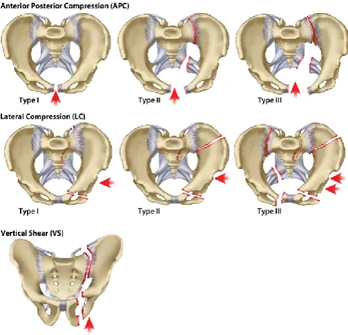
Figure 1. (Monticelli et al., 1998) Tile classification
The Young-Burgess system links the direction of the force that created the lesion to the fracture type observed in radiographs. The Young-Burgess system, when combined with a clinical examination, provides valid information about the overall condition of the patient and the risk of associated lesions. The Young-Burgess system identifies 4 types of pelvic ring injury
based on radiographic image interpretation: lateral
compression, antero-posterior compression, vertical cutting force, and combined mechanical injury.
Figure 2. (http://www.clinorthop.org/articles/10.1007/s11999-014-3693-8) Young-Burgess classification
Clinical and radiographic approach
1) Diagnosis and Primary Evaluation
History
The lesion mechanism can provide clues to the type of pelvic fracture and can help in immediate treatment. Careful examination of accident records, pre-hospital recordings and accurate patient interrogation regarding the mechanism of injury, can help the surgeon to understand the mechanism of trauma. Often the interview with the patients is not possible because they usually get intubated at the first aid station. Examination: A pelvic fracture is one of the few bone injuries that can lead to the death of the patient, even within hours of trauma. Diagnostic evaluation of traumatized patients follows the guidelines set by the American College of Surgeons (ACS) and begins with the ABCD (airways, breath and circulatory evaluation). The purpose of this primary exam is to identify and treat injury that can immediately cause the death of the patient.
The secondary examination includes an analysis of the patient according to the following scheme:
Inspection:
Deformity research: asymmetry of the limbs, abnormal
posture;
Presence of wounds: wounds involving only soft
tissues, exposed fractures (the presence of a pelvic fracture and a genital bleeding should consider this fracture as exposed fracture);
Soft tissue injuries;
Scrotum, labial, lateral and lumbosacral hematomas;
Urethral inspection.
Palpation
Deep tissue lesions;
Continuous bone solutions;
Rotational instability evaluation;
Vertical instability assessment;
Digital, rectal and gynecological examination.
2) Radiographic Approach
Conventional Radiographs: Among conventional radiographs, the most useful for diagnostic purposes and choice of treatment are the following projections:
Antero-posterior projection: initial radiographic examination of patients with closed trauma, projection which allows us to have a picture of the pelvic lesions, evaluating:
Frontal lesions (pubic limb injuries and diastasis of the
symphysis, iliac fractures, apophysis avulsions, lesions of lumbar transverse processes, pelvic ring injuries);
Pelvic symmetry;
Any dislocation of the coxo-femoral joints.
Inlet projection:
Highlights the pelvic entry;
Evaluates the presence of dislocation of sacral-iliac
joints;
Evaluates the internal rotation of the ileum;
Evaluates the presence of impact lesions of the sacumd
(shortening of one side of the sacrum);
Evaluates the presence of fractures-avulsions of
ischiatic spines.
Outlet projection:
Highlights the pelvic entry;
Evaluates the higher rotations of the hemipelvis;
Evaluates the presence of pelvic dislocation in the
vertical direction.
CT whit 2D and 3D reconstructions: CT is used to collect more information on fracture features and can also detect the size and location of a pelvic hematoma. This is an indispensable exam that allows us to evaluate:
the exact nature of the posterior lesions and the
involvement of the rear interlining ligaments;
the nature of the sacral lesion (direct impact or
compression);
[image:2.595.39.284.50.128.2] [image:2.595.37.289.274.516.2] the presence, location and size of a pelvic hematoma and / or any bleeding in progress. Therefore, the 2D CT and 3D reconstruction allow us to better understand the type and location of the lesions present.
3) Selective Examinations:
Abdominal ultrasound;
Direct abdominal rx;
Urethrography;
Angiography.
Treatment
Conservative treatment
Conservative treatment is indicated in the case of stable and composite lesions or in minimal displaced lesions. In these cases a cautious mobilization and a subsequent secure partial load guarantee a correct healing process; it is recommended to perform a radiographic check after giving a partial load. Less and less space is given to classical treatment with plump devices and long rest in bed, while traction is reserved for those patients with injuries of surgical interest but inoperable for various reasons.
Surgical treatment
For all patients hospitalized as politraumatized, a standardized protocol for the first treatment is applied.
If the pelvic fracture causes hemodynamic instability, this protocol is expanded with a form for complex pelvic fractures. It is based on two simple decisions to be taken within minutes of hospitalization in first aid:
Unstable or stable pelvic ring;
Stable or unstable hemodynamic of the patient.
While rare cases of pelvic bleeding should lead to
immediate surgery, most patients will undergo a primary diagnostic evaluation.
If hemodynamic instability is caused by a pelvic ring instability, immediate emergency stabilization is performed. The pelvic emergency clamp or the simple external fixator allows for effective stabilization 10-15 minutes. If these
instruments are unavailable, emergency non-invasive
techniques (traction and closure of the pelvic ring with a sheet or pelvic belt) are used for emergency stabilization. Often mechanical stabilization reduces the amount of bleeding, but does not provide complete hemostasis. If the hemodynamic situation of the patient remains unstable after 10-15 minutes from the application of these measures, an immediate surgical hemostasis should be performed. In patients with unstable pelvic ring lesion but hemodynamic stability, a detailed assessment of the nature of the pelvic lesion is necessary before deciding on the indications and choice of suitable stabilization techniques.
The decision on surgical stabilization or conservative treatment is based on the type of fracture (Tile classification):
Type A (stable lesion): normally no surgical
stabilization is required, functional treatment will not
cause further breaks. The treatment consists of a few days of bed rest, pharmacological therapy (deep venous thrombosis -DVT- prophylaxis and analgesics) and subsequent walking with partial progressive load. Indications for surgical reduction and synthesis are exceptional (eg open fractures or avulsion fractures in young professional athletes);
Type B (rotational instability with partial back
stability): the stabilization of the anterior pelvic ring can usually allow a partial load; the differentiation between type B lesions and type C lesions may be unclear during the first evaluation, especially in the minimum break-down side compression fractures. Therefore, it is necessary to perform radiological controls at 8 and 14 days after the injury or after the beginning of the walk, to check that there has not been any posterior displacement;
Type C (front and back instability): the pelvic ring
requires both back and front stabilization to restore anatomic relationships, providing the possibility of giving an early load and avoiding complications. Any part of the pelvic ring in which a real instability is to be diagnosed should be subjected to surgical stabilization, to provide both stability and safety to allow early partial load and then complete load.
Although pelvic fractures remain among the most
difficult orthopedic lesions to treat, the ability to treat them has improved, and recent developments indicate that further improvement is possible by perfecting minimally invasive techniques, the aid of highly selective instrumentation, improved techniques of intraoperative imaging.
Aim of the study
The aim of this study is to expose the benefits obtained with the application of CT-3D reconstruction and the production of 3D models in 1:1 scale in a case of diastasis of the pubic symphysis.
The parameters that we considered are as follows:
Surgical timing;
Exposure to ionizing radiations (intraoperative
fluoroscopy time);
Blood loss/transfusions;
Any local complication, such as postoperative
infections, surgical wound healing problems, fracture healing problems (consolidation delay or nonunion);
Any general complications;
Patient compliance;
Harris Hip Score (http://www.orthopaedicscore.com/
scorepages/harris_hip_score.html), SF-12 (http://crc. marionegri.it/qdv/downloads/SF12%20Manuale.pdf; http://crc.marionegri.it/qdv/downloads/SF12_Questiona rio.pdf), EQ-5D (EuroQuol) (Di Novi).
Case report
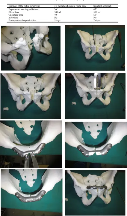
Table 1. Differences between 3D model and custom made plate and standard approach to diastasis of pubic symphysis. Discussion (Philippe, 2013; Yu et al., 2015; Bizzotto et al., 2015; Brown et al., 2003; Bortolotto et al., 2016; Bizzotto et al., 2016; Krishnan et al., 2012; Garrett et al., 2012;
Giannetti et al., 2016; Battiato et al., 2017)
Diastasis of the pubic symphysis 3D model and custom made plate Standard approach Exposure to ionizing radiations 30’’ 45’’
Blood loss 300 ml 500 ml
Operating time 40’ 60’
Infections No No
Postoperative hospitalization 5 days 7 days
Figures 3.1 – 3.5. Intraoperative images
According to Tile, this injury was classified as B1 or open book. The time to get the 3D model was 48 hours. We sent the CT images with the 3D reconstructions to the reference company, who sent back the 3D model in 1:1 scale and the custom made plate in time to perform the surgery. The Senior Surgeon and his assistants performed the simulation (Figures 1.1 – 1.8) of diastasis reduction and synthesis maneuvers, so both the plate and the 3D model were sent to sterilization. The next day we performed surgery. We placed the patient in supine position on the operating table. After sterile field setting, the surgical access was performed according to Pfannenstiel. After soft tissue incision, we detected the diastasis of the pubic symphysis of about 3 cm. the bladder appeared intact and not imprisoned in the diastasis; the application of bladder catheter ensured the bladder empty. Using fluoroscopy support and dedicated instrumentation, we performed the reduction of the diastasis and its syntesis by a two-component plate. In particular, we positioned and fixed the first plate without removing the Jungbluth instrument (Figure 2) that we used for the reduction.
We fixed the Jungbluth instrument to the pubic bone with two screws (one for each side). The holes for the Jungbluth screws corresponded to the holes for the second plate. This was possible because we had a polyamide model made with CAD during 3D planning. In fact, we had two polyamide targeting devices (one for each side) to center the perfect position and direction of the drill (Figure 1.1). So, once the first plate positioned, we removed the Jungbluth instrument because we reduced the diastasis of the pubic symphysis with the first plate, and then we placed the second plate with the screws corresponding to the holes for the Jungbluth screws (Figures 3.1 – 3.5). At the end of the surgery (Figures 4.1 – 4.2), we put an under fascial drainage and we performed the suture for anatomic planes. The patient went to the Intensive Care Unit for the one night and the day after he returned to the Orthopedics and Traumatology Unit for the remaining days of hospitalization.
On postoperative week 1, the patient was resigned with the following indications:
Dressing every 3-4 days;
Half seated position and autonomous lower limb
mobilization;
Blood tests for hemoglobin level and platelet counts
every 7 days;
DVT prophylaxix.
The patient returned to the following controls:
Suture point removal in fifteenth postoperative day;
One month postoperative clinical and radiographic
control: sitting position with the legs off the bed and wheelchair-bed passage permitted;
Two months postoperative clinical and radiographic
control: progressive weight bearing to full recovery, DVT prophylaxix continued to full recovery;
Six months postoperative clinical and radiographic
control: return to normal daily activities and quiet sport activity.
Currently the patient is at twenty-two months of follow-up. The patient has resumed all the daily activities prior to trauma, does not complain about any symptoms, there have been no late complications.
All scores scored very well with Harris Hip Score, SF-12 and EQ-5D. Compared to a patient treated according to the standard approach, all the parameters evaluated were better in the 3D case patient: operating time, exposure to ionizing radiation and blood loss were reduced, no infection episode occurred, postoperative hospitalization days were lower in confront of a standard approach (Table 1). Pelvic fractures are the most difficult fractures to treat due to complex anatomy of the pelvis, difficult access to surgical sites, and the relatively low incidence of such cases. Proper evaluation and accurate surgical planning are necessary to achieve the symmetry of the
pelvic ring and a stable fracture reduction. Medical, surgical
and dental specialties that use the 3D printing method in the diagnostic-therapeutic approach to the various pathologies are
more frequently used. The aim of this study is to present the
usefulness of this method in trauma surgery, and in particular in pelvic trauma and the possibility of using “modular” plates that do not require the removal of the reduction devices. Rapid prototyping, 3D model printing on 1:1 scale and the ability to reduce operating times, blood loss, exposure to ionizing radiations and postoperative hospidalization days.
Conclusion
3D reconstruction and rapid prototyping, starting from CT, are promising technologies that can facilitate preoperative
programming and intraoperative implementation, thus
obtaining better results in complex fractures. Unfortunately, this approach is only at the beginning and is therefore an expensive method and not applicable as a standardized approach. A favourable sign not only to cut costs rapidly but also production times, so it can be applied in simple fracture, even for educational purpose.
REFERENCES
Bagaria V, Deshpande S, Rasalkar DD, Kuthe A, Paunipagar BK. 2011. Use of rapid prototyping and three-dimensional reconstruction modeling in the management of complex
fractures. Eur J Radiol., 80: 814–820.
Battiato C, Basiglini L, Giovannini F. 2017. Traumatological application of 3D model in complex acetabular fractures.
International Journal of Current Research, vol. 9, issue 60,
pp. 52636 – 52638, June, 2017, ISSN: 0975-833X. Bizzotto N, Sandri A, Regis D, Romani D, Tami I, Magnan B
2015. Three-Dimensional Printing of Bone Fractures: A New Tangible Realistic Way for Preoperative Planning and
Education. Surg Innov., 22: 548-51.
Bizzotto N, Tami I, Tami A, Spiegel A, Romani D, Corain M, Adani R, Magnan B. 2016. 3D Printed models of distal
radius fractures. Injury, 47: 976-8.
Bortolotto C, Eshja E, Peroni C, Orlandi MA, Bizzotto N, Poggi P 2016. 3D Printing of CT Dataset: Validation of an
Open Source and Consumer-Available Workflow. J Digit
Imaging, 29: 14-21.
Brown GA, Firoozbakhsh K, DeCoster TA, Reyna JR Jr, Moneim M. 2003. Rapid prototyping: the future of trauma
surgery? J Bone Joint Surg Am., 85: 49–55.
Campbell, S. Terry Canale: “Chirurgia ortopedica”.
Colton C.L., Fernandez Dell’Oca A., Holz U., Kellam J.F., Ochsner P.E., Ruedi T.P., Marphy W.M.: “Principi AO per il trattamento delle fratture” .
Garrett J, Halvorson J, Carroll E, Webb LX. 2012. Value of 3-D CT in classifying acetabular fractures during orthopedic
residency training. Orthopedics, 35: 615–620.
Giannetti S, Bizzotto N, Stancati A, Santucci A. 2016. Minimally invasive fixation in tibial plateau fractures using an pre-operative and intra-operative real size 3D printing.
Injury, 1383: 30753-7.
http://crc.marionegri.it/qdv/downloads/SF12%20Manuale.pdf http://crc.marionegri.it/qdv/downloads/SF12_Questionario.pdf http://www.clinorthop.org/articles/10.1007/s11999-014-3693-8
http://www.ipasvi.it/ecm/rivista-linfermiere/rivista-linfermiere-page-24-articolo-286.htm
http://www.orthopaedicscore.com/scorepages/harris_hip_ score.html
Krishnan SP, Dawood A, Richards R, Henckel J, Hart AJ 2012. A review of rapid prototyped surgical guides for
patient-specific total knee replacement. J Bone Joint Surg
Br. 94: 1457–1461.
Monticelli G., Bocchi L., Letizia G., Mollica Q., Romanini L., Spinelli R.R. 1998. “Ortopedia e traumatologia”.
Philippe B 2013. Custom-made prefabricated titanium miniplates in le Fort I osteotomies: principles, procedure
and clinical insights. Int J Oral Maxillofac Surg., 42:
1001–1006.
Rockwood e Green: “Traumatologia dell’adulto”.
Yu AW, Duncan JM, Daurka JS, Lewis A, Cobb J. 2015. A Feasibility Study into the Use of Three-Dimensional
Printer Modelling in Acetabular Fracture Surgery. Adv
Orthop., 617046.