DISSIPATION OF TEMPERATURE DURING ELECTRO THERMAL
1
Dr. Yogesh G.,
2Dr. Vikram S.,
and
1
Department of Orthodontic, RKDF Dental College, Bhopal, India
2
Department of Oral Medicine and Radiology, RKDF Dental College, Bhopal, India
3
Department of public health dentistry, Pananeeya institute of dental sciences and research centre, Hyderabad,
4
Oral and maxillofacial radiologist, department of oral medicine and radiology, Goa University, India
5
Department of oral medicine and radiology, Bhabh
6
Professor of Specialization Course in Endodontics at Brazilian Dental Association
Professor of specialization course in Endodontics at Santos Dumont Air Force
ARTICLE INFO ABSTRACT
Introduction:
The present study has been done on extracted teeth to compare three Electrothermal Debonding (ETD) Techniques. The results were evaluated statistically and conclusions we
Objective: pulp with ETD. Material & Method:
Occlusal side ‘A’ and gingival side ‘B’ of the wings were marked. An ET debonding machine was used. An electronic thermometer was used to measure the intrapulpal temperature. For co
the brackets were held with pliers in three different methods. Results:
1.50oC respectively. Discussion:
and different bracket system. This study has been evaluated three ways Group 1 by holding it mesio
by holding it ging
Brackets debonded by holding it gingivo the archwire.
The results were statistically evaluated and conclusions were Conclusion:
statistically, no significant change in intrapulpal temperature due to ETD was seen among all groups.
Copyright © 2016 Yogesh et al. This is an open access
distribution, and reproduction in any medium, provided the original work is properly cited.
INTRODUCTION
Once case is clinically and radiographically complete, we proceed with steps required for debonding the fixed appliance. Secondly, preadjusted appliances demand a high degree of precision in bracket placement.
*Corresponding author: Sankalp Verma,
Bhabha College of Dental Sciences, India.
ISSN: 0975-833X
Article History:
Received 28th December, 2015
Received in revised form 30th January, 2016
Accepted 20th February, 2016
Published online 16th March,2016
Key words:
Brackets,
Electrothermal Debonding Machine, Electronic Thermometer.
Citation: Dr. Yogesh G., Dr. Vikram S., Dr. Shruti S., Dr.
temperature during electro thermal Debonding- an in vitro study
RESEARCH ARTICLE
ERATURE DURING ELECTRO THERMAL DEBONDING
Dr. Vikram S.,
3Dr. Shruti S.,
4Dr. Aarathi S.,
*5Dr. Sankalp V.
and
6Dr. Marilia marceliano-alves
Department of Orthodontic, RKDF Dental College, Bhopal, India
Department of Oral Medicine and Radiology, RKDF Dental College, Bhopal, India
health dentistry, Pananeeya institute of dental sciences and research centre, Hyderabad,
India
Oral and maxillofacial radiologist, department of oral medicine and radiology, Goa University, India
Department of oral medicine and radiology, Bhabha college of dental sciences, Bhopal, MP, India
Professor of Specialization Course in Endodontics at Brazilian Dental Association
Professor of specialization course in Endodontics at Santos Dumont Air Force \ Dental Clinic
Brazil
ABSTRACT
Introduction: The risk of pulpal injury has been a matter of concern with electrothermal debonding. The present study has been done on extracted teeth to compare three Electrothermal Debonding (ETD) Techniques. The results were evaluated statistically and conclusions we
Objective: To find out the best method of debonding the brackets with least possible damage to the pulp with ETD.
Material & Method: 30 standard edgewise brackets were bonded on to the 30 extracted teeth. Occlusal side ‘A’ and gingival side ‘B’ of the wings were marked. An ET debonding machine was used. An electronic thermometer was used to measure the intrapulpal temperature. For co
the brackets were held with pliers in three different methods.
Results: The mean increase in intrapulpal temperature with groups 1, 2 & 3 were 2.34oc, 1.17oC & 1.50oC respectively.
Discussion: In most of the studies, different techniques were used with different bonding materials and different bracket system. This study has been evaluated three ways Group 1
by holding it mesio-distally and rotating it in anti-clockwise direction. Group 2
by holding it gingivo-occlusally and rotating it gingivo-occlusally without archwire. Group 3 Brackets debonded by holding it gingivo-occlusally and rotating it gingivo
the archwire.
The results were statistically evaluated and conclusions were made.
Conclusion: The mean increase in intrapulpal temperature was minimum with group 3 but statistically, no significant change in intrapulpal temperature due to ETD was seen among all groups.
is an open access article distributed under the Creative Commons Attribution License, which distribution, and reproduction in any medium, provided the original work is properly cited.
Once case is clinically and radiographically complete, we proceed with steps required for debonding the fixed appliance. Secondly, preadjusted appliances demand a high degree of
With preadjusted brackets, the position of the bracket on to the tooth determines its final. Poor bracket positioning can render even the most customized prescription ineffective. During active orthodontic treatment, sometimes we feel that the position of a bracket should be improved. This needs debonding and rebonding of the bracket. Thus, it is important to have a simple debonding technique which does not harm the tooth as well as does not change the in
bracket. For orthodontists, the quest for a better technique still continues. The traditional bracket debonding is achieved by
International Journal of Current Research
Vol. 8, Issue, 03, pp. 27747-27751, March, 2016
INTERNATIONAL
Dr. Yogesh G., Dr. Vikram S., Dr. Shruti S., Dr. Aarathi S., Dr. Sankalp V. and Dr. Marilia marceliano an in vitro study”, International Journal of Current Research, 8, (03), 2774
DEBONDING- AN IN VITRO STUDY
Dr. Sankalp V.
Department of Orthodontic, RKDF Dental College, Bhopal, India
Department of Oral Medicine and Radiology, RKDF Dental College, Bhopal, India
health dentistry, Pananeeya institute of dental sciences and research centre, Hyderabad,
Oral and maxillofacial radiologist, department of oral medicine and radiology, Goa University, India
a college of dental sciences, Bhopal, MP, India
Professor of Specialization Course in Endodontics at Brazilian Dental Association - Niterói - RJ;
Dental Clinic - Rio de Janeiro,
The risk of pulpal injury has been a matter of concern with electrothermal debonding. The present study has been done on extracted teeth to compare three Electrothermal Debonding (ETD) Techniques. The results were evaluated statistically and conclusions were made.
To find out the best method of debonding the brackets with least possible damage to the
30 standard edgewise brackets were bonded on to the 30 extracted teeth. Occlusal side ‘A’ and gingival side ‘B’ of the wings were marked. An ET debonding machine was used. An electronic thermometer was used to measure the intrapulpal temperature. For comparison,
The mean increase in intrapulpal temperature with groups 1, 2 & 3 were 2.34oc, 1.17oC &
ed with different bonding materials and different bracket system. This study has been evaluated three ways Group 1 - Brackets debonded clockwise direction. Group 2 - Brackets debonded occlusally without archwire. Group 3 - occlusally and rotating it gingivo-occlusally with holding
made.
The mean increase in intrapulpal temperature was minimum with group 3 but statistically, no significant change in intrapulpal temperature due to ETD was seen among all groups.
ribution License, which permits unrestricted use,
With preadjusted brackets, the position of the bracket on to the tooth determines its final. Poor bracket positioning can render even the most customized prescription ineffective. During active orthodontic treatment, sometimes we feel that the a bracket should be improved. This needs debonding and rebonding of the bracket. Thus, it is important to have a simple debonding technique which does not harm the tooth as well as does not change the in-built features of the e quest for a better technique still continues. The traditional bracket debonding is achieved by
INTERNATIONAL JOURNAL OF CURRENT RESEARCH
applying a sufficiently large force to break the bond. But there is possibility of adhesive remnants on the tooth and subsequent damage to the enamel. Various other debonding techniques have been reported including electro thermal debonding or electrothermic debracketing (ETD). ETD is the removal of bracket from the adhesive composite by heat, generated by a battery. When the heat applied to the bracket is transferred to deform the adhesive-bracket interface, the bracket can be gently lifted up from the tooth surface without exerting excessive forces. Thus there are less chances for tooth damage and/or bracket deformation. When teeth are debonded with ETD, bond failure is usually induced at the bracket-resin interface. There is possibility of injury to the pulp with ETD. This is the reason why it has not been easily taken up by most of the practitioners (Brinkmann-Jost et al., 1992). According to Christopher (2007), as a low-compliance system, pulp tissue is vulnerable to temperature changes. According to Crooks et al., 1997, tolerance of heat by human teeth without irreversible pulp changes are not known. Different levels of tolerance of temperature have been shown by a number of investigators.
Objective
This study was done to know the amount of temperature reaching the pulp while using ETD, so that a proper method of debonding the brackets from the tooth surface with minimal damage to the pulp could be established.
MATERIALS AND METHODS
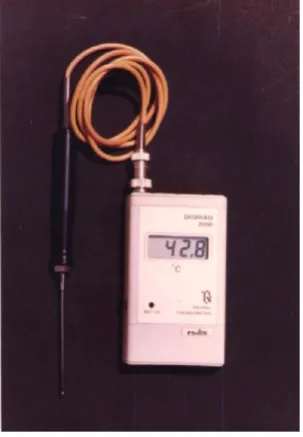
30 extracted premolars of orthodontic patients were taken in this study irrespective of the quadrants. Teeth were divided into three groups. Each group had 10 teeth. All the teeth were without caries. 30 standards edgewise .022" x .030" brackets with mesh base (80/linear inch) were taken. No mix adhesive was used to bond the brackets on to the teeth. A pt. 2000 digital thermometer which is accurate to 0.1oC (range 0.0-100oC) with tip of diameter 2.2mm (Fig.1), was used to measure intrapulpal temperature before and after debonding. An Electro thermal Debonding Unit was used (Fig.2). A piece of .022" x .028" straight length stainless steel arch wire was used to fill bracket slot. 0.010 stainless steel ligature wire was used to tie the archwire to the bracket.
All the 30 teeth were cleaned and numbered from 1 to 30. Brackets numbered from 1 to 30 were bonded on to the buccal surface of the teeth. A hole was prepared on to the lingual surface of each tooth to put the tip of probe of the thermometer into the pulp chamber. Teeth were fixed in plaster of paris and separate plaster blocks were made to debond brackets by different methods. White correction fluid was used to put mark on one side of base to identify occlusal (A) side and gingival (B) side of the wings.
Group 1: (No. 1–10). Brackets were debonded by holding it mesio-distally at the base of the bracket and rotating it in anti-clockwise direction. (Fig. 3)
Group 2: (No. 11-20). Brackets were debonded by holding it gingivo-occlusally and rotating it gingivo-occlusally (without archwire into the slot. (Fig 4)
Group 3: (No. 21-30). Bracket slot were filled with archwire piece and then debonded by holding it gingivo-occlusally and rotating it gingivo-gingivo-occlusally. (Fig. 5)
The intrapulpal temperature was measured before and after debonding the brackets one by one and results were noted.
RESULTS
Table shows intrapulpal temperature before and after debonding and the differences between the two when 30 brackets were debonded with ETD.
Results were evaluated individually and statistically.
The mean increase in intrapulpal temperature while electro thermal debonding with groups 1, 2 & 3 was 2.34oc, 1.17oC &
1.50oC respectively.
DISCUSSION
[image:2.595.364.516.340.559.2]A number of studies have been done to describe the effects of conventional debonding techniques on the tooth surface. With the advent of electro thermal debonding technique in 1986
[image:2.595.319.558.584.762.2]Fig. 1. Electronic Thermometer
(Sheridan et al.,1986), most of the studies have been done to evaluate the temperature reaching the pulp due to ETD or the effects of the ETD on the dental pulp. Lisanti and Zander (1952) conducted a vivo study on dogs and shown that enamel and dentin have capacity to dissipate heat efficiently to the pulp chamber and despite increase in temperature, the pulp showed healing capacity. Postle et al., in 1959 investigated unaltered
dog teeth exposed for 20 sec. to heat at temperature of 102oc,
201oc and 482oc. In the teeth exposed to 102o c and 201oc, the
pulps showed no signs of pathosis. In the teeth exposed to
482o c, there was evidence of abscesses or necrosis. The most
definite study on the thermal thresholds of the pulps in unaltered teeth was done by 2ach and Cohen (1965) on a primate Macaca rhesus monkeys. When the pulpal temperature
increase was assessed at 5.5oc, reversible changes occurred in
85% of the teeth and when the temperature increase was 11.1oc,
abscess formation occurred in 60% of the teeth. When there
was a 16.6oc elevation in pulpal temperatures, pulpal necrosis
occurred in all teeth. Kraut et al. (1991) did not find any evidence of pulpal injury during ETD. Further research in vivo has demonstrated that minimal pulpal inflammation occurs without any loss or damage to odontoblasts (Brinkmann-Jost
et al., 1997) Initially, studies that define the thermal limitations
of human pulp on unaltered teeth were lacking. Previous thermal studies on human pulp have measured the effect of heat generated by frictional heat during dental procedures (Bashkar and Lilly, 1965; Langeland and Langeland, 1968; Peyton, 1958 (Sheridan et al., 1986), or the thermal effects of restorative filling materials (Robinson and Lefkowitz, 1962; Stanley, 1971; Wolcott et al., 1951; Zander, 1946) (Sheridan et al., 1986). These investigations on altered teeth have been confined to exper¬imental animals. Odegaard & Segner 1988 said that Orthodontists want to remove the appliance at the end of treatment without exerting excessive forces. Electro thermal debonding was introduced by Sheridan et al. in 1986. ETD was defined as the controlled application of heat to the bracket bulk. The heat deforms the bracket–adhesive interface, melting the resin component of the adhesive. This leads to a reduction in the force required to remove the bracket. From primary samples (25 extracted teeth), he concluded that the mean
temperature at the time of debracketing is 0.8oC (Range 0o –
1.9oC). The mean residual temperature, after removal of
bracket was 3.2oC (Range 1.1o – 5.1oC). In secondary samples
(5 extracted teeth). Cool water spray of 20oC was used after debonding, the mean residual temperature increase at the pulpal
wall was 0.7oC. An in vivo study, Sheridan et al. (1986)
examined the teeth, histologically, which were extracted 2 weeks after ETD. It revealed no evidence or indication of any pathosis due to thermal insult. All the patients reported that the ETD procedure did not elicit any feeling of discomfort or any feeling that the bracket was being forcefully removed from the tooth.
Tikku et al. (1989) did a vivo study, involving the pulps of 27 human premolar teeth, subjected to electro thermal debonding of orthodontic brackets. They concluded that:
(a) ETD is better method of removal of brackets.
(b) As the temperature applied to bracket increased, time taken for debonding decreased.
In further study, Gupta et al. (1989) did not find any signs of pulpal pathosis due to ETD. Sylvester Edwin (1991) suggested the application of dry heat to the bonded bracket with a Handi Dry tooth dryer (M/s Lancer Orthodontics, Carlsbad, 2018). It is suggested to hold the dryer 3-4 mm from the tooth and direct the heated air at the bracket for 10-15 seconds and then remove the brackets as per manufacturer's instructions. He said "the maximum temperature of the dry air stream is 65oc which is less than that of a hot cup of coffee and is well tolerated by patients. Patients did not report for residual sensitivity or discomfort after debonding". He concluded from his study that this method could be used for removal of stubborn metal brackets. Brinkmann et al. (1992) debonded metal brackets by squeezing the bracket wings and by ETD technique.
Fig.3. Debonding Pliers holding the bracket mesio-distally
Fig.4. Debonding Pliers holding the bracket occluso-gingivally
They found no pathologic alterations in pulp after debonding. The only difference related to the debonding techniques was that debracketing by squeezing the bracket wings together led to severe bracket deformations. Wool, in 1992, used a small
wood burning pen (maximum temperature 600oC) for
debonding ceramic brackets. He found that 7-10 sec. are enough to debond a bracket. Carter (2003) recommended hot water bath to facilitate debonding of ceramic brackets. Todd Lee Knight et al. (1997) in their study concluded that ETD provides predictable debonding of ceramic brackets with no veneer damage and minimal risk to the pulp. Chiyako et al. (2011) worked on experimentally produced easily debondable orthodontic adhesive (EDA) containing heat expandable microcapsules. The temperature of the pulp wall increased
1.8-3.6oC after heating less than that required to induce pulp
damage. They concluded that heating for 8sec. is optimum while using electothermal debonding.
The present study has done to know the best way to remove the brackets in terms of change in intrapulpal temperature when ETD technique was used. The results found in the study are as follows-
Group 1 - The mean increase in intrapulpal temperature with group 1 was 2.34oc, when bracket was debonded by holding it mesio-disally and rotating it in anti-clockwise direction.
Group 2 - The mean increase in intrapulpal temperature with group 2 was 1.17oC when bracket debonded by holding it gingivo-occlusally and rotating it gingivo-occlusally without holding archwire.
Group 3- The mean increase in intrapulpal temperature with group 3 was 1.50oC when bracket debonded by holding it gingivo-occlusally and rotating it gingivo-occlusally with holding archwire.
Table
Group-1 Group-2 Group-3
Temperature in 0 C Temperature in 0 C Temperature in 0 C
Pre Post Difference Pre Post Difference Pre Post Difference
27.5 30.5 3.0 27.8 28.8 1.0 28.0 29.6 1.6
27.5 28.3 0.8 27.6 27.9 0.3 29.0 30.1 1.1
27.3 28.0 0.7 27.5 28.5 1.0 29.0 31.1 2.1
27.2 30.6 3.4 27.5 28.8 1.3 28.4 29.9 1.5
27.0 30.8 3.8 27.6 29.0 1.4 28.4 29.6 1.2
26.6 28.6 2.0 28.0 30.0 2.0 28.3 29.4 1.1
26.6 28.2 1.6 27.4 28.3 0.9 28.5 29.5 1.0
26.8 30.8 4.0 27.4 28.7 1.3 27.0 29.2 2.2
26.8 27.4 0.6 27.6 29.1 1.5 26.6 27.8 1.2
26.7 30.2 3.5 27.5 28.5 1.0 26.6 28.6 2.0
Conclusion
After the active Orthodontic treatment, the brackets are removed by various debonding techniques. Thermal debonding was developed to overcome the problems of enamel damage and high forces produced when removing brackets mechanically. Among electrotharmal debonding techniques, all the methods were found satisfactory to debond brackets as far as change in intrapulpal temperature was concerned but the mean increase in intrapulpal temperature was minimum when brackets debonded by holding it gingivo-occlusally and rotating it gingivo-occlusally without holding archwire. Further research is needed to reduce the change in intrpulpal temperature furthermore.
REFERENCES
Bashkar, SN, Lilly, GE. 1985. Intrapulpal temperature during
cavity preparation. J. Dent. Res., 44 : 644-647.
Brinkmann, PCJ et al. 1992. Histological investigations of the human pulp after thermo debonding of metal & ceramic brackets. AJO, 102: 410-417.
Carter RN. 2003. Hot water bath facilitate ceremic debonding.
JCO, 37:620
Chiyako et al. 2011. The use of easily debondable orthodontic adhesives with ceramic brackets. Dental Material Journal, 30(5):642-647.
Christopher Millen et al. 2007. A study of temperature rise in the pulp chamber during composite polymerization with different light curing units. J Conte. Dent Prac., Nov (8)7:29-37.
Crooks M, Hood J, Harkness M. 1997. Thermal debonding of ceramic brackets: an in vitro study. American Journal of
Orthodontics and Dentofacial Orthopedics, 111:163-172.
Gupta DS et al. 1989. A histological evaluation of dental pulp after ETD: JIOS, 20.3: 111-115.
Jost-Brinkmann, PG et al. 1997. Risk of pulp damage due to temperature increase during thermodebonding of ceramic brackets. European Journal of Orthodontics, 19: 623-628. Kraut J, Radin S, Trowbridge HI, Emling RC, Yankell SC.
1991. Clinical evaluations on thermal versus mechanical debonding of ceramic brackets. Journal of Clinical
Dentistry, 2:92-96.
Langeland, K and Langeland, LK. 1968. Cutting procedures with minimized trauma. J. Am. Dent. Assoc., 76: 991-1005. Lisanti VF and Zander HA. 1952. Thermal injury to normal
dog teeth: In view measurements of pulp temperature increases and their effect on the pulp tissue. J. Dent. Res., 31: 548-58.
Odegaard, J. & Segner, D. 1988. Shear bond strength of metal brackets compared with a new ceramic bracket. AJO, 94 : 201-206.
Peyton, FA. 1958. Effectiveness of water coolants with rotary cutting instruments. J. Am. Dent. Assoc.1958; 56 : 664-675. Postle HH et al. 1959. Pulp response to heat. J Dent. Res., 38:
740-751.
Robinson HDG, Lefkowitz, W. 1962. Operative dentistry and the pulp: Journal of Prosthetic Dentistry, 12:985-1001. Sheridan, John J et al. 1986. Electrothermal debracketing. Part
I. An in vitro study. AJO, 89: 21-27.
Sheridan, John J et al. 1986. Electrothermal debracketing. Part II. An in vitro study: AJO, 89:141-145.
Stanley, HR. 1971. Pulpal response to dental techniques and materials. Dent. Clin. North Am.., 15: 115-126.
Sylvester, Edwin. 1991. Thermal debonding of cermaic brackets: JCO, 25:748.
Tikku, T et al. 1989. An evaluation of the ETD technique.
JIOS, 20.2: 54-59.
Todd Lee Knight et al, AJO & DO. 1997. Mechanical and electro thermal debonding effect on ceramic veneers and dental pulp: AJO & DO, vol.112 issue 3 :263-270.
Wolcott, RB et al. 1951. Direct resinous filling materials: temprature rise during polymerization. J. Am. Dent. Assoc., 42: 253-263.
Wool, A.L. 1992. A better debonding procedure: AJO, 102: 84-86.
Zach L & Cohen G. 1965. Pulp response to externally applied heat. Oral surg. Oral Med. oral Patho., 19:515-530. Zander, H.A. 1946. The reactions of dental pulps to silicate
cements. J. Am. Dent. Assoc., 33: 1233-1243.