Effect of Heat Treatment on Characteristics of Plasma Sprayed
Hydroxyapatite Coatings
Chun-Cheng Chen
1and Shinn-Jyh Ding
2 1Dental Department, Chung-Shan Medical University Hospital, Taichung 402, Taiwan, R. O. China
2Institute of Oral Materials Science, Chung-Shan Medical University, Taichung 402, Taiwan, R. O. China
Bioactive hydroxyapatite (HA)-coated implants plasma sprayed on Ti6Al4V substrates have been widely used in load-bearing applications because of their biocompatibility and their intimate contact with bone. The improvement of the characteristics of HA coatings is concerned. The purpose of this work was to evaluate corrosion behavior and bond strength of HA coatings after post-deposition heat treatment at 500–700C. The results indicated that the heat treatment led to recrystallization of amorphous calcium phosphate of as-sprayed HA coatings. The reduction of layer defects associated with plasma-sprayed coatings and the enhancement of the resistance to corrosion took place after heat treatment. Bond strength of the heat-treated coatings was sensitive to the treatment temperature. It is concluded that the heat treatment at 600C for 1 h in air, endowing with increased crystallinity and the reduced defects without significantly reduced bond strength, provided a better corrosion protection than the other two treatment temperatures.
(Received December 12, 2005; Accepted January 30, 2006; Published March 15, 2006)
Keywords: plasma spray, hydroxyapatite, heat treatment, coating, corrosion, bond strength
1. Introduction
The chemical and phase composition of hydroxyapatite (HA) is similar to the mineral component of bone and tooth.1,2)Over the past 15 years, HA coatings plasma sprayed on metallic substrates have been widely used in clinical load-bearing implant applications because of their biocompati-bility and their intimate contact with bone.2–6) Plasma spraying is the most widely used technique to deposit HA coatings due to its process feasibility. However, during plasma spraying process HA coating crystallinity decreases, phase composition of the coating is subjected to change, and residual stresses occur within the coating as a result of high temperatures.7–10) Hence, plasma-sprayed HA-coated
im-plants are essentially composed of a mixture of crystalline, amorphous, and non-apatite phases such as Ca3(PO4)2(TCP),
Ca4(PO4)2O (TTCP) or even CaO. The presence of TCP and
TTCP phases would enhance the resorption process of HA coating leading to implant instability.11)In addition, the thick coatings produced by plasma spray often exhibited porosity that weakened the interfacial strength and provided an easy fracture path for adhesion failure.11)Pores and other coating imperfections reduced the corrosion resistance.12,13)
As far as the longevity and safety of coating implants are concerned, the coating with higher crystallinity and fewer defects may be preferentially considered. HA coatings with higher crystallinity yielded a decreased dissolution rate14,15)
and enhanced rate of cell proliferation.16) As suggested by
Darimontet al., the implants in trabecular or cancellous bone would require coatings with a very high crystallinity.17)For
improving the quality of plasma sprayed coatings, the heat treatment is sometimes a used method to increase coating crystallinity and reduce the residual stress of HA-based coatings,5,6,15,18–23) aside from optimizing plasma spray processing and altering material composition.14,24,25)Caulier et al.confirmed in a goat animal model that heat-treated HA plasma-sprayed implant showed less reduction in coating thickness than as-sprayed HA coatings.6)In a recent paper,26)
we found that the HA coatings heat-treated at 650C gave rise
to recrystallization of amorphous calcium phosphate and the conversion of non-apatite phases into apatite as well as showing a higher corrosion resistance. Liet al.demonstrated that the complete crystallization of the amorphous phase of high-velocity oxy-fuel sprayed HA coatings occurred at approximately 700C.27)
So far, most studies have concentrated on the changes in purity and crystallinity of plasma-sprayed HA coatings, relatively few data on the changes in the mechanical properties and corrosion behavior were reported. The purpose of this study is to study the characteristics of plasma sprayed HA coatings, such as coating crystallinity, corrosion behav-ior, and bond strength, when subjected to heat treatment at different temperatures.
2. Experimental Procedure
2.1 Plasma spraying processing
Commercial HA powder (AMDRY 6021, Plasma Technik A.G., Wohlen, Switzerland) with particle size between 44 and 149mmwas used in this study. Commercially available plates of Ti6Al4V alloy (100103mm thick) were used as the substrate material. Prior to plasma spray, the substrate surface was mechanically polished to #1200 grit level, cleaned and sandblasted with 450mmSiC particles. To obtain a uniform coating, the substrate was mounted on a disk that could rotate during spraying in air using a Plasma-Technik A-3000 system (Plasma Technik A.G., Wohlen, Switzerland). The parameters used included a plasma power of 37 kW and plasma gas of Ar and H2 with the flow rate of 45 and 11 L/
min, respectively. A spray distance of 130 mm and spray angle of 90 were chosen. The powder was transported at a
feed rate of 0.02 kg/min with Ar as a carrier gas. Monolithic HA coating of about 100mmthick was fabricated.
2.2 Heat treatment
In order to study the effect of heat treatment, the
sprayed HA coatings were heat-treated at 500, 600 and 700C for 1 h each at a heating rate of 5C/min in an air-circulated
furnace, and then the furnace cooling to room temperature.
2.3 Phase composition and microstructure analyses
Phases of all coatings were analyzed by an X-ray diffractometer (Shimadzu XD-D1, Kyoto, Japan) operated at 30 kV and 30 mA. The crystallinity was estimated by the area ratio of three major peaks, (211), (112) and (300), of the coated samples to that of the HA powder used for the plasma spray. A Fourier transform infrared (FTIR) spectroscopy (Bomem DA8.3, Hartman & Braun, Canada) in reflection absorption mode with a spectral resolution of 1 cm1, was
used to characterize the various functional groups on the coating surface. The microstructure was characterized under field emission scanning electron microscope (FESEM) (Hitachi S-4200, Hitachi, Tokyo, Japan). Specimens for cross-sectional microscopy were prepared by mounting the coated specimens in epoxy resin, followed by polishing through 1mmalumina.
2.4 Electrochemical test
The commonly-used Hanks’ balanced salt solution (HBSS), recommended by Pourbaix,28)has an ionic
compo-sition similar to that of human plasma, which adjusted to an initial pH of 7.4. The electrochemical measurements carried out on the coated samples with a surface area of 1 cm2 were open circuit potential (OCP)-time measurements and poten-tiodynamic polarization in HBSS, using a conventional three-electrode cell connected with a CHI 660A electrochemical system (CH Instrument, Austin, Texas). For OCP measure-ment, only two electrodes (working electrode and reference electrode) were involved, while for potentiodynamic polar-ization method a conventional three-electrode cell was used. A saturated calomel reference electrode (SCE) and a platinum counter electrode were employed. The sample surface was cleaned by distilled water. Deaerated conditions under N2 gas purging and a temperature of 37C were used
forin vitroexperiments. The evaluation of potentiodynamic polarization was started after immersion in HBSS for 1 h. The scanned potential range varied from1V up to 2 V toward the anodic direction at a sweep rate of 0.5 mV/s in the Tafel mode. Six measurements were performed for each group. Corrosion potential, corrosion current and polarization resistance were provided after being analyzed by the built-in software.
2.5 Bond strength measurement
Bond strength (or adhesive strength) of the HA coatings before and after heat treatment was used to represent the present pull-out test results using an EZ-Test machine (Shimadzu, Kyoto, Japan) at a loading rate of 0.5 mm/min. In doing the testing, a 2.7 mm dia. aluminum pull stud (Quad Group, Spokane, WA) was bonded to the coated surface with a solid epoxy and then cured at 150C for 1 h in oven. After
the coated specimen/stud assembly was gripped on a platen, the stud was then pulled down against the platen until failure. The maximum fracture force can be recorded and averaged to obtain the mean value and standard derivation. The number of measurements is sixteen for each group.
2.6 Statistical analysis
One-way ANOVA statistical analysis was used to evaluate the statistical significance of the bond strength and electro-chemical data. A Scheffe’ multiple comparison test was used to determine the significance of the deviations in bond strength and electrochemical data of the coated samples without and with heat treatment. In all cases, results were considered statistically different when p<0:05.
3. Results and Discussion
3.1 Phase composition and microstructure
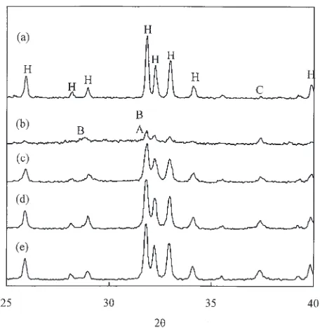
Figure 1 shows the XRD patterns of the HA powder, as-sprayed coating and heat-treated coatings. The as-as-sprayed coating was the characteristic of an apatitic structure of lower crystallinity than HA powders with a reduction of 63%. Besides the broadening of apatite peaks and increased intensity of CaO phase, TCP phases were observed in the as-sprayed sample. The high temperature involved in plasma spray process had obviously enhanced the decomposition of apatite as well as chemical reactions within HA phase. Compared with the as-sprayed HA coating, heat-treated coatings had a higher apatite crystallinity and a smaller amount of TCP phases but the retention of CaO. Using the integrated area of (211), (112) and (300) peaks of apatite phase, the crystallinity of heat-treated coatings was about three-fold greater than that of the as-sprayed coating and increased with the increasing treatment temperature.
Figure 2 shows FTIR reflection spectra of all coatings. The broad bands at 970–1130 cm1 in FTIR reflection spectra of
as-sprayed coatings, probably resulted from overlaping apatite, -TCP and -TCP signals, were attributed to HPO4/PO4 functional groups.29,30) The bands between 603
and 574 cm1 were suggested to arise from the vibrational mode of PO4groups.30)After post-deposition treatment in air
[image:2.595.313.541.69.303.2]at 500–700C, the sharper PO
4 stretching mode at 603 and
574 cm1and the two more appreciable adsorption bands at
970–1130 cm1demonstrated an increased crystallinity. The
appearance of the absorption band at 630 cm1 and sharper
band at 3572 cm1 with an indication of the vibrational
modes of OH groups29)suggested the reestablishment of a hydroxylated HA structure when the as-sprayed HA coating was heat-treated in air. The atmospheric moisture can react with amorphous oxyapatite so that OH groups recovered and promoted the reconstitution of amorphous oxyapatite into crystalline oxyhydroxyapatite.18,19)
In addition to enhancing coating crystallinity, the heat treatment could effectively convert non-apatite phases (TCP) into apatite, consistent with a previous study.20,26)Like other
amorphous phases,18,19)the amorphous calcium phosphate in
the as-sprayed samples is thermodynamically metastable and an appropriate thermal treatment could induce recrystalliza-tion of them. Although higher-temperature treatment can help hasten the amorphous-crystalline phase transformation, it is practical to use lower heat-treatment temperatures to avoid serious irreversible structural and property changes to the Ti alloy substrate.31)In this study 600C seems to be ideal
as almost complete crystallization of HA (86% of HA powder) was achieved.
SEM micrographs showed that the appearance of the coating surfaces after heat treatment were similar to those of as-sprayed coatings, which had well-flattened splats and shiny glassy films and irregularly-shaped particles, due to that the crystallization of amorphous phases did not influence the surface morphology under low magnification.24)
Ran-domly distributed pores of different sizes and microcracks were also observed. During plasma spraying, small size powder was completely melted and formed a glassy phase during fast cooling, while larger size powder was only partially melted resulting in a rough, irregularly-shaped surface morphology.32,33)This observation is consistent with
the earlier-discussed XRD results (e.g.broadening in apatite peaks). In Fig. 3 the cross-sectional SEM micrographs showed that the thickness of all coatings was roughly 100mm. The ‘‘flake-type’’ of layer defects between the splats within coatings are observed tending to be parallel to the coating surface [Fig. 3(a)]. Besides, there were a lot of perpendicular defects inside the coatings, e.g., pores and cracks. These plasma spray-induced layer defects were Fig. 2 FTIR spectra of the as-sprayed coating (a) and heat-treated coatings
at 500C (b), 600C (c), and 700C (d).
(d)
50
µ
m
50
µ
m
(c)
50
µ
m
(b)
(a)
50
µ
m
[image:3.595.55.284.70.275.2] [image:3.595.128.469.491.768.2]frequently observed in plasma sprayed HA and other coatings.25,34) The ‘‘flake-type’’ regions were examined to be amorphous with micro-Raman spectroscopy and nano-indentation techniques.35) After post-deposition heat treat-ment, such microstructure of plasma-sprayed coatings were improved. It seems that change in porosity can be deduced from SEM pictures between as-sprayed and crystallized coatings. The changes in CTE (coefficient of thermal expansion) caused by the annealing might more or less contribute to microstructure changes because different phases possess different CTEs (HA: 11:5106, TCP: 14:2
106C1).9,36)In addition, this can be explained in terms of
the heat treatment during which the atoms gained high kinetic energy and diffused much faster than at room temperature.19)
The faster diffusion of the atoms speeded up the phase transition and affected the flake-type appearance.
3.2 Electrochemical test
The OCP-time plots for the coating samples heat-treated at 500, 600 and 700C as a function of time along with the sprayed coating are shown in Fig. 4. It seems that the as-sprayed HA coating is in a steady state possibly due to the apatite precipiatation,26) although having a more negative
initial potential of0:198V. On the contrary, all heat-treated samples showed a higher initial OCP, tending to a decreased potential, in particular, for the coating heat-treated at 700C.
Figure 5 shows the typical potentiodynamic polarization curves of the plasma-sprayed coatings without and with heat treatment in deaerated HBSS at 37C. It can be seen all
coatings indicated a breakdown potential in the range between 0.5 and 1.0 V. All electrochemical parameters, including corrosion potential (Ecorr), current density (icorr), and polarization resistance (Rp), of coating samples are also compiled in Table 1. As corrosion potential was concerned, there are significant differences (p<0:05) among all test samples. After heat treatment at 500, 600 and 700C,
corrosion potentials of the heat-treated samples were found to be 0:61, 0:48 and 0:62V (vs. SCE), respectively. Moreover,via Scheffe’ multiple comparison test the differ-ence in corrosion potential between 600C-sample and
500C-sample is found (p<0:05), also between 600
C-sample and 700C-sample (p<0:05). On the contrary, there
is no significant difference (p>0:05) in corrosion potential between as-sprayed coating and heat-treated coating at 600C. The results confirmed that 600C-treatment HA coatings exhibited a significantly better corrosion-resistant ability than all the other coatings by virtue of more noble corrosion potential, although all polarization curves were characterized by a very similar trend.
The corrosion current density and polarization resistance of the samples were determined from the potentiodynamic polarization curves using Tafel extrapolation method. As for current density, it could be found that the average values of the heat-treated coatings of between 1.12 and1:94103
A/m2, which lower than that of the as-sprayed coating
(2:22103A/m2), were dependent on treatment
temper-ature, revealing the significant difference (p<0:05). Pre-vious studies have reported similar behaviour.26)In contrast to current density, there was an increase in the Rp values about two times, representing that heat treatment favored the coatings a better resistance to corrosion. The polarization Fig. 4 Open circuit potential-time measurements in deaerated HBSS of the
as-sprayed coating (a) and heat-treated coatings at 500C (b), 600C (c), and 700C (d).
[image:4.595.53.281.68.292.2]Fig. 5 Typical polarization curves of the as-sprayed coating (a) and heat-treated coatings at 500C (b), 600C (c), and 700C (d) in deaerated HBSS at 37C.
Table 1 Mean and standard derivation values of electrochemical param-eters of the as-sprayed coating and heat-treated HA coatings at different temperatures after electrochemical test.
Coating Ecorr(V) icorr(103Am2) Rp(m2)
As-sprayed 0:570:06 2:220:60 369
500C 0:610:08 1:500:69 6110 Heat-treated 600C 0:480:06 1:120:33 819
[image:4.595.310.541.70.301.2] [image:4.595.305.549.395.459.2]resistance is a parameter correlated to the corrosion rate. The higher the polarization resistance, the lower the corrosion rate was on the coating when exposed to physiological solution.37)
The critical factors affecting the corrosion characteristics of coatings are their quality (crystallinity, purity, residual stress and ion substitution in the apatite lattice) and structure.37–42) The porosity is a characteristic of plasma
sprayed coatings and strongly affects their corrosion behav-iours. Generally speaking, the corrosion rate increases with increasing porosity in the coatings.41) The electrolyte infiltrates into the inner portion of the coating through the structural imperfections such as pores and cracks or pinholes existing in the coating and comes into contact with the deeper portion of the coating,42)causing corrosion. The polarization resistance (Rp) obtained can be used to determine the porosity that corresponds simply to the ratio of the polarization resistance of the uncoated substrates and the coated sam-ples.38,39) Using a modified equation, the ratio ofP
s=Phs¼ Rp,hs=Rp,scan be used to represent the porosity change, where PsandPhsare the porosity of the as-sprayed and heat-treated coatings, Rp,s an Rp,hs the polarization resistance of the as-sprayed and heat-treated coatings, respectively. To substitute the obtainedRpvalues into the above-mentioned equation, it is obvious thatPs=Phs is approximately two, indicating that the positive effect on the reduction of the porosity occurred in the heat-treated samples.25,34)This was because that the heat treatment apparently reduced the plasma spray-induced flake-type structures, as described earlier in morphology change, resulting in heat-treated coating possessing a higher corrosion-resistant ability. More importantly, the in vitro electrochemical test results indicated that 600C-treatment
coatings had a more beneficial and desired effect on corrosion behavior than the as-sprayed and the other two hea-treated samples at 500 and 700C from theR
pand corrosion potential
points of view.
3.3 Bond strength
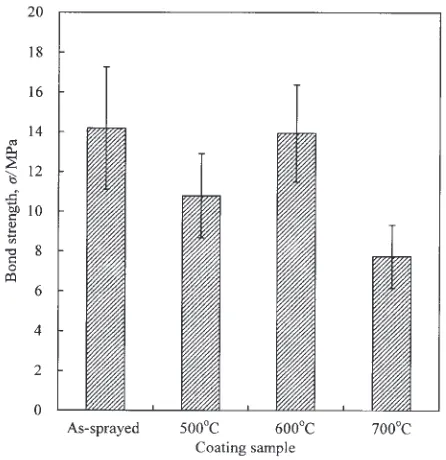
The bonding strength of the HA coating to metallic substrate and the quality of the coating itself are always concerned in determining the performance and reliability of HA-coated implants. Bonding strength test results of the as-sprayed coating and the three heat-treated coatings are shown in Fig. 6. It can be seen that bond strength values of the heat-treated coatings depended on treatment temperature. The bond strength of the as-sprayed coatings was 14.2 MPa. After heat treatment at 500, 600 and 700C, the value became 10.8,
13.9 and 7.7 MPa, respectively. The differences in bond strength values between as-sprayed and heat-treated coatings were statistically significant (p<0:05). The statistical analysis using Scheffe’s multiple comparison test showed that bond strength of 700C-treatment coatings significantly
declined by about 46%, but there is not significantly different between the as-sprayed and 600C-treatment coatings.
Many investigations have reported that bond strength of the coatings was apparently related to different deposition techniques and post-treatment parameters.5,21,24,27)For exam-ple, Tsuiet al.21)found that heat treatment at 700C for 1 h was effective in enhancing crystallinity, the OHion content and the purity of plasma sprayed HA coatings. However, a
significant drop in adhesion occurred on heat-treated coatings sprayed at high powers of 36 kW. Similarly, Lynn and DuQuesnay reported that heat treatment of plasma sprayed HA coatings at 400C for 90 h significantly reduced their
fatigue resistance due to stress relief in the titanium substrate.22) In contrast, Liet al.27)suggested that the heat treatment at 750C for 30 min is beneficial for the improved strength, as well as fracture toughness of high-velocity oxy-fuel sprayed HA coatings, because the crystallization of the amorphous phase and the chemical reaction at the interface induced by the heat treatment. According to Burgesset al.,5)
their hydrothermal post-plasma spray process did not affect the bond strength of HA coating to the metal substrate. As suggested by Brossaet al.,43)the variations in bond strength
can be ascribed to different CTE of Ti6Al4V (8:9
106C1) and of hydroxyapatite (11:5106C1),9)and
a progressive oxidation of the metallic substrate caused by traces of oxygen, which has been proven in the literature.22) As for the present study, in the case of heat-treated samples at 600C, we suggested that the microstructure changes in the reduction of the porosity and the splats bonding within the coating layer may positively contribute to the bond strength. On the contrary, when crystallization and phase transforma-tions occur, the stresses caused by the associated volume change orinigating from the transformation of various non-apatite phases to non-apatite is superimposed on the residual stresses already present, which can induce cracking inside the deposit.21,44)Many studies claimed that the coatings adjacent
to substrate have more amorphous phase.8,35) When
heat-treated at higher temperature such as 700C, the regions near
the substrate may result in a larger amount of cracks caused by the large volume shrinkage during the crystallization and phase transformation, in turn, detrimental to the bonding strength of the coating, although possessing a higher crystallinity. Hence, in this work, the differences in the bonding strengths can be explained in terms of the stress relief and microstructure changes.
[image:5.595.311.534.70.300.2]4. Conclusions
The microstructural and chemical inhomogeneity in plasma-sprayed HA coating may affect its properties includ-ing interfacial bond strength and dissolution behavior. The results of the comparative study indicated that the heat-treated HA coatings obtained had a higher crystallinity by a factor of 3–4 but temperature-sensitive bond strength. In addition, the heat-treated HA coatings had a better corrosion-resistant ability with an increased polarization resistance value by approximately two times as compared to as-sprayed samples. Improved corrosion resistance was due to a coating surface modification with higher crystallinity and less dissoluble non-apatite phases (TCP), as well as a reduction of coating defects when plasma-sprayed coatings subjected to post-deposition heat treatment. The presently used treatment temperature at 600C may play a predominant role in
enhancing the characteristics of plasma-sprayed HA coat-ings.
Acknowledgments
The work was supported by National Science Council of the Republic of China under the contract No. NSC 89-2218-E-040-002.
REFERENCES
1) R. Holmes, V. Mooney, R. Bucholz and A. Tencer: Clin. Orthop. Relat. Res.188(1984) 252–262.
2) J. E. Lemons: Clin. Orthop. Relat. Res.235(1988) 220–223. 3) K. de Groot, R. Geesink, C. P. A. T. Klein and P. Serekian: J. Biomed.
Mater. Res.21(1987) 1375–1381.
4) J. E. Dalton and S. D. Cook: J. Biomed. Mater. Res.29(1995) 239–245. 5) A. V. Burgess, B. J. Story, D. La, W. R. Wagner and J. P. LeGeros:
Clin. Oral Impl. Res.10(1999) 245–256.
6) H. Caulier, J. P. C. M. van der Waerden, Y. C. G. J. Paquay, J. G. C. Wolke, W. Kalk, I. Naert and J. A. Jansen: J. Biomed. Mater. Res.29 (1995) 1061–1069.
7) J. Weng, X. Liu, X. Zhang and Z. Ma: Biomaterials14(1993) 225–228. 8) K. A. Gross, C. C. Berndt and H. Herman: J. Biomed. Mater. Res.39
(1998) 407–414.
9) V. Sergo, O. Sbaizero and D. R. Clarke: Biomaterials18(1997) 477– 482.
10) C. W. Yang, T. M. Lee, T. S. Lui and E. Chang: Mater. Trans.46(2005) 709–715.
11) S. R. Radin and P. Ducheyne: J. Mater. Sci. Mater. Med.3(1992) 33– 42.
12) S. H. Ahn, Y. S. Choi, J. G. Kim and J. G. Han: Surf. Coat. Technol. 150(2002) 319–326.
13) M. S. Ali, S. Song and P. Xiao: J. Eur. Ceram. Soc.22(2002) 101–107. 14) S. H. Maxian, J. P. Zawadsky and M. G. Dunn: J. Biomed. Mater. Res.
27(1993) 717–728.
15) C. P. A. T. Klein, J. G. C. Wolke, J. M. A. de Blieck-Hogervorst and K. de Groot: J. Biomed. Mater. Res.28(1994) 961–967.
16) L. Chou, B. Marek and W. R. Wagner: Biomaterials20(1999) 977– 985.
17) G. L. Darimont, R. Cloots, E. Heinen, L. Seidel and R. Legrand: Biomaterials23(2002) 2569–2575.
18) J. Weng, T. Cal, J. Chen and X. Zhang: J. Mater. Sci. Lett.14(1995) 211–213.
19) J. Chen, W. Tong, Y. Cao, J. Feng and X. Zhang: J. Biomed. Mater. Res.34(1997) 15–20.
20) S. J. Ding, T. H. Huang and C. T. Kao: Surf. Coat. Technol.165(2003) 248–257.
21) Y. C. Tsui, C. Doyle and T. W. Clyne: Biomaterials19(1999) 2031– 2043.
22) A. K. Lynn and D. L. DuQuesnay: Biomaterials23(2002) 1947–1953. 23) C. W. Yang, T. S. Lui, T. M. Lee and E. Chang: Mater. Trans.45(2004)
2922–2929.
24) Z. Zyman, J. Weng, X. Liu, X. Li and X. Zhang: Biomaterials15(1994) 151–155.
25) Z. E. Erkmen: J. Biomed. Mater. Res.48(1999) 861–868.
26) Y. P. Lee, C. K. Wang, T. H. Huang, C. C. Chen, C. T. Kao and S. J. Ding: Surf. Coat. Technol.197(2005) 367–374.
27) H. Li, K. A. Kohr and P. Cheang: Biomaterials23(2002) 2105–2112. 28) M. Pourbaix: Biomaterials5(1984) 122–134.
29) B. O. Flowler, E. C. Moreno and W. E. Brown: Arch Oral Biol.11 (1966) 477–492.
30) J. Arends, J. Christoffersen, M. R. Christoffersen, H. Eckert, B. O. Fowler, J. C. Heughebaert, G. H. Nancollas, J. P. Yesinowski and S. J. Zawacki: J. Crystal Growth84(1987) 515–532.
31) Z. L. Dong, K. A. Khor, C. H. Quek, T. J. White and P. Cheang: Biomaterials24(2003) 97–105.
32) J. G. C. Wolke, C. P. A. T. Klein and K de Groot: Bioceramics for maxillofacial applications. InBioceramics and the human body, ed. by A. Ravaglioli and A. Krajewski (Elsevier, London, UK, 1992) pp. 166– 180.
33) H. Ji, C. B. Ponton and P. M. Marquis: J. Mater. Sci. Mater. Med.3 (1992) 283–287.
34) J. Weng, X. Liu, X. Zhang and X. Ji: J. Mater. Sci. Lett.13(1994) 159– 161.
35) J. Wen, Y. Leng, J. Chen and C. Zhang: Biomaterials21(2000) 1339– 1343.
36) M. Milosevski, J. Bossert, D. Milosevski and N. Gruevska: Ceram. Int. 25(1999) 693–696.
37) Y. Li, L. Qu and F. Wang: Corros. Sci.45(2003) 1367–1381. 38) T. M. Sridhar, U. K. Mudali and M. Subbaiyan: Corros. Sci.45(2003)
237–252.
39) K. H. W. Seah, R. Thampuran and S. H. Teoh: Corros. Sci.40(1998) 547–556.
40) B. Matthes, E. Broszeit, J. Aromaa, H. Ronkainen, S. P. Hannula, A. Leyland and A. Mathews: Surf. Coat. Technol.49(1991) 489–495. 41) E. Celik, I. Ozdemir, E. Avci and Y. Tsunekawa: Surf. Coat. Technol.
193(2005) 297–302.
42) Y. Cao, J. Weng, J. Chen, J. Feng, Z. Yang and X. Zhang: Biomaterials 17(1996) 419–424.
43) F. Brossa, A. Cigada, R. Chiesa, L. Paracchini and C. Consonni: J. Mater. Sci. Mater. Med.5(1994) 855–857.