ORIGINAL RESEARCH
ADULT BRAIN
APOE*E4
Is Associated with Gray Matter Loss in the Posterior
Cingulate Cortex in Healthy Elderly Controls Subsequently
Developing Subtle Cognitive Decline
XS. Haller,XM.-L. Montandon,XC. Rodriguez,XM. Ackermann,XF.R. Herrmann, andXP. Giannakopoulos
ABSTRACT
BACKGROUND AND PURPOSE:The presence ofapolipoprotein E4(APOE*E4) is the strongest currently known genetic risk factor for Alzheimer disease and is associated with brain gray matter loss, notably in areas involved in Alzheimer disease pathology. Our objective was to assess the effect ofAPOE*E4on brain structures in healthy elderly controls who subsequently developed subtle cognitive decline.
MATERIALS AND METHODS: This prospective study included 382 community-dwelling elderly controls. At baseline, participants under-went MR imaging at 3T, extensive neuropsychological testing, and genotyping. After neuropsychological follow-up at 18 months, partic-ipants were classified into cognitively stable controls and cognitively deteriorating controls. Data analysis included whole-brain voxel-based morphometry and ROI analysis of GM.
RESULTS: APOE*E4-related GM loss at baseline was found only in the cognitively deteriorating controls in the posterior cingulate cortex. There was noAPOE*E4-related effect in the hippocampus, mesial temporal lobe, or brain areas not involved in Alzheimer disease pathology. Controls in the cognitively deteriorating group had slightly lower GM concentration in the hippocampus at baseline. Higher GM densities in the hippocampus, middle temporal lobe, and amygdala were associated with a decreased risk for cognitively deteriorating group status at follow-up.
CONCLUSIONS: APOE*E4-related GM loss in the posterior cingulate cortex (an area involved in Alzheimer disease pathology) was found only in those elderly controls who subsequently developed subtle cognitive decline but not in cognitively stable controls. This finding might explain the partially conflicting results of previous studies that typically did not include detailed neuropsychological assessment and follow-up. Most important,APOE*E4status had no impact on GM density in areas affected early by neurofibrillary tangle formation such as the hippocampus and mesial temporal lobe.
ABBREVIATIONS:AD⫽Alzheimer disease;APOE⫽apolipoprotein E; dCON⫽cognitively deteriorating controls; MCI⫽mild cognitive impairment; sCON⫽ cognitively stable controls
T
he apolipoprotein E gene (APOE) has 3 different alleles,APOE*E2,APOE*E3, andAPOE*E4. The presence ofAPOE*E4
is the strongest currently known genetic risk factor for Alzheimer disease (AD).1,2APOE*E3is the most common variant in the
general population, while theAPOE*E2variant is associated with a lower risk of AD. TheAPOEvariant modifies not only the risk of AD but also the age of onset of cognitive symptoms.3,4
Cross-sectional structural MR imaging studies indicated re-duced gray matter in elderlyAPOE*E4carriers including healthy controls, subjective memory impairment, mild cognitive impair-ment (MCI), and AD.5-14In MCI and AD, theAPOE*E4-related
GM decrease seems to affect areas involved in AD pathology, no-tably the hippocampus, amygdala, and mesial temporal cor-tex14-17but also the left occipital, frontal, and anterior cingulate
cortices.14,18In healthy elderly controls, theAPOE*E4effect on
brain structure is less clear. Decreased GM volume in the caudate nuclei14and the right cingulate gyrus and decreased white matter Received September 25, 2016; accepted after revision February 17, 2017.
From the Affidea Centre de Diagnostic Radiologique de Carouge (S.H.), Geneva, Switzerland; Faculty of Medicine (S.H., M.-L.M., F.R.H., P.G.), University of Geneva, Switzerland; Departments of Surgical Sciences and Radiology (S.H.), Uppsala Uni-versity, Uppsala, Sweden; Department of Neuroradiology (S.H.), University Hospital Freiburg, Freiburg, Germany; and Department of Mental Health and Psychiatry (M.-L.M., M.A.), Division of Institutional Measures, Medical Direction (C.R., P.G.), and Division of Geriatrics, Department of Internal Medicine, Rehabilitation and Geriat-rics (F.R.H.), University Hospitals of Geneva, Geneva, Switzerland.
This work is supported by Swiss National Foundation grants SNF 3200B0-1161193 and SPUM 33CM30-124111 and an unrestricted grant from the Assocation Suisse pour la Recherche Alzheimer.
Please address correspondence to Sven Haller, MD, MSc, Affidea Centre de Diag-nostic Radiologique de Carouge CDRC, Geneva, Switzerland; Faculty of Medicine of the University of Geneva, Geneva, Switzerland; Departments of Surgical Science and Radiology, Uppsala University, Uppsala, Sweden; Department of Neuroradiol-ogy, University Hospital Freiburg, Freiburg, Germany; e-mail: [email protected]
Indicates open access to non-subscribers at www.ajnr.org
Indicates article with supplemental on-line table.
integrity in right parahippocampal gyrus19were found. However,
negative data were also reported.15,18,20In youngerAPOE*E4
car-riers, the results are also more ambiguous. Some studies demon-strated reduced GM in middle-aged21and youngAPOE*E4
carri-ers22,23compared with the otherAPOEallele carriers, whereas
others reported noAPOE*E4effect throughout adulthood.20
Longitudinal studies also indicated that among MCI convert-ers, those with a positiveAPOE*E4status displayed increased GM atrophy in AD-related brain regions.24,25
The current investigation goes 1 step earlier in the degenera-tive process and assesses the effect ofAPOEallele status in healthy controls who subsequently developed subtle cognitive decline. To this end, we performed MR imaging and cognitive assessment at baseline in 382 community-dwelling elderly controls. Extensive cognitive assessment was repeated at 18-month follow-up to de-fine a subsample of 181 individuals with a stable condition and 201 with a deteriorating condition. We demonstrated a gradually progressive GM loss in the posterior cingulate cortex as a function ofAPOEalleles (E2⬍E3⬍E4) only in deteriorating groups, with preserved GM densities in cognitively stable groups.
MATERIALS AND METHODS
Study Protocol and ParticipantsAll data used in the preparation of this article were obtained from a large, population-based study of community-dwelling older adults who have undergone an 18-month follow-up. These com-munity-dwelling subjects were recruited via advertisements in local newspapers and media. All participants had normal or cor-rected-to-normal visual acuity, and none reported a history of neurologic or psychiatric disorders or alcohol or drug abuse. To avoid contamination by reversible forms of cognitive decline, subjects with vitamin B12 or folic acid deficiency and those with infectious diseases were excluded. Subjects with regular use of neuroleptics, antidepressants, mood stabilizers, anticonvulsant drugs, or psychostimulants and those with contraindications to MR imaging were excluded. Initially, 433 patients were screened. Thirty-seven refused to continue after the first evaluation (no death or illness, but for personal reasons). Fourteen were not con-sidered due to the above-mentioned exclusion criteria. Five among them had substantial abnormal findings on MR imaging at baseline.
The education level was defined according to the Swiss scholar system, in which level 1⫽ ⬍9 years (primary school), level 2⫽ between 9 and 12 years (high school), and level 3⫽ ⬎12 years (university). All participants provided written informed consent after formal approval by the local ethics committee.
Neuropsychological Assessment
At baseline, all individuals were evaluated with an extensive neuro-psychological battery, including the Mini-Mental State Examina-tion,26the Hospital Anxiety and Depression Scale,27and the Lawton
Instrumental Activities of Daily Living.28Cognitive assessment
included the following: 1) attention (Digit-Symbol-Coding,29
Trail-Making Test A,30); 2) working memory (verbal: Digit Span
Forward31; visuospatial: visual memory span [Corsi]32); 3)
epi-sodic memory (verbal: RI-48 Cued Recall Test33; visual: Shapes
Test34); 4) executive functions (Trail-Making Test B,30Wisconsin
Card Sorting Test, and Phonemic Verbal Fluency Test); 5) lan-guage (Boston Naming Test35); 6) visual gnosis (Ghent
Overlap-ping Figures); and 7) apraxia: ideomotor,36reflexive,37and
con-structional (Consortium to Establish a Registry for Alzheimer Disease; figure copy test38). All individuals were also evaluated
with the Clinical Dementia Rating scale.39
Those who met dementia theDiagnostic and Statistical Manual of Mental Disorders, 4th edition, diagnostic criteria based on the neuropsychological and clinical assessments were excluded. In agreement with the criteria of Petersen et al,40participants with a
Clinical Dementia Rating scale score of 0.5 but no dementia and a score of⬎1.5 SDs below the age-appropriate mean in any of the previously mentioned tests were also excluded from the present study. Participants with neither dementia nor MCI were classified as cognitively healthy older controls and underwent full neuro-psychological assessment at follow-up by the same neuropsychol-ogist, on average 18 months later. Those whose cognitive scores remained stable and those whose performance at follow-up was at least 0.5 SD lower compared with the baseline evaluation in at least 2 cognitive tests were classified as stable (sCON) and deteri-orating (dCON), respectively. Two neuropsychologists clinically assessed all individuals independently. The final classification of sCON versus dCON was made by a trained neuropsychologist who took into account both the neuropsychological test results and overall clinical assessment.41In her final appreciation, the
trained neuropsychologist considered the most relevant test per-formances, usually altered in the course of Alzheimer disease (ver-bal episodic memory: RI-48 Cued Recall Test; attention: Trail-Making Test A; executive functions: Trail-Trail-Making Test B30and
Phonemic Verbal Fluency Test). Among the 2 tests needed for dCON classification, altered cognitive scores in at least 1 of these tests were mandatory.
MR Imaging Acquisition
Imaging data were acquired on a clinical routine whole-body 3T MR scanner (Tim Trio; Siemens, Erlangen, Germany). The struc-tural 3D T1 sequence was performed with the following funda-mental parameters: 256⫻256 matrix; 176 sections; 1⫻1⫻1 mm3; TE, 2.3 ms; TR, 2300 ms. Additional sequences (T2WI, susceptibility-weighted imaging, diffusion tensor imaging) were used to exclude incidental brain lesions.
Genetic Testing
Whole-blood samples were collected at baseline for all subjects for
APOEgenotyping. Standard DNA extraction was performed by using either 9-mL ethylenediaminetetraacetic acid tubes (Sarstedt AG, Nu¨mbrecht, Germany) or the Oragene Saliva DNA Kit (DNA Genotek; Kanata, Ontario, Canada); tubes were stored at⫺20°C.
APOEgenotyping was performed on the LightCycler (Roche Di-agnostics, Basel, Switzerland) as described previously.42
Statistical Analysis
segmenta-tion with the FMRIB Automated Segmentasegmenta-tion Tool (http:// fsl.fmrib.ox.ac.uk/fsl/fslwiki/fast), nonlinear transformation into Montreal Neurological Institute reference space, and creation of a study-specific GM template to which the native GM images were then nonlinearly reregistered. The modulated segmented images were then smoothed with an isotropic Gaussian kernel with aof 2 mm. Finally, the voxelwise FSL General Linear Model (http:// fsl.fmrib.ox.ac.uk/fsl/fslwiki/GLM) was applied by using permu-tation-based nonparametric testing with the FSL Randomize tool (http://fsl.fmrib.ox.ac.uk/fsl/fslwiki/Randomise/) with threshold-free cluster enhancement correction for multiple comparisons,43
considering fully correctedPvalues⬍.05 as significant. The anal-ysis was performed to compare regions andAPOEgenotypes with the percentage of GM as a dependent variable with age, sex, and education as potential confounders. Furthermore, we created a mask for the bilateral mesial temporal cortex, posterior cingulate cortex, hippocampus, amygdala, caudate nuclei, and parietal and occipital lobes that was then applied to the GM image of the study-specific template.
Demographic and neuropsychological data were compared among groups by using regression models with group (stable ver-sus deteriorating),APOEgenotype (3/2, 3/3, 4/3), and a group⫻ genotype interaction term as predictors. Logistic, ordered logistic, and linear regression models were used for binary, ordinal, and respectively continuous variables. Group effects (stable versus de-teriorating) were compared by using theZ-test for plain and or-dered logistic regressions and thettest for linear regression.
Logistic regression models were built to assess the relationship between cognitive status (dependent variable) andAPOE geno-typing, GM densities in the 3 areas of interest (posterior cingulate, hippocampus, mesial temporal lobe), age, sex, and education lev-els (independent variables). Because GM density values were all ranging between 0.32 and 0.87, we transformed them by usingz
scores (subtracting the overall mean and dividing by the overall SD) and thus calculated the standardized odds ratio, which rep-resents the decrease in estimated risk for a 1-SD change in GM density.
RESULTS
Demographic and Neuropsychological Data
The final sample included 382 subjects with 3D T1 scans available andAPOEgenotyping. We grouped these subjects into 3 groups: 43 withAPOE*E2(mean age, 74.1⫾3.8 years; 22 with sCON and
21 with dCON), 274 withAPOE*E3(mean age, 74.1⫾4.1 years; 132 with sCON and 142 with dCON), and 65 withAPOE*E4
(mean age, 73.6⫾4.1 years; 27 with sCON and 38 with dCON) genotypes. Demographic data and neuropsychological perfor-mances at baseline did not differ between sCON and dCON groups (data not shown). As expected, there were group differ-ences at follow-up, with worse cognitive performances of dCON for the Trail-Making Test B (P ⫽.015), Verbal Fluency (P ⫽
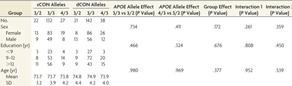
.045), and the Wisconsin Card Sorting Test (number of catego-ries) (P⫽.034). TheAPOE*E4allele had a negative impact on verbal fluency performance at follow-up (P⫽.011) with a signif-icant group⫻APOEinteraction (P⫽.003 for dCON). There was no other significant association betweenAPOEgenotyping and neuropsychological performances at follow-up (Table 1and On-line Table).
Whole-Brain Voxel-Based Morphometry
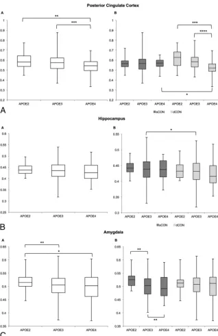
In a first step, we performed a voxelwise analysis across the entire brain. The posterior cingulate cortex demonstrated a significant difference betweenAPOE*E3⬎APOE*E4for the dCON group (Fig 1).
ROI Analysis:APOEEffect on GM Densities in dCON and sCON
In a second step, we additionally performed an ROI analysis in 7 regions of particular interest in the context of cognitive decline (posterior cingulate cortex, hippocampus, mesial temporal lobe, parietal lobe, amygdala, and caudate nucleus) with the occipital lobe as a control region.
In the posterior cingulate, the GM concentration decreased fromE2toE3toE4across all participants. When we separated the patients into stable-versus-deteriorating controls, this decrease in GM concentration was present in only the dCON group(Fig 2A andTable 2). Moreover, we found a significant difference be-tween sCON versus dCON in APOE*E4-positive individuals. Concerning the hippocampus, patients with dCON had lower GM concentrations compared with those with sCON, which was significant only in theAPOE*E3group, presumably due to the large sample size (Fig 2BandTable 3). In the amygdala, there was a decrease in GM concentration fromAPOE*E2toAPOE*E3to
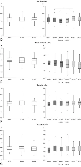
APOE*E4in all participants, and this effect was present only in the sCON but not in the dCON subgroups (Fig 2CandTable 3). In the parietal lobe, the pattern was inverse, with increased GM
con-Table 1: Essential demographic data of the included study groups of stable and deteriorating elderly control participantsa
Group
sCON Alleles dCON Alleles APOEAllele Effect 3/3 vs 3/2 (PValue)
APOEAllele Effect 4/3 vs 3/2 (PValue)
Group Effect (PValue)
Interaction 1 (PValue)
Interaction 2 (PValue) 3/2 3/3 4/3 3/2 3/3 4/3
No. 22 132 27 21 142 38
Sex .734 .411 .172 .261 .359
Female 13 83 19 8 86 26
Male 9 49 8 13 56 12
Education (yr) .466 .324 .676 .808 .450
⬍9 3 23 4 3 27 3
9–12 8 53 14 9 72 20
⬎12 11 56 9 9 43 15
Age (yr) .980 .969 .377 .952 .539
Mean 73.7 73.7 73.8 74.8 74.9 73.9
SD 3.2 3.9 4.2 4.4 4.2 4.0
[image:3.594.57.534.56.196.2]centration in those with dCON, notably in those withAPOE*E3andAPOE*E4
(Fig 2DandTable 3). No significant dif-ferences were found in the mesial tem-poral lobe, occipital lobe, or caudate nu-clei (Fig 2E–GandTable 3).
Logistic Regression Models
Higher GM densities in the hippocampus, middle temporal lobe, and amygdala were all associated with a decreased risk for dCON status at follow-up (hippocam-pus: standardized OR⫽0.75; 95% CI, 0.61– 0.92;P⫽ .006; middle temporal lobe: standardized OR⫽0.80; 95% CI, 0.65– 0.98; P ⫽ .028; and amygdale: standardized OR⫽0.75; 95% CI, 0.61– 0.93,P ⫽ .008). Although significant, these associations explained⬍1.5% of cognitive variability.APOEgenotyping, age, sex, and duration did not predict dCON status at follow-up. When cate-gorizing GM densities into quintiles, we confirmed the assumption of a linearity of their association with the log odds, because the odds ratios displayed a gra-dient that is statistically significant for quintile 4 (thresholdⱖ0.4426118) and quintile 5 (threshold ⱖ 0.48496512), with ORs of 0.51 (P⫽ .044) and 0.44 (P⫽.013).
DISCUSSION
Several studies demonstrated that the presence of theAPOE*E4allele modu-lates the expression of brain atrophy in MCI and clinically overt AD, increasing the vulnerability of the areas prone to early neurodegeneration such as the hip-pocampus, amygdala, and mesial tem-poral lobe.15-17Although higher cortical
amyloidload and decreased metabo-lism in the above-mentioned areas were reported in theAPOE*E4allele, cross-sectional MR imaging studies failed to identify consistent GM decreases associ-ated with this genotype in elderly con-trols (for a review see Fouquet et al44).
The finding in the current investigation thatAPOE*E4was related to GM loss in only the subsequently deteriorating but not in the cognitively stable groups might explain these partially conflicting results of previous studies, which typi-cally do not include detailed neuropsy-chological assessment and follow-up.
The present data show that the
APOE*E4 genotype is not associated
FIG 1. Whole-brain voxel-based morphometry analysis demonstrating higher GM density for the comparison ofAPOE*E3⬎APOE*E4in the posterior cingulate cortex.P⬍.05 corrected.
[image:4.594.55.377.48.161.2] [image:4.594.56.375.208.700.2]with an increased risk of dCON status at follow-up. Consistent with previous reports in elderly controls,15,18,20noAPOE
geno-typing–related effect was identified in the hippocampus and
me-sial temporal cortex in subjects with both sCON and dCON. In agreement with previous observations in this field, these observations indicate that the
APOE*E4 allele detrimental effect in terms of structural changes and clinical progression becomes evident only in el-derly individuals with significant cogni-tive deterioration (MCI) or clinically overt symptoms of dementia (early AD).14-17
The posterior cingulate cortex is known to be affected early in the AD process with significant hypometabo-lism in cognitively healthy individuals and those with MCI (both converters and nonconverters; for review see Tei-pel and Grothe45). In more advanced
stages of the degenerative process, this area exhibits subtle atrophy and hypo-metabolism in subjects with amnestic amyloid-negative AD.46Rare
cross-sec-tional studies addressed the impact of
APOEgenotyping on the structural and functional integrity of the posterior cin-gulate cortex. Early contributions from the Cardiovascular Health Study indi-cated anAPOE*E4-independent age-re-lated atrophy in the hippocampus and posterior cingulate cortex in healthy el-derly controls.47More recently, an
al-tered energy metabolism was reported in this area in young adult APOE*E4
carriers.48In the same line, Lu et al49
re-ported cortical atrophy in the right cingu-late gyrus in cognitively intactAPOE*E4
carriers. Our observations parallel these findings, suggesting that the presence of this allele may have a detrimental effect on GM density in this vulnerable area.
The strengths of the present study in-clude its longitudinal design, large num-ber of community-dwelling subjects, and detailed neuropsychological testing at inclusion and follow-up. However, some limitations should also be con-sidered. First, in line with recent core clinical criteria for MCI,50the
identi-fication of deteriorating controls was based on the objective decline in cogni-tive functions measured by using serial, comprehensive neuropsychological as-sessments. However, in the absence of longer follow-up and AD biomarker characterization at baseline (including PET amyloid scans or the CSF/amyloid 42 ratio), the cognitive fate of these individuals remains uncer-tain so that they cannot be a priori considered as subjects with incipient AD. Second, the rarity ofAPOE*E4homozygotes
[image:5.594.58.371.48.688.2]cluded a detailed analysis of gene-dose effect on GM densities. Third, handedness was not considered as a covariate in our MR imaging analysis, which included both left- and right-handed controls.
CONCLUSIONS
Our data reveal that the presence of theAPOE*E4allele is associ-ated with decreased GM density in the posterior cingulate cortex in dCON, a community-based group of elderly controls who sub-sequently had subtle cognitive decline at 18-month follow-up. ThisAPOEeffect was not identified in cognitively stable controls. Most important, theAPOE*E4allele has no impact on GM den-sity in areas affected early by neurofibrillary tangle formation such as the hippocampus and mesial temporal lobe. These observa-tions suggest that decreased GM density in the posterior cingulate cortex should be systematically detected amongAPOE*E4 con-trols because it could represent a structural marker preceding sub-tle cognitive deficits in the very early stages of the degenerative process.
Disclosures: Sven Haller—RELATED:Grant: Swiss National Foundation,Comments: grant SNF 3200B0 –1161193 and SPUM 33CM30 –124111* and an unrestricted grant from the Assocation Suisse pour la Recherche Alzheimer*. *Money paid to the institution.
REFERENCES
1. Corder EH, Saunders AM, Strittmatter WJ, et al.Gene dose of apoli-poprotein E type 4 allele and the risk of Alzheimer’s disease in late onset families.Science1993;261:921–23CrossRef Medline
2. Strittmatter WJ, Saunders AM, Schmechel D, et al.Apolipoprotein E: high-avidity binding to beta-amyloid and increased frequency of type 4 allele in late-onset familial Alzheimer disease.Proc Natl Acad Sci U S A1993;90:1977– 81CrossRef Medline
3. Jack CR Jr, Therneau TM, Wiste HJ, et al.Transition rates between amyloid and neurodegeneration biomarker states and to dementia: a population-based, longitudinal cohort study.Lancet Neurol2016; 15:56 – 64CrossRef Medline
4. Jack CR Jr, Wiste HJ, Weigand SD, et al.Age-specific population frequencies of cerebral-amyloidosis and neurodegeneration among people with normal cognitive function aged 50 – 89 years: a cross-sectional study.Lancet Neurol2014;13:997–1005
CrossRef Medline
5. Lee YM, Ha JK, Park JM, et al.Impact of apolipoprotein E4 poly-morphism on the gray matter volume and the white matter integ-rity in subjective memory impairment without white matter hyperintensities: voxel-based morphometry and Tract-Based Spa-tial Statistics Study under 3-Tesla MRI.J Neuroimaging2016;26: 144 – 49CrossRef Medline
6. Li B, Shi J, Gutman BA, et al.Influence of APOE genotype on hip-pocampal atrophy over time: an Nⴝ1925 surface-based ADNI study.PLoS One2016;11:e0152901CrossRef Medline
7. Shi J, Lepore´ N, Gutman BA, et al.Genetic influence of apolipopro-Table 2: ROI analysis of GM concentration in the 7 regions for the comparison of sCON versus dCONa
sCON versus dCON Posterior
Cingulate Hippocampus
Mesial Temporal Cortex
Parietal Lobe
Occipital
Lobe Amygdala
Caudate Nuclei
APOE*E2(n⫽22/21) .0524 .3890 .4189 .8146 .4223 .3146 .2863
APOE*E3(n⫽132/142) .0813 .0261b .0756 .0349b .2425 .2567 .1384
APOE*E4(n⫽27/38) .0155b .1916 .4076 .0316b .2433 .5410 .3960
aData arePvalues. b
[image:6.594.53.532.58.125.2]P⬍.05.
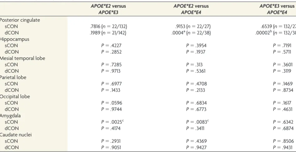
Table 3: ROI analysis of GM concentration in the 7 regions for the comparison ofAPOE*E2versusAPOE*E3andAPOE*E2versus
APOE*E4andAPOE*E3versusAPOE*E4
APOE*E2versus
APOE*E3
APOE*E2versus
APOE*E4
APOE*E3versus
APOE*E4
Posterior cingulate
sCON .7816 (n⫽22/132) .9153 (n⫽22/27) .6539 (n⫽132/27)
dCON .1989 (n⫽21/142) .0004a(n⫽22/38) .00002b(n⫽132/38)
Hippocampus
sCON P⫽.4227 P⫽.3954 P⫽.7191
dCON P⫽.2852 P⫽.1937 P⫽.5711
Mesial temporal lobe
sCON P⫽.7285 P⫽.313 P⫽.3601
dCON P⫽.9713 P⫽.5361 P⫽.3119
Parietal lobe
sCON P⫽.6977 P⫽.4708 P⫽.1469
dCON P⫽.1433 P⫽.2133 P⫽.8734
Occipital lobe
sCON P⫽.0596 P⫽.6834 P⫽.1617
dCON P⫽.9744 P⫽.6773 P⫽.4631
Amygdala
sCON P⫽.0025c P⫽.0083c P⫽.6342
dCON P⫽.4174 P⫽.3411 P⫽.6874
Caudate nuclei
sCON P⫽.2931 P⫽.4369 P⫽.8506
dCON P⫽.9051 P⫽.9427 P⫽.9431
a
P⬍.001.
b
P⬍.0001.
c
[image:6.594.59.532.171.413.2]tein E4 genotype on hippocampal morphometry: an Nⴝ725 sur-face-based Alzheimer’s disease neuroimaging initiative study.Hum Brain Mapp2014;35:-3903–18CrossRef Medline
8. Gon˜i J, Cervantes S, Arrondo G, et al.Selective brain gray matter atrophy associated with APOE4 and MAPT H1 in subjects with mild cognitive impairment. J Alzheimers Dis 2013;33:1009 –19
CrossRef Medline
9. Lu PH, Thompson PM, Leow A, et al.Apolipoprotein E genotype is associated with temporal and hippocampal atrophy rates in healthy elderly adults: a tensor-based morphometry study.J Alzheimers Dis
2011;23:433– 42CrossRef Medline
10. Honea RA, Vidoni E, Harsha A, et al.Impact of APOE on the healthy aging brain: a voxel-based MRI and DTI study.J Alzheimers Dis
2009;18:553– 64Medline
11. Agosta F, Vossel KA, Miller BL, et al.Apolipoprotein E epsilon4 is associated with disease-specific effects on brain atrophy in Alzhei-mer’s disease and frontotemporal dementia.Proc Natl Acad Sci U S A2009;106:2018 –22Medline
12. Wishart HA, Saykin AJ, McAllister TW, et al.Regional brain atrophy in cognitively intact adults with a single APOE epsilon4 allele. Neu-rology2006;67:1221–24CrossRef Medline
13. Pennanen C, Testa C, Boccardi M, et al.The effect of apolipoprotein polymorphism on brain in mild cognitive impairment: a voxel-based morphometric study.Dement Geriatr Cogn Disord2006;22: 60 – 66CrossRef Medline
14. Liu Y, Paajanen T, Westman E, et al.Effect of APOE4 allele on cortical thicknesses and volumes: the AddNeuroMed study.J Alz-heimers Dis2010;21:947– 66CrossRef Medline
15. Lupton MK, Strike L, Hansell NK, et al.The effect of increased ge-netic risk for Alzheimer’s disease on hippocampal and amygdala volume.Neurobiol Aging2016;40:68 –77CrossRef Medline
16. Wang X, Wang J, He Y, et al.Apolipoprotein E4 modulates cogni-tive profiles, hippocampal volume, and resting-state functional connectivity in Alzheimer’s disease.J Alzheimers Dis2015;45:781–95
CrossRef Medline
17. Susanto TA, Pua EP, Zhou J, et al.Cognition, brain atrophy, and cerebrospinal fluid biomarkers changes from preclinical to demen-tia stage of Alzheimer’s disease and the influence of apolipoprotein e.J Alzheimers Dis2015;45:-253– 68CrossRef Medline
18. Chen J, Shu H, Wang Z, et al.The interaction of APOE genotype by age in amnestic mild cognitive impairment: a voxel-based morpho-metric study.J Alzheimers Dis2015;43:657– 68CrossRef Medline
19. Chen Y, Chen K, Zhang J, et al.Disrupted functional and struc-tural networks in cognitively normal elderly subjects with the APOE 4 allele. Neuropsychopharmacology 2015;40:1181–91
CrossRef Medline
20. Habes M, Toledo JB, Resnick SM, et al.Relationship between APOE genotype and structural MRI measures throughout adulthood in the study of health in Pomerania population-based cohort.AJNR Am J Neuroradiol2016;37:1636 – 42CrossRef Medline
21. Matura S, Prvulovic D, Jurcoane A, et al.Differential effects of the ApoE4 genotype on brain structure and function.Neuroimage2014; 89:81–91CrossRef Medline
22. Shaw P, Lerch JP, Pruessner JC, et al.Cortical morphology in children and adolescents with different apolipoprotein E gene polymorphisms: an observational study.Lancet Neurol2007;6:494 – 500CrossRef Medline
23. O’Dwyer L, Lamberton F, Matura S, et al.Reduced hippocampal volume in healthy young ApoE4 carriers: an MRI study.PLoS One
2012;7:e48895CrossRef Medline
24. Ha¨ma¨la¨inen A, Grau-Olivares M, Tervo S, et al.Apolipoprotein E epsilon 4 allele is associated with increased atrophy in progressive mild cognitive impairment: a voxel-based morphometric study.
Neurodegener Dis2008;5:186 – 89CrossRef Medline
25. Risacher SL, Shen L, West JD, et al; Alzheimer’s Disease Neuroimag-ing Initiative (ADNI).Longitudinal MRI atrophy biomarkers: rela-tionship to conversion in the ADNI cohort.Neurobiol Aging2010; 31:1401–18CrossRef Medline
26. Folstein MF, Folstein SE, McHugh PR.“Mini-mental state”: a prac-tical method for grading the cognitive state of patients for the clini-cian.J Psychiatr Res1975;12:189 –98CrossRef Medline
27. Zigmond AS, Snaith RP.The hospital anxiety and depression scale.
Acta Psychiatr Scand1983;67:361–70CrossRef Medline
28. Barberger-Gateau P, Commenges D, Gagnon M, et al.Instrumental activities of daily living as a screening tool for cognitive impairment and dementia in elderly community dwellers.J Am Geriatr Soc1992; 40:1129 –34CrossRef Medline
29. Wechsler DA.Wechsler Memory Scale.3rd ed. San Antonio: Psycho-logical Corporation; 1987
30. Reitan RM.Validity of the Trail Making Test as an indicator of or-ganic brain damage.Percept Mot Skills1958;8:271–76
31. Wechsler D.Manual for the Wechsler Adult Intelligence Scale.New York: Psychological Corporation; 1995
32. Milner B.Interhemispheric differences in the localization of psy-chological processes in man.Br Med Bull1971;27:272–77CrossRef Medline
33. Buschke H, Sliwinski MJ, Kuslansky G, et al.Diagnosis of early de-mentia by the Double Memory Test: encoding specificity improves diagnostic sensitivity and specificity. Neurology 1997;48:989 –97
CrossRef Medline
34. Baddley A, Emslie H, Nimmo-Smith I.Doors and People. A Test of Visual and Verbal Recall and Recognition.Bury St. Edmunds: Thames Valley Test Company; 1994
35. Kaplan EF, Goodglass H, Weintraub S.The Boston Naming Test.2nd ed. Philadelphia: Lea & Febiger; 1983
36. Schnider A, Hanlon RE, Alexander DN, et al.Ideomotor apraxia: behavioral dimensions and neuroanatomical basis. Brain Lang
1997;58:125–36CrossRef Medline
37. Poeck K.Clues to the nature of disruption to limb Praxis.In: Roy EA, ed.Neuropsychological Studies of Apraxia and related Disorders.
Amsterdam: North-Holland; 1985:99 –109
38. Welsh KA, Butters N, Mohs RC, et al.The Consortium to Establish a Registry for Alzheimer’s Disease (CERAD), Part V: a normative study of the neuropsychological battery.Neurology1994;44:609 –14
CrossRef Medline
39. Hughes CP, Berg L, Danziger WL, et al.A new clinical scale for the staging of dementia.Br J Psychiatry1982;140:566 –72CrossRef Medline
40. Petersen RC, Doody R, Kurz A, et al.Current concepts in mild cognitive impairment. Arch Neurol 2001;58:1985–92 CrossRef Medline
41. Xekardaki A, Rodriguez C, Montandon ML, et al.Arterial spin label-ing may contribute to the prediction of cognitive deterioration in healthy elderly individuals.Radiology2015;274:490 –99 CrossRef Medline
42. Nauck M, Hoffmann MM, Wieland H, et al.Evaluation of the apo E genotyping kit on the LightCycler.Clin Chem2000;46:722–24
Medline
43. Smith SM, Nichols TE. Threshold-free cluster enhancement: addressing problems of smoothing, threshold dependence and localisation in cluster inference. Neuroimage 2009;44:83–98
CrossRef Medline
44. Fouquet M, Besson FL, Gonneaud J, et al.Imaging brain effects of APOE4 in cognitively normal individuals across the lifespan. Neu-ropsychol Rev2014;24:290 –99CrossRef Medline
45. Teipel S, Grothe MJ; Alzheimer⬘s Disease Neuroimaging Initiative.
Does posterior cingulate hypometabolism result from disconnec-tion or local pathology across preclinical and clinical stages of Alz-heimer’s disease. Eur J Nucl Med Mol Imaging 2016;43:526 –36
CrossRef Medline
46. Che´telat G, Ossenkoppele R, Villemagne VL, et al.Atrophy, hypo-metabolism and clinical trajectories in patients with amyloid-negative Alzheimer’s disease.Brain2016;139:2528 –39CrossRef Medline
gray matter volume in cognitively normal elders.Neurobiol Aging
2012;33:-834.e7–16CrossRef
48. Perkins M, Wolf AB, Chavira B, et al.Altered energy metabolism pathways in the posterior cingulate in young adult apolipoprotein E4 carriers.J Alzheimers Dis2016;53:95–106CrossRef Medline
49. Lu H, Fung AW, Chan SS, et al.Disturbance of attention net-work functions in Chinese healthy older adults: an
intra-indi-vidual perspective. Int Psychogeriatr2016;28:291–301CrossRef Medline
50. Albert MS, DeKosky ST, Dickson D, et al.The diagnosis of mild cognitive impairment due to Alzheimer’s disease: recommenda-tions from the National Institute on Aging-Alzheimer’s Associa-tion workgroups on diagnostic guidelines for Alzheimer’s disease.