ORIGINAL RESEARCH
Brain Volume and Diffusion Markers as
Predictors of Disability and Short-Term Disease
Evolution in Multiple Sclerosis
P.G. Sa¨mann M. Knop E. Golgor S. Messler M. Czisch F. Weber
BACKGROUND AND PURPOSE: MRI markers of neuroaxonal damage in MS have emerged as critical long-term predictors of MS-related disability. Here we investigated the potential of whole-brain diffusivity and brain volume for the prediction of cross-sectional disability and short- to medium-term clinical evolution.
MATERIALS AND METHODS: In this multimodal prospective longitudinal MRI study of 54 patients with MS (87% under immunomodulatory therapy, baseline and follow-up at a median of 12 months), ADC histogram analysis, WM lesion load, BPF, whole-brain atrophy rate, MSFC score, and EDSS score were obtained. A total of 44 patients with no relapse at both time points were included.
RESULTS: At both time points, ADC histogram analysis provided robust predictors of the MSFC scores (maximalR2⫽0.576,P ⬍.001), incorporated cognition and fine-motor skill subscores, and EDSS scores. Significant changes beyond physiologic age-related changes at follow-up were noted for ADC histogram markers and BPF. Stronger diffusivity alterations and brain volume at baseline predicted MSFC decline, as demonstrated by multiple linear regression analysis (mean ADC,R2⫽0.203;P⫽ .003) and lower baseline BPF in patients with declined compared with stable MSFC scores (P⫽.001). Results were independent of intercurrent relapses.
CONCLUSIONS:Diffusion histogram analysis provided stable surrogates of disability in MS and proved sensitive for monitoring disease progression during a median of 12 months. Advanced neuroaxonal pathology at baseline was indicative of an increased risk for sustained progression during a median of 12 months, independent of intercurrent relapses.
ABBREVIATIONS:BPF⫽brain parenchyma fraction; CI⫽confidence interval; EDSS⫽Expanded Disability Status Scale; GM⫽gray matter; MSFC⫽MS Functional Composite; 9-HPT⫽9-Hole Peg Test; PASAT⫽Paced Auditory Serial Addition Test; PBVC⫽percentage brain volume change; TWT⫽timed walk test; WMLLperc⫽WM lesion load volume as percentage of total WM volume
A
cute inflammation and demyelination, secondary neu-roaxonal pathology, and additional neurotrophic distur-bances conjointly lead to clinical impairment in MS.1,2Among these factors, the cumulative neuroaxonal damage is a partic-ularly strong determinant of disability.3Exceeding a certain threshold of neuroaxonal damage might accelerate a patient’s transition to secondary-progressive MS or “sustained progres-sion.”3 Therefore, MRI techniques that are sensitive to the cumulative neuroaxonal damage such as volumetry2,4 and DWI5 warrant further investigation to improve the clinical management of MS.Brain-volume loss in MS is a multifactorial process that originates from inflammatory focal axonal damage and de-pletion of myelin sheaths, secondary neuroaxonal
degenera-tion, and other immunologically triggered neurotrophic dis-turbances.2,4It can be observed across all MS subtypes,6even in early stages,7and its relation to physical and cognitive dis-ability is generally recognized.8-10In relapsing-remitting MS, between 47% and 81% of brain atrophy was ascribed to the previous cumulative gadolinium enhancement.11Other stud-ies suggest that brain atrophy is a consequence of diffuse pa-thology rather than focal lesions.4,12In fact, signs of strong tissue destruction may occur during the course of MS despite low cumulative inflammatory activity.13The strong clinical relevance of brain volumetry in MS is supported by correla-tions between baseline brain volume and disability occurring 8 years later14and associations between early brain atrophy rates and clinical deterioration.15
DWI detects alterations of microscopic diffusion processes in MS due to a variety of factors, including loss of myelin sheaths, loss of axonal membranes, neuronal apoptosis, and gliosis formation.5It is now well-established that diffusivity measurements are sensitive to MS-related pathology in brain areas that appear normal on conventional T2- and T1-weighted images.5,16-18Notably, diffusivity changes parallel grades of axonal pathology in animal models19and in hu-mans,20,21 which might explain correlations of diffusivity markers with patient disability status.22-24Serial application of DWI revealed progressive microstructural GM changes in untreated relapsing-remitting MS,25and a good prediction Received July 12, 2011; accepted after revision October 16.
From the Neuroimaging Research Group (P.G.S., E.G., M.C.) and Inflammatory Disorders of the Central Nervous System Research Group (M.K., F.W.), Max Planck Institute of Psychi-atry, Munich, Germany; and Department of Statistics (S.M.), Ludwig-Maximilians-Univer-sita¨t, Munich, Germany. Dr Golgor is currently affiliated with the Institute of Diagnostic Radiology, University Hospital Carl Gustav Carus, Technical University Dresden, Dresden, Germany. Dr Messler is currently affiliated with INC Research GmbH, Munich, Germany.
The authors declare no competing interests.
Please address correspondence to P.G. Sa¨mann, MD, Kraepelinstr 2-10, 80804 Munich, Germany; e-mail: [email protected]
Indicates article with on-line appendix.
Indicates article with supplemental on-line figure.
of the potential for the clinical status after 5 years in primary-progressive MS.26
Despite the sensitivity of DWI, however, there are scant serial data on longitudinal DWI measures, particularly in treated relapsing-remitting MS and in combination with sen-sitive clinical monitoring instruments such as the MSFC score.27 Furthermore, serial studies have either focused on DWI25,26,28,29 or brain volume measurements,6,9,11,14,30-36 with only 1 serial study on primary-progressive MS and secondary-progressive MS using both techniques.37 In this prospective, longitudinal, and multimodal MRI study on pa-tients with MS under treatment, we investigated the potential of whole-brain diffusivity and brain volume for the prediction of cross-sectional disability and short-to-medium-term clini-cal evolution.
Materials and Methods
Patients with MS, Clinical Evaluation, and Controls Patients were consecutively recruited from the outpatient clinic and the neurologic ward of the Max Planck Institute of Psychiatry, Mu-nich. They fulfilled the criteria of definite MS according to McDonald et al,38with the major proportion classified as relapsing-remitting MS (47 patients) (secondary-progressive MS, 5 patients; primary-progressive MS, 2 patients). Patients with relapses at baseline or at follow-up (n⫽10) were excluded from clinicoradiologic correlation analysis to avoid confounding influences from transient clinical exac-erbation,39leaving 44 patients for the final analysis (Table 1). Disease duration was estimated from a detailed clinical history and file review. Thirty-seven of 44 patients (84%) received immunomodulatory ther-apy at study entry (Table 1). At baseline and follow-up after a median of 12 months (median, 371 days; range 308 –702 days), the EDSS40 and MSFC scores,27comprising the TWT, 9-HPT, and a 3-second version of the PASAT, were obtained. Patients withⱖ1 relapse during the observation interval were identified for post hoc analyses as spec-ified below. The number of patients with MSFC scores available at both time points varied between 38 and 40 due to (disease-related) dropouts in subtests.
Follow-up MSFC scores were interpolated to a 12-month interval (annualized scores). Clinical progression was parameterized as the difference between baseline and annualized follow-up scores. For the
MSFC sum score and subscores, patients with a negative annual change value were assigned to the respective progression group in a first step. Second, to reduce false classification into the MSFC-progression group, we classified 20% of the patients with the lowest progression rates as stable. For the EDSS, an increase ofⱖ0.5 point between baseline and follow-up with confirmation 3 months later was classified as EDSS progression.
For proof-of-concept comparisons and estimation of age effect, an age and sex-matched control group free of neurologic or psychi-atric disease underwent the same MRI protocol once (n⫽54, Table 1).
The study followed the principles of the Declaration of Helsinki and was approved by the local ethics committee. All participants gave their written informed consent.
MR Imaging Acquisition and Postprocessing: Overview Images were acquired on a clinical 1.5T scanner (Signa Excite; GE Healthcare, Milwaukee, Wisconsin). Sequence details and postpro-cessing steps are described in the on-line supplemental material. In brief, we extracted the following MRI markers: 1) Whole-brain ADC histograms were calculated; mean, variance, skew, and peak-height values were extracted.18,412) The BPF (brain parenchyma volume divided by total intracranial volume42) was calculated at baseline and follow-up from T2-weighted images with high in-plane resolution and CSF/parenchyma contrast by using. 3) The brain-volume change between baseline and follow-up (PBVC) was calculated by using the SIENA algorithm of the FSL software (http://www.fmrib.ox.ac.uk, version 3.2).434) For WM lesion load quantification, multispectral image segmentation based on an expectation maximization algo-rithm was used.445) Axial and coronal postgadolinium images of both time points were screened by 2 raters (F.W., P.G.S.) blinded to patient identity and time points.
Statistical Analysis
Baseline MRI measures of all patients were compared with those of controls by using univariate multivariate analysis of covariance based on Wilks, covarying for age. Group⫻age interaction effects were explored, and the term was removed from the model if not significant (P⬎.05).
Follow-up MRI variables (ADC histogram metrics, BPF, and WMLLperc) as well as follow-up clinical scores were interpolated to a 12-month interval. Paired tests were used to compare baseline and annualized MRI and parametric clinical markers (Wilcoxon signed rank test for EDSS;ttest for other variables). The patients’ annual atrophy rates as calculated by the SIENA algorithm were compared against zero by using a 1-samplettest. Annual change rates and 95% CIs for MRI variables of the control group were estimated by linear regression analysis.
For cross-sectional clinico-radiologic correlations, the Spearman rank correlation tests (for EDSS) and the Pearson partial correlation tests corrected for age (for MSFC scores) were applied to baseline and follow-up values. For the 28 MSFC- and 7 EDSS-related tests, Bonferroni-adjusted significance thresholds were defined (0.05/28⬃ 0.0018 and 0.05/7⬃0.0071, respectively) to adjust for explorative testing. For baseline and follow-up MSFC and EDSS, stepwise linear regression analysis (variable entry atP⬍.05, variable removal atP⬎
.10) was appended to identify independent predictors among the MRI variables. ReportedR2values represent the proportion of explained variance, adjusted for the entry of multiple regressors.
Prediction of clinical progression was analyzed in 2 ways: 1)
Base-Table 1: Demographic, clinical, and therapeutic characteristics of patients and controls
Patients Controls
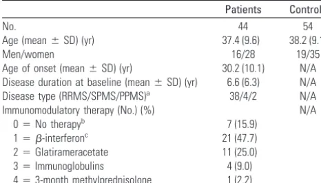
No. 44 54
Age (mean⫾SD) (yr) 37.4 (9.6) 38.2 (9.1) Men/women 16/28 19/35 Age of onset (mean⫾SD) (yr) 30.2 (10.1) N/A Disease duration at baseline (mean⫾SD) (yr) 6.6 (6.3) N/A Disease type (RRMS/SPMS/PPMS)a 38/4/2 N/A
Immunomodulatory therapy (No.) (%) N/A 0⫽No therapyb 7 (15.9)
1⫽-interferonc 21 (47.7)
2⫽Glatirameracetate 11 (25.0) 3⫽Immunoglobulins 4 (9.0) 4⫽3-month methylprednisolone 1 (2.2)
Note:—NA indicates not applicable; PPMS, primary-progressive MS; RRMS, relapsing-remitting MS; SPMS, secondary-progressive MS.
aNo significant differences between patients with RRMS and SPMS/PPMS were detected
for age, age of onset, and disease duration.
bOne patient changed to interferon treatment during the study period. c
Three patients changed from interferon to mitoxantrone therapy during the study period.
BRAIN
ORIGINAL
[image:2.594.52.285.70.203.2]line MRI markers were compared between patients with and without MSFC progression by using analysis of covariance (2-level group fac-tor, age as a covariate). The analysis was repeated for patients strati-fied according to EDSS progression. 2) Stepwise linear regression, by using the annual change of MSFC sum score and subscores as depen-dent variables and baseline MRI variables (ADC histogram markers, BPF, WMLLperc) as predictor variables, was applied. To exclude any influence of relapses during the observation interval, we performed the following post hoc analyses: 1) The proportion of patients with intercurrent relapses was compared between the MSFC progression and nonprogression groups (Fisher exact test). 2) MSFC prediction analyses were repeated after exclusion of patients with intercurrent relapses and for patients with relapsing-remitting MS only.
Results
Clinical Characteristics and Disease Progression
Clinical and demographic sample information is given in Table 1. Table 2 shows comparisons of clinical baseline and annualized follow-up scores. Subtle yet nonsignificant progression of the MSFC sum score, TWT, and 9-HPT and improvement of the PASAT were noted. Twenty percent of patients showed a confirmed EDSS increase ofⱖ0.5 points. The MSFC threshold that stratified patients into MSFC-progression and MSFC-nonMSFC-progression groups was⫺0.047 (13 progressive, 25 nonprogressive patients).
Patient/Control Comparison of ADC Histograms and BPF Significant differences between patients and controls were de-tected for ADC histogram markers (multivariate analysis of covariance,F⫽13.895,P⬍10⫺6) with an age covariate effect (F⫽4.840,P⫽.001). Similarly, mean BPF differed signifi-cantly between patients (0.815⫾0.043) and controls (0.837⫾ 0.041) (analysis of covariance,F⫽6.592,P⫽.004) with a covariate effect of age (F ⫽12.580,P⫽.001) and a trend group⫻age interaction (F⫽3.667,P⫽.059) (On-line Table 1). BPF correlated with age in the control group (r2⫽0.269, P⬍10⫺4) but not in the patient group (r2⫽0.024,P⫽.314).
Longitudinal Changes of MRI Markers
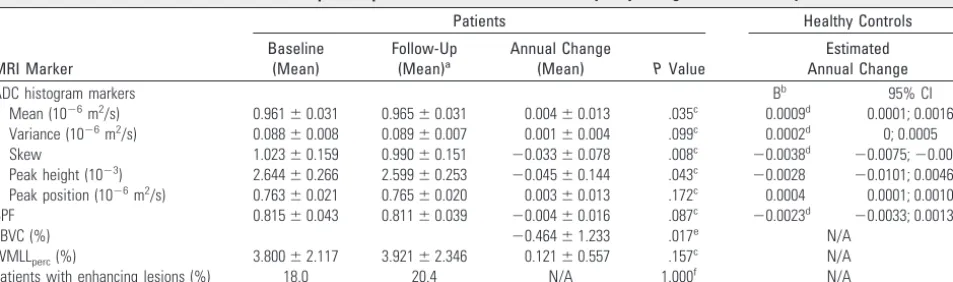
Changes were most explicit for ADC histogram skew, followed by mean ADC and peak height (Table 3). Mean BPF decreased by 0.42% (P ⫽ .087), with a similar yet significant mean change calculated by SIENA (PBVC,⫺0.46%;P⫽.017), con-firming that SIENA is more sensitive to change compared with subtraction of 2 BPF measurements.43 WMLL
[image:3.594.53.532.60.124.2]perc did not change significantly. Annual change rates of all markers in the patient group were outside the 95% CI of the estimated coef-ficients of controls and exceeded control values by factors 1.7 (BPF) to 8.7 (ADC histogram skew). Gadolinium enhance-ment was found in 8 patients (18.2%; median gadolinium-enhancement score, 1.0 [range, 1–2]) at baseline and in 9 pa-tients (20.4%; median gadolinium-enhancement score, 2.0 [range, 1–10]) at follow-up. At both baseline and follow-up,
Table 3: MRI markers at baseline and follow-up of all patients with MS and estimated yearly changes of control subjects
MRI Marker
Patients Healthy Controls Baseline
(Mean)
Follow-Up (Mean)a
Annual Change
(Mean) PValue
Estimated Annual Change
ADC histogram markers Bb 95% CI
Mean (10⫺6m2/s) 0.961⫾0.031 0.965⫾0.031 0.004⫾0.013 .035c 0.0009d 0.0001; 0.0016
Variance (10⫺6m2/s) 0.088⫾0.008 0.089⫾0.007 0.001⫾0.004 .099c 0.0002d 0; 0.0005
Skew 1.023⫾0.159 0.990⫾0.151 ⫺0.033⫾0.078 .008c ⫺0.0038d ⫺0.0075;⫺0.0002
Peak height (10⫺3) 2.644⫾0.266 2.599⫾0.253 ⫺0.045⫾0.144 .043c ⫺0.0028 ⫺0.0101; 0.0046
Peak position (10⫺6m2/s) 0.763⫾0.021 0.765⫾0.020 0.003⫾0.013 .172c 0.0004 0.0001; 0.0010
BPF 0.815⫾0.043 0.811⫾0.039 ⫺0.004⫾0.016 .087c ⫺0.0023d ⫺0.0033; 0.0013
PBVC (%) ⫺0.464⫾1.233 .017e N/A
WMLLperc(%) 3.800⫾2.117 3.921⫾2.346 0.121⫾0.557 .157
c N/A
Patients with enhancing lesions (%) 18.0 20.4 N/A 1.000f N/A
Note:—N/A indicates not applicable.
aAnnualized values are given except for assessment of gadolinium enhancement. bUnstandardized coefficient of linear regression.
cTwo-sided pairedttest.
dSignificant linear correlation with age in the control group (Pⱕ.05).
ePBVC as estimated by the SIENA method. One-samplettest for comparison against zero. fFisher exact test.
Table 2: Change of neurologic scores during a median of 12 months
Score No. Baseline Follow-Upa PValueb Split Thresholdc Progression Nonprogression EDSS (median) (range) 44 2.0 (0–5.5) 2.0 (0–5.5) 0.950 ⱖ0.5c 9 35
MSFC (mean) (SD) 38 0.550 (0.415) 0.549 (0.475) 0.969 ⬍⫺0.047 13 25 TWT (mean) (SD)d 39 ⫺0.431 (0.082) ⫺0.412 (0.098) 0.105 ⬎0.013 16 23
9-HPT (mean) (SD)e 40 0.780 (0.735) 0.736 (0.837) 0.382 ⬍⫺0.113 15 25
PASAT (mean)f 39 0.460 (0.693) 0.531 (0.800) 0.414 ⬍⫺0.101 8 31
aAnnualized values for MSFC, raw values for EDSS (for details see “Materials and Methods”). b
Two-sided Wilcoxon signed rank test (EDSS) and paired Studentttest (MSFC, annualized follow-up values), respectively.
cSee “Materials and Methods” section for details on threshold definition. d
TWT.
e9-HPT. f
[image:3.594.57.534.197.338.2]the gadolinium-enhancement score had no effect on ADC his-togram markers or BPF (P⬎.05).
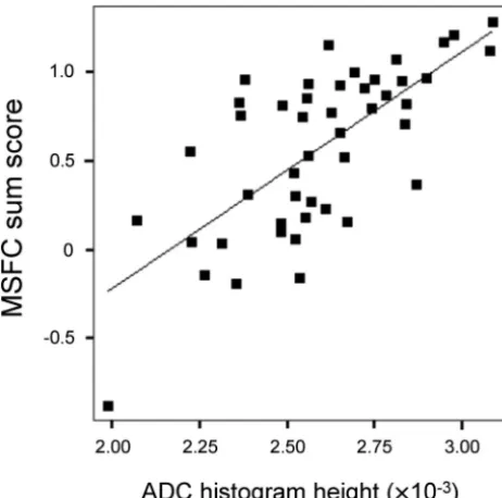
Interrelation between MRI Markers and Disability Interrelations between MRI and disability were highly similar at baseline and follow-up (see On-line Table 2 for details on correlations at follow-up). At follow-up, interrelations with diffusion markers were more distinct (r⫽0.759 [ADC histo-gram skew, P ⬍ .001], Fig 1) than interrelations with WMLLperc(maximumr⫽ ⫺0.487 for MSFC sum score) or BPF (maximumr⫽0.438 for 9-HPT). EDSS correlated with 4 of 5 ADC histogram markers and with WMLLperc, but not with BPF.
At baseline, linear regression analysis identified ADC his-togram variance as an independent predictor of EDSS (R2⫽ 0.137,P⫽.008), MSFC (R2⫽0.186,P⫽.003), and 9-HPT (R2⫽0.204,P⫽.001). At follow-up, ADC histogram variance was again identified as a predictor of EDSS (R2⫽0.118,P⫽ .013); ADC histogram skew, as a predictor of MSFC (R2⫽ 0.533,P⬍.001); and PASAT (R2⫽0.364,P⬍.001) and ADC histogram height, as a predictor of TWT (R2⫽0.090,P⫽ .032) and 9-HPT (R2⫽0.371,P⬍.001).
Prediction of Disease Progression from Baseline MRI Markers
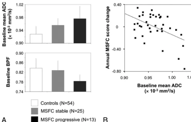
Among the baseline MRI markers, ADC and BPF allow a dif-ferentiation between future MSFC progression versus non-progression (P⫽.042 andP⫽.001, respectively, Table 4). The latter result was robust toward the Bonferroni correction (P⬍ .05/7⬃.007) (Fig 2A). The 2 patient subgroups did not differ with regard to age, age at onset, disease duration, baseline MSFC, baseline EDSS, and the proportion of patients with a relapse during the observation interval (24% and 22%, Fisher exact test,P⫽.709). Baseline BPF also differentiated between patients with and without EDSS progression (P⫽
.038). No significant covariate effect of age was found in any comparison.
When we modeled the MSFC subscore changes as con-tinuous variables, mean ADC emerged as an independent predictor of the annual change of MSFC (F⫽10.40,R2⫽ 0.203,P⫽.003, Fig 2B), 9-HPT (F⫽12.40,R2⫽0.226,P⫽ .001), and PASAT (F⫽4.36,R2⫽0.081,P⫽.044). For a change of TWT, baseline WMLLpercemerged as a predictor (F⫽5.81,P⫽.021,R2⫽0.112).
Post hoc, an effect of intercurrent relapses on these results was excluded as follows: 1) Patients with intercurrent relapses showed a lower (absolute) mean annual MSFC decrease (n⫽ 4, ⫺0.19 ⫾ 0.11) than patients without relapses (n ⫽ 9,
⫺0.28⫾0.26), excluding the finding that MSFC decline was driven by intercurrent relapses. 2) After exclusion of patients with intercurrent relapses, results were stable for the linear regression on the annual MSFC change (baseline mean ADC, r⫽0.487,P⫽.007) and for the comparison of baseline BPF between progressive and nonprogressive patients (P⫽.0009). 3) Both results were also stable after restriction to patients with relapsing-remitting MS only (n⫽38).
Discussion
Neuroaxonal damage in MS is a strong mediator of clinical impairment1 and critical for the development of sustained progression.3This clinico-radiologic study on putative neu-roaxonal markers obtained 3 main results: 1) Diffusivity his-togram metrics were robust predictors of current disability (MSFC and EDSS). 2) Longitudinal analysis revealed patho-logically accelerated changes of diffusivity histogram metrics and whole-brain volume during a median of 12 months, mostly in treated patients with relapsing-remitting MS. 3) Ad-vanced brain atrophy and diffusivity alterations at baseline were associated with MSFC decline, independent of intercur-rent relapses. More advanced brain atrophy at baseline was also associated with EDSS decline.
ADC histogram parameters of patients with MS distinctly differed from those of controls, with control values found in the range reported in an earlier normative study.18 In MS, Fig 1.Relationship between ADC histogram peak height and MSFC at follow-up (n⫽46,
[image:4.594.54.285.44.273.2]r⫽0.699, age-corrected partialr⫽0.713,P⬍.001). Patients with acute relapses at the time of assessment were excluded due to potential distortion of their clinical scores.
Table 4: Baseline MRI and clinical markers of patients with and without MSFC progression
MSFC Nonprogression
(Mean)
MSFC Progression
(Mean) P No. of patients 25 13
ADC histogram markersa
Mean (10⫺6m2/s) 0.956⫾0.021 0.976⫾0.039 .042
Variance (10⫺6m2/s) 0.088⫾0.008 0.092⫾0.008 n.s.
Skew 1.035⫾0.116 0.951⫾0.193 n.s. Peak height (10⫺3) 2.662⫾0.241 2.539⫾0.282 n.s.
Peak position (10⫺6m2/s) 0.758⫾0.017 0.768⫾0.018 n.s.
BPF 0.828⫾0.040 0.783⫾0.027 .001b
WMLLperc(%) 3.365⫾1.673 4.435⫾2.228 n.s.
Clinical variablesc
Age 36.9⫾9.2 39.5⫾11.0 n.s. Disease duration 6.6⫾6.6 7.5⫾7.0 n.s. Baseline MSFC 0.53⫾0.46 0.59⫾0.33 n.s.
Note:—n.s. indicates not significant.
a
Analysis of covariance with 2-level group factor and age as covariate for MRI variables.
bPvalue lower than Bonferroni-corrected .05/7⬃.007. c
[image:4.594.300.532.67.235.2]abnormal diffusivity is an established finding detectable in fo-cal lesions (ie, WM areas of T2 hyperintensity), whole WM, whole brain, and normal-appearing WM and GM.5,18,22,45 Comparisons of ADC values among normal-appearing WM, T1 isointense, and T1 hypointense lesions suggest that ADC might parallel different degrees of axonal pathology.20,21 Sim-ilar interrelations between diffusivity and axonal pathology were reported in animal models of MS.19Because axonal in-jury is considered a key factor of disability in MS,1,3diffusion imaging might serve as a source of particularly useful surro-gate markers. In this study, ADC histogram metrics indeed correlated with the patient’s current disability status both at baseline and follow-up (see On-line Table 2 for cross-sectional correlations). Mean ADC proved robust across most clinical measures, while even higher correlations were obtained by his-togram distribution measures (eg, skew), suggesting that dis-tribution measures detect diffuse or multifocal diffusivity changes most sensitively.16,46 As expected, the relationship between the MSFC sum score and WMLLperc was weaker, likely because focal T2 hyperintensity is histopathologically unspecific and does not capture diffuse pathology.47 Fine motor skills (9-HPT) and cognition (PASAT) also correlated more strongly with diffusivity measures than with BPF and WMLLperc. Interrelations with ambulatory function were gen-erally weaker, most likely due to the lack of a spinal marker.
Brain volume was the second marker on which we focused. Patients showed a reduced baseline BPF compared with matched controls, as reported by Kalkers et al6and Rudick et al.48The physiologic negative relationship between BPF and age was disrupted in patients because younger patients already showed a low BPF. Most interesting, younger patients (me-dian, 36.4 years) exhibited a shorter disease duration (4.6⫾ 4.0 versus 9.4⫾6.8 years,P⫽.003) compared with the older patients, suggesting that young age at onset could increase the risk of developing brain atrophy. This hypothesis, however, needs support from larger samples. BPF shared only a low proportion of variance with WMLLperc(⬃22%), as reported by Guttman et al49and Simon et al,50confirming that both the
pathology of the normal WM and cortical processes contrib-ute substantially to brain atrophy.31,34,51 Moderate correla-tions between BPF and the MSFC sum scores, as described by Kalkers et al,52and with the 9-HPT could be established. No correlation with the PASAT was found, as shown in a pre-vious negative report.53So far, significant BPF/PASAT corre-lations have only been reported for 82 patients with MS of different clinical subtypes54and for 45 patients with primary-progressive MS10; however, these patient samples showed a more advanced atrophy.
The longitudinal analysis served to probe whether the posed markers are suitable to monitor and predict disease pro-gression. In contrast to the clinical ratings, diffusivity mea-sures and BPF showed a significant annual progression that exceeded the values estimated for normal aging. An average annual BPF decrease of approximately 0.4%– 0.5% is in line with 0.45% reported for treated relapsing-remitting MS,33,35 larger than the reported rates in healthy subjects (0.1%– 0.3%),4and slightly above 0.36% observed in patients with relapsing-remitting MS with optimally suppressed inflamma-tory activity.11
Eventually, the longitudinal analysis revealed that more ad-vanced brain volume loss and higher mean ADC at baseline were associated with short-term progression, as primarily de-fined from the MSFC score. When we modeled this progres-sion as a continuous variable, avoiding arbitrary thresholding, baseline mean ADC emerged as an independent predictor. Changes of the 9-HPT and PASAT subscores that generally contribute strongly to the MSFC score55were also predictable from baseline mean ADC. The same result pattern emerged when the analysis was restricted to patients with relapsing-remitting MS and when patients with intercurrent relapses were excluded. Contrary to BPF and diffusivity measures, WMLLpercas a focal disease marker showed no progression and proved a weak predictor of current disability and no pre-dictor of disease progression.
[image:5.594.134.450.43.245.2]pathology cannot be claimed. Postmortem data, for example, have revealed a significant contribution of myelin content to mean diffusivity,56and also in our study, mean ADC and BPF shared approximately 34% of variance. Diffuse demyeli-nation, through its effects on myelin volume and microscopic diffusion, might therefore influence both volume and ADC measurements. For brain-volume loss in MS, axonal pathol-ogy has been proposed to play a prior role because WM volume comprises more axonal (46%) than myelin volume (24%).4In turn, higher sensitivity of diffusivity toward demy-elination is suggested by larger covariation between WMLLperc as an indicator of focal demyelination and ADC markers (av-erageR2⬃32%) compared with BPF (R2⫽21%). Statistically, results pointed to a higher sensitivity of whole-brain mean ADC compared with BPF in linear prediction models. This effect was robust toward covarying for BPF and remained sig-nificant after recalculation of the histograms at a stricter CSF threshold of 1.5⫻10⫺3mm2/s (data not shown). The corre-lation between baseline BPF and disease progression was more robust in that changes of EDSS and MSFC could be predicted, yet it was more nonlinear. Most interesting, such a nonlinear relationship between axonal pathology and secondary pro-gression was hypothesized earlier.3
Taken together, we propose that longitudinal results add new evidence for the hypothesis that accumulation of diffuse axonal pathology may (gradually) increase the risk for clinical progression3whereby the contribution of other nonfocal pro-cesses, including diffuse demyelination, remains to be clari-fied. With regard to brain-volume loss, reaching the stage of secondary progression is particularly critical because slowing brain atrophy rates by immunomodulatory therapy then be-comes difficult.57Results also demonstrate that subtle clinical progression can be detected by the MSFC score before conven-tional criteria of secondary disease progression58apply. No predictive value of central atrophy for EDSS progression dur-ing 14 months was found in a larger study36; however, central atrophy is different from BPF used in this study.
Several limitations of the present study need to be consid-ered. Foremost, the small sample size and the observational design impose limitations on result generalizability. In partic-ular, results are not representative of the spontaneous course of MS because treatment was applied as clinically required. Furthermore, intrinsic to the study design, EDSS or MSFC score progression may reflect a combination of sustained pro-gression as attributable to the natural course of disease and nonresponse to therapy. Technically, higher spatial resolution and fully automated repositioning tools would be preferred, particularly for optimal lesion volumetry. Last, for the gener-ation of ADC histograms, CSF masking was based on a previ-ously reported41fixed ADC threshold, which leads to partial volume effects by macroscopic CSF and hampers a definite attribution of ADC effects to microscopic tissue properties. Indeed, covariation between BPF and mean ADC decreased about linearly when the ADC clipping threshold was lowered. So far, however, it has not been systematically defined which threshold best reflects the true biological correlation between the 2 markers. To categorically avoid partial volume effects, fluid-attenuated DWI may be useful.59
Conclusions
Whole-brain diffusivity and whole-brain volume measure-ments provide clinically valid and sensitive integral markers to monitor cerebral disease burden in MS during a clinically short time interval of approximately 1 year. The association of advanced brain volume loss and diffusivity changes at baseline with short-term disease progression further suggests that ad-vanced neuroaxonal damage represents a risk for sustained progression.
Acknowledgments
We thank Reinhold Borschke and Elke Schreiter for their expertise in clinical MRI acquisitions and Rosa Hemauer for supporting image postprocessing. We also gratefully acknowl-edge Kemal Ates for informatics support and software main-tenance; and Emma Bauer, Dr Sandra Lutz, and Dr Felix Mu¨ller-Sarnowski for clinical rating. We further thank Dr Erina Schumann-Spa¨th for critical discussion of radiologic as-pects of the study and Dorothea Skottke for kind revision of the manuscript.
Disclosures: Susanne Messler—RELATED:Grant: Deutsche Forschungsgemeinschaft, Com-ments: SFB 386 (Sonderforschungsbereich),*UNRELATED: Employment: Definiens AG,
Comments: full-time employee in the field of image analysis in the life sciences area. Frank Weber—UNRELATED:Consultancy: Pfizer-Pharma, Merck-Serono, Orion Pharma;Grants/ Grants Pending: Bayer-Schering AG,* Merck-Serono,* TEVA Pharma GmbH,*Payment for Lectures (including service on speakers bureaus): Bayer-Schering AG, Biogen Idec, Orion Pharma,Comments: honoraria for lectures;Travel/Accommodations/Meeting Expenses Unrelated to Activities Listed: Biogen Idec, Merck-Serono. *Money paid to the institution.
References
1. Hauser SL, Oksenberg JR.The neurobiology of multiple sclerosis: genes, in-flammation, and neurodegeneration.Neuron2006;52:61–76
2. Anderson VM, Fox NC, Miller DH.Magnetic resonance imaging measures of brain atrophy in multiple sclerosis.J Magn Reson Imaging2006;23:605–18 3. Trapp BD, Ransohoff R, Rudick R.Axonal pathology in multiple sclerosis:
relationship to neurologic disability.Curr Opin Neurol1999;12:295–302 4. Miller DH, Barkhof F, Frank JA, et al.Measurement of atrophy in multiple
sclerosis: pathological basis, methodological aspects and clinical relevance.
Brain2002;125:1676 –95
5. Rovaris M, Gass A, Bammer R, et al.Diffusion MRI in multiple sclerosis. Neu-rology2005;65:1526 –32
6. Kalkers NF, Ameziane N, Bot JC, et al.Longitudinal brain volume measure-ment in multiple sclerosis: rate of brain atrophy is independent of the disease subtype.Arch Neurol2002;59:1572–76
7. Chard DT, Griffin CM, Parker GJ, et al.Brain atrophy in clinically early relaps-ing-remitting multiple sclerosis.Brain2002;125:327–37
8. De Stefano N, Matthews PM, Filippi M, et al.Evidence of early cortical atrophy in MS: relevance to white matter changes and disability.Neurology2003;60: 1157– 62
9. Zivadinov R, Sepcic J, Nasuelli D, et al.A longitudinal study of brain atrophy and cognitive disturbances in the early phase of relapsing-remitting multiple sclerosis.J Neurol Neurosurg Psychiatry2001;70:773– 80
10. Sastre-Garriga J, Ingle GT, Chard DT, et al.Grey and white matter atrophy in early clinical stages of primary progressive multiple sclerosis.Neuroimage
2004;22:353–59
11. Richert ND, Howard T, Frank JA, et al.Relationship between inflammatory lesions and cerebral atrophy in multiple sclerosis.Neurology2006;66:551–56 12. De Stefano N, Iannucci G, Sormani MP, et al.MR correlates of cerebral atrophy
in patients with multiple sclerosis.J Neurol2002;249:1072–77
13. Bielekova B, Kadom N, Fisher E, et al.MRI as a marker for disease heterogene-ity in multiple sclerosis.Neurology2005;65:1071–76
14. Fisher E, Rudick RA, Simon JH, et al.Eight-year follow-up study of brain atrophy in patients with MS.Neurology2002;59:1412–20
15. Horakova D, Dwyer MG, Havrdova E, et al.Gray matter atrophy and disability progression in patients with early relapsing-remitting multiple sclerosis: a 5-year longitudinal study.J Neurol Sci2009;282:112–19
16. Ciccarelli O, Werring DJ, Wheeler-Kingshott CA, et al.Investigation of MS normal-appearing brain using diffusion tensor MRI with clinical correla-tions.Neurology2001;56:926 –33
white matter damage in patients with early-onset multiple sclerosis.J Neurol
2006;253:903– 07
18. Cercignani M, Inglese M, Pagani E, et al.Mean diffusivity and fractional an-isotropy histograms of patients with multiple sclerosis.AJNR Am J Neurora-diol2001;22:952–58
19. Budde MD, Kim JH, Liang HF, et al.Axonal injury detected by in vivo diffusion tensor imaging correlates with neurological disability in a mouse model of multiple sclerosis.NMR Biomed2008;21:589 –97
20. Phuttharak W, Galassi W, Laopaiboon V, et al.ADC measurements in various patterns of multiple sclerosis lesions.J Med Assoc Thai2006;89:196 –204 21. Nusbaum AO, Lu D, Tang CY, et al.Quantitative diffusion measurements in
focal multiple sclerosis lesions: correlations with appearance on TI-weighted MR images.AJR Am J Roentgenol2000;175:821–25
22. Vrenken H, Pouwels PJ, Geurts JJ, et al.Altered diffusion tensor in multiple sclerosis normal-appearing brain tissue: cortical diffusion changes seem re-lated to clinical deterioration.J Magn Reson Imaging2006;23:628 –36 23. Tavazzi E, Dwyer MG, Weinstock-Guttman B, et al.Quantitative diffusion
weighted imaging measures in patients with multiple sclerosis.Neuroimage
2007;36:746 –54
24. Wilson M, Morgan PS, Lin X, et al.Quantitative diffusion weighted magnetic resonance imaging, cerebral atrophy, and disability in multiple sclerosis.
J Neurol Neurosurg Psychiatry2001;70:318 –22
25. Oreja-Guevara C, Rovaris M, Iannucci G, et al.Progressive gray matter damage in patients with relapsing-remitting multiple sclerosis: a longitudinal diffu-sion tensor magnetic resonance imaging study.Arch Neurol2005;62:578 – 84 26. Rovaris M, Judica E, Gallo A, et al.Grey matter damage predicts the evolution
of primary progressive multiple sclerosis at 5 years.Brain2006;129:2628 –34 27. Fischer JS, Rudick RA, Cutter GR, et al.The Multiple Sclerosis Functional
Composite Measure (MSFC): an integrated approach to MS clinical outcome assessment—National MS Society Clinical Outcomes Assessment Task Force.
Mult Scler1999;5:244 –50
28. Cassol E, Ranjeva JP, Ibarrola D, et al.Diffusion tensor imaging in multiple sclerosis: a tool for monitoring changes in normal-appearing white matter.
Mult Scler2004;10:188 –96
29. Gallo A, Rovaris M, Riva R, et al.Diffusion-tensor magnetic resonance imag-ing detects normal-appearimag-ing white matter damage unrelated to short-term disease activity in patients at the earliest clinical stage of multiple sclerosis.
Arch Neurol2005;62:803– 08
30. Bermel RA, Puli SR, Rudick RA, et al.Prediction of longitudinal brain atrophy in multiple sclerosis by gray matter magnetic resonance imaging T2 hypoin-tensity.Arch Neurol2005;62:1371–76
31. Tiberio M, Chard DT, Altmann DR, et al.Gray and white matter volume changes in early RRMS: a 2-year longitudinal study.Neurology2005;64: 1001– 07
32. Chard DT, Griffin CM, Rashid W, et al.Progressive grey matter atrophy in clinically early relapsing-remitting multiple sclerosis.Mult Scler2004;10: 387–91
33. Paolillo A, Pozzilli C, Giugni E, et al.A 6-year clinical and MRI follow-up study of patients with relapsing-remitting multiple sclerosis treated with interfer-on-beta.Eur J Neurol2002;9:645–55
34. Quarantelli M, Ciarmiello A, Morra VB, et al.Brain tissue volume changes in relapsing-remitting multiple sclerosis: correlation with lesion load. Neuroim-age2003;18:360 – 66
35. Lukas C, Minneboo A, de Groot V, et al.Early central atrophy rate predicts 5 year clinical outcome in multiple sclerosis.J Neurol Neurosurg Psychiatry2010; 81:1351–56. Epub 2010 Sep 8
36. Mesaros S, Rocca MA, Sormani MP, et al.Clinical and conventional MRI pre-dictors of disability and brain atrophy accumulation in RRMS: a large scale, short-term follow-up study.J Neurol2008;255:1378 – 83
37. Rovaris M, Gallo A, Valsasina P, et al.Short-term accrual of gray matter pa-thology in patients with progressive multiple sclerosis: an in vivo study using diffusion tensor MRI.Neuroimage2005;24:1139 – 46
38. McDonald WI, Compston A, Edan G, et al.Recommended diagnostic criteria
for multiple sclerosis: guidelines from the International Panel on the Diagno-sis of Multiple ScleroDiagno-sis.Ann Neurol2001;50:121–27
39. Garaci FG, Colangelo V, Ludovici A, et al.A diffusion longitudinal MR imaging study in normal-appearing white matter in untreated relapsing-remitting multiple sclerosis.AJNR Am J Neuroradiol2007;28:475–78
40. Kurtzke JF.Rating neurological impairment in multiple sclerosis: an ex-panded disability status scale (EDSS).Neurology1983;33:1444 –52 41. Chabriat H, Pappata S, Poupon C, et al.Clinical severity in CADASIL related to
ultrastructural damage in white matter: in vivo study with diffusion tensor MRI.Stroke1999;30:2637– 43
42. Chard DT, Parker GJ, Griffin CM, et al.The reproducibility and sensitivity of brain tissue volume measurements derived from an SPM-based segmentation methodology.J Magn Reson Imaging2002;15:259 – 67
43. Smith SM, Zhang Y, Jenkinson M, et al.Accurate, robust, and automated lon-gitudinal and cross-sectional brain change analysis.Neuroimage2002;17: 479 – 89
44. Van Leemput K, Maes F, Vandermeulen D, et al.Automated segmentation of multiple sclerosis lesions by model outlier detection.IEEE Trans Med Imaging
2001;20:677– 88
45. Cercignani M, Bozzali M, Iannucci G, et al.Magnetisation transfer ratio and mean diffusivity of normal appearing white and grey matter from patients with multiple sclerosis.J Neurol Neurosurg Psychiatry2001;70:311–17 46. Griffin CM, Chard DT, Ciccarelli O, et al.Diffusion tensor imaging in early
relapsing-remitting multiple sclerosis.Mult Scler2001;7:290 –97
47. van Waesberghe JH, Kamphorst W, De Groot CJ, et al.Axonal loss in multiple sclerosis lesions: magnetic resonance imaging insights into substrates of dis-ability.Ann Neurol1999;46:747–54
48. Rudick RA, Fisher E, Lee JC, et al.Use of the brain parenchymal fraction to measure whole brain atrophy in relapsing-remitting MS. Multiple Sclerosis Collaborative Research Group.Neurology1999;53:1698 –704
49. Guttmann CR, Meier DS, Holland CM.Can MRI reveal phenotypes of multiple sclerosis?Magn Reson Imaging2006;24:475– 81
50. Simon JH, Jacobs LD, Campion MK, et al.A longitudinal study of brain atro-phy in relapsing multiple sclerosis: the Multiple Sclerosis Collaborative Re-search Group (MSCRG).Neurology1999;53:139 – 48
51. Kalkers NF, Vrenken H, Uitdehaag BM, et al.Brain atrophy in multiple sclerosis: impact of lesions and of damage of whole brain tissue.Mult Scler
2002;8:410 –14
52. Kalkers NF, Bergers E, Castelijns JA, et al.Optimizing the association between disability and biological markers in MS.Neurology2001;57:1253–58 53. Hildebrandt H, Hahn HK, Kraus JA, et al.Memory performance in multiple
sclerosis patients correlates with central brain atrophy.Mult Scler2006;12: 428 –36
54. Lazeron RH, Boringa JB, Schouten M, et al.Brain atrophy and lesion load as explaining parameters for cognitive impairment in multiple sclerosis.Mult Scler2005;11:524 –31
55. Kalkers NF, de Groot V, Lazeron RH, et al.MS functional composite: relation to disease phenotype and disability strata.Neurology2000;54:1233–39 56. Schmierer K, Altmann DR, Kassim N, et al.Progressive change in primary
progressive multiple sclerosis normal-appearing white matter: a serial diffu-sion magnetic resonance imaging study.Mult Scler2004;10:182– 87 57. Molyneux PD, Kappos L, Polman C, et al.The effect of interferon beta-1b
treatment on MRI measures of cerebral atrophy in secondary progressive multiple sclerosis: European Study Group on Interferon Beta-1b in Second-ary Progressive Multiple Sclerosis.Brain2000;123(pt 11):2256 – 63 58. Rovaris M, Confavreux C, Furlan R, et al.Secondary progressive multiple
sclerosis: current knowledge and future challenges.Lancet Neurol2006;5: 343–54