Acta Cryst.(2001). E57, o335±o337 DOI: 101107/S160053680100438X Jarno Kansikaset al. C20H24O2S2
o335
organic papers
Acta Crystallographica Section E
Structure Reports
Online ISSN 1600-5368
(1
S
*,2
R
*,2
000R
*)-2-Phenyl-1-phenylthio-1-(tetrahydro-pyran-2-ylthio)propan-2-ol at 193 K
Jarno Kansikasa* and Kaija SipilaÈb
aDepartment of Chemistry, Laboratory of
Inor-ganic Chemistry, PO Box 55, 00014 University of Helsinki, Finland, andbDepartment of
Chemistry, Laboratory of Organic Chemistry, PO Box 55, 00014 University of Helsinki, Finland
Correspondence e-mail: jarno.kansikas@helsinki.fi
Key indicators Single-crystal X-ray study
T= 193 K
Mean(C±C) = 0.004 AÊ
Rfactor = 0.046
wRfactor = 0.107
Data-to-parameter ratio = 15.8
For details of how these key indicators were automatically derived from the article, see http://journals.iucr.org/e.
#2001 International Union of Crystallography Printed in Great Britain ± all rights reserved
2-Phenyl-1-phenylthio-1-(tetrahydropyran-2-ylthio)propan-2-ol, C20H24O2S2, was synthesized as a mixture of four diastereoisomers. An isomer which crystallizes in the centro-symmetric monoclinic space group P21/c with the relative con®guration (1S*,2R*,20R*) is presented. Intermolecular
hydrogen bonds between the hydroxyl group and the O atom of the tetrahydropyran ring of a neighbouring molecule [O O 2.751 (2) AÊ] form zigzag chains in thec-axis direction. The tetrahydropyran ring has a chair conformation with an axial S-side chain.
Comment
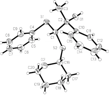
According to the crystallographic atomic labelling of 2-phenyl-1-phenylthio-1-(tetrahydropyran-2-ylthio)propan-2-ol (Fig. 1), the C atom bearing the S-side chain in the tetra-hydropyran ring is C16 and thus the relative con®guration of the title compound may also be named (1S*,2R*,16R*), (I).
We have earlier reported the structure of another diaster-eoisomer (1S*,2S*,16R*), (II) (Kansikaset al., 1995). A very closely related compound 1-phenyl-2-phenylthio-2-(tetra-hydropyran-2-ylthio)ethanol, (C19H22O2S2), where the methyl group is replaced by an H atom, also forms four diastereoi-somers. Applying the above labelling scheme, atom C15 is now in the tetrahydropyran ring bonded to S. Three of the ethanol diastereoisomers crystallize in non-centrosymmetric space groups as conglomerates of enantiomeric crystals with the con®gurations (1S,2S,15R), (III) (Kansikas et al., 1996), (1R,2S,15S), (IV), and (1S,2S,15S), (V) (Kansikas & SipilaÈ, 2000). The fourth diastereoisomer crystallizes in a centro-symmetric space group with the relative con®guration (1R*,2S*,15R*), (VI) (SipilaÈ et al., 2001). Some interatomic distances, angles and torsion angles for (I) are listed in Table 1. The SÐC distances fall well within the values found in the compounds (II)±(VI), but the C1ÐS1ÐC4 angle of 99.21 (1)
is the smallest among them. Torsion angles around the S atoms in compounds (I)±(VI) vary considerably and that gives rise to
organic papers
o336
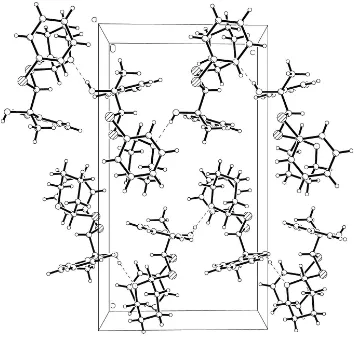
Jarno Kansikaset al. C20H24O2S2 Acta Cryst.(2001). E57, o335±o337very different appearances of these otherwise rather similar molecules. In the compound (I) the hydroxyl group forms an intermolecular hydrogen bond to the O atom of the tetra-hydropyran ring of the neighbouring molecule at the equiva-lent position (x, 1
2ÿy, zÿ12) (Fig. 2 and Table 2). A comparable intermolecular hydrogen bond, but between the molecules with the symmetry code (x,y, zÿ1), is found in the ethanol derivative (V). The O O distances are 2.751 (2) AÊ for (I) and 2.764 (3) AÊ for (V). The title isomer (I) crystallizes in the space groupP21/c(No. 14), whereas the ethanol deri-vative (IV) with the same relative con®guration crystallizes in the non-centrosymmetric space group P212121 (No. 19) and possesses an intramolecular hydrogen bond between the hydroxyl group and the O atom of the tetrahydropyran ring similar to (II) and (III).
Experimental
2-Phenyl-1-phenylthio-1-(tetrahydropyran-2-ylthio)propan-2-ol was synthesized as a mixture of four diastereoisomers according to a procedure reported previously (Kansikas et al., 1995). The di-astereoisomers (1S*,2R*,20R
*)-2-phenyl-1-phenylthio-1-(tetrahydro-pyran-2-ylthio)propan-2-ol, (I), and (1S*,2S*,20R
*)-2-phenyl-1-phenylthio-1-(tetrahydropyran-2-ylthio)propan-2-ol, (II) (Kansikas et al., 1995), were separated by high-pressure liquid chromatography (HPLC) from the crude product and recrystallized from absolute ethanol. The relative amounts of the diastereoisomers in elution order were: 31% (II) and 19% (I). The remaining two diastereoi-somers (50% of the total amount) could not be separated by this procedure. HPLC separation was performed with an ISCO 2350 liquid chromatograph equipped with a Shimadzu SPD-6A UV spec-trophotometric detector and a Shimadzu C-R6A Chromatopac. Components were monitored measuring the absorption at 254 nm. The column used was Merck Lichrocart Si 60 (25010 mm ID), 5mm, the mobile phase 2% ethyl acetate in dichloromethane; ¯ow rate 7 ml minÿ1. The NMR spectra were recorded on a Varian Gemini
200 spectrometer using tetramethylsilane as an internal standard. The assignments are based on chemical-shift data and DEPT measure-ments.1H NMR (200 MHz, CDCl
3):1.4±1.9 (6H,m; CH2), 1.80 (s, CH3), 3.4±3.6 and 3.6±3.7 (2H,m; OCH2), 4.7 (1H,s, SCHS), 4.9 (1H, s, OH), 5.2±5.3 (1H,m, OCHS), 7.2±7.6 (10H,m; aromatic H).13C NMR (50 MHz; CDCl3):21.7 and 25.1 and 30.7 (CH2), 29.5 (CH3), 65.1 (OCH2), 68.5 (SCHS), 78.2 (CPh), 80.1 (OCS), 125.1±147.3 (aromatic C).
Crystal data
C20H24O2S2 Mr= 360.51
Monoclinic,P21/c a= 9.2440 (18) AÊ
b= 19.239 (4) AÊ
c= 10.682 (2) AÊ = 93.37 (3)
V= 1896.5 (6) AÊ3 Z= 4
Dx= 1.263 Mg mÿ3
MoKradiation Cell parameters from 25
re¯ections = 4±10
= 0.29 mmÿ1 T= 193 (2) K Prismatic, colourless 0.360.300.25 mm
Data collection
Rigaku AFC-7Sdiffractometer !/2scans
3637 measured re¯ections 3418 independent re¯ections 2438 re¯ections withI> 2(I)
Rint= 0.029
max= 25.3
h= 0!11
k= 0!23
l=ÿ12!12 3 standard re¯ections
every 100 re¯ections intensity decay: <0.1%
Re®nement
Re®nement onF2 R[F2> 2(F2)] = 0.046 wR(F2) = 0.107 S= 1.05 3418 re¯ections 217 parameters
H-atom parameters constrained
w= 1/[2(F
o2) + (0.0501P)2
+ 0.1099P]
whereP= (Fo2+ 2Fc2)/3
(/)max= 0.001 max= 0.28 e AÊÿ3 min=ÿ0.29 e AÊÿ3
Figure 1
View of (I) showing the crystallographic atom labelling. Displacement
ellipsoids are drawn at the 50% probability level. Figure 2
Table 1
Selected geometric parameters (AÊ,).
S1ÐC4 1.778 (3)
S1ÐC1 1.831 (2)
O1ÐC2 1.423 (3)
C1ÐC2 1.546 (3)
C1ÐS2 1.819 (2)
S2ÐC16 1.828 (2)
O2ÐC16 1.424 (3)
O2ÐC20 1.431 (3)
C4ÐS1ÐC1 99.20 (11)
C2ÐC1ÐS2 115.16 (17)
C2ÐC1ÐS1 108.91 (16)
S2ÐC1ÐS1 107.30 (12)
C1ÐS2ÐC16 101.66 (11)
C16ÐO2ÐC20 114.32 (19)
O1ÐC2ÐC1 104.76 (19)
O2ÐC16ÐS2 112.52 (16)
C4ÐS1ÐC1ÐC2 ÿ162.48 (17)
C4ÐS1ÐC1ÐS2 72.25 (15)
C2ÐC1ÐS2ÐC16 96.02 (18) S1ÐC1ÐS2ÐC16 ÿ142.57 (12)
S2ÐC1ÐC2ÐO1 66.0 (2)
S1ÐC1ÐC2ÐO1 ÿ54.5 (2) S2ÐC1ÐC2ÐC3 ÿ176.06 (17)
S1ÐC1ÐC2ÐC3 63.4 (2)
S1ÐC1ÐC2ÐC10 ÿ173.97 (16) C1ÐS2ÐC16ÐO2 67.04 (18)
Table 2
Hydrogen-bonding geometry (AÊ,).
DÐH A DÐH H A D A DÐH A
O1ÐH1A O2i 0.84 1.92 2.751 (2) 168
Symmetry codes: (i)x;1 2ÿy;zÿ12.
Data collection: AFC-7Ssoftware; cell re®nement: AFC-7S soft-ware; data reduction: TEXSAN(Molecular Structure Corporation, 1993); program(s) used to solve structure: SHELXS97 (Sheldrick, 1997); program(s) used to re®ne structure:SHELXL97 (Sheldrick, 1997); molecular graphics:SHELXTL(Bruker, 1997); software used to prepare material for publication:SHELXL97.
References
Bruker (1997). SHELXTL. Release 5.1. Bruker AXS Inc., Madison, Wisconsin, USA.
Kansikas, J., LeskelaÈ, M., SipilaÈ, K. & Hase, T. (1995).Acta Chem. Scand.49, 809±812.
Kansikas, J. & SipilaÈ, K. (2000).Acta Cryst.C56, 1383±1385.
Kansikas, J., SipilaÈ, K. & Hase, T. (1996). Acta Chem. Scand. 50, 1147± 1152.
Molecular Structure Corporation (1993).TEXSAN.Version 1.6b. MSC, 3200 Research Forest Drive, The Woodlands, TX 77381, USA.
Sheldrick, G. M. (1997).SHELXS97 andSHELXL97. University of GoÈtt-ingen, Germany.
SipilaÈ, K., Hase, T., Kansikas, J. & Koskimies, J. (2001). Unpublished results.
supporting information
sup-1
Acta Cryst. (2001). E57, o335–o337
supporting information
Acta Cryst. (2001). E57, o335–o337 [doi:10.1107/S160053680100438X]
(1
S
*,2
R
*,2
′
R
*)-2-Phenyl-1-phenylthio-1-(tetrahydropyran-2-ylthio)propan-2-ol
at 193
K
Jarno Kansikas and Kaija Sipil
ä
S1. Comment
According to the crystallographic atomic labelling of 2-phenyl-1-phenylthio-1-(tetrahydropyran-2-ylthio)propan-2-ol
(Fig. 1), the C atom bearing the S-side chain in the tetrahydropyran ring is C16 and thus the relative configuration of the
title compound may also be named (1S*,2R*,16R*), (I). We have earlier reported the structure of another diastereoisomer
(1S*,2S*,16R*), (II) (Kansikas et al., 1995). A very closely related compound
1-phenyl-2-phenylthio-2-(tetrahydro-pyran-2-ylthio)ethanol, (C19H22O2S2), where the methyl group is replaced by an H atom, also forms four diastereoisomers.
Applying the above labelling scheme, atom C15 is now in the tetrahydropyran ring bonded to S. Three of the ethanol
diastereoisomers crystallize in non-centrosymmetric space groups as conglomerates of enantiomeric crystals with the
configurations (1S,2S,15R), (III) (Kansikas et al., 1996), (1R,2S,15S), (IV), and (1S,2S,15S), (V) (Kansikas and Sipilä,
2000). The fourth diastereoisomer crystallizes in a centrosymmetric space group with the relative configuration
(1R*,2S*,15R*), (VI) (Sipilä et al., 2001). Some interatomic distances, angles and torsion angles for (I) are listed in Table
1. The S—C distances fall well within the values found in the compounds (II)–(VI), but the C1—S1—C4 angle of
99.21 (1)° is the smallest among them. Torsion angles around the S atoms in compounds (I)–(VI) vary considerably and
that gives rise to very different appearances of these otherwise rather similar molecules. In the compound (I) the hydroxyl
group forms an intermolecular hydrogen bond to the O atom of the tetrahydropyran ring of the neighbouring molecule at
the equivalent position (x, 1/2 - y, z - 1/2) (Fig. 2 and Table 2). A comparable intermolecular hydrogen bond, but between
the molecules with the symmetry code (x, y, z - 1), is found in the ethanol derivative (V). The O···O distances are
2.751 (2) Å for (I) and 2.764 (3) Å for (V). The title isomer (I) crystallizes in the space group P21/c (No. 14), whereas the
ethanol derivative (IV) with the same relative configuration crystallizes in the non-centrosymmetric space group P212121
(No. 19) and possesses an intramolecular hydrogen bond between the hydroxyl group and the O atom of the
tetrahydro-pyran ring similar to (II) and (III).
S2. Experimental
2-Phenyl-1-phenylthio-1-(tetrahydropyran-2-ylthio)propan-2-ol was synthesized as a mixture of four diastereoisomers
according to a procedure reported previously (Kansikas et al., 1995). The diastereoisomers (1S*,2R*,2′R
*)-2-phenyl-1-phenylthio-1-(tetrahydropyran-2-ylthio)propan-2-ol, (I), and (1S*,2S*,2′R
*)-2-phenyl-1-phenylthio-1-(tetrahydropyran-2-ylthio)propan-2-ol, (II) (Kansikas et al., 1995), were separated by high-pressure liquid chromatography (HPLC) from the
crude product and recrystallized from absolute ethanol. The relative amounts of the diastereoisomers in elution order
were: 31% (II) and 19% (I). The remaining two diastereoisomers (50% of the total amount) could not be separated by this
procedure. HPLC separation was performed with an ISCO 2350 liquid chromatograph equipped with a Shimadzu SPD-6
A UV spectrophotometric detector and a Shimadzu C—R6A Chromatopac. Components were monitored measuring the
supporting information
sup-2
Acta Cryst. (2001). E57, o335–o337
acetate in dichloromethane; flow rate 7 ml min-1. The NMR spectra were recorded on a Varian Gemini 200 spectrometer
using tetramethylsilane as an internal standard. The assignments are based on chemical-shift data and DEPT
measurements. 1H NMR (200 MHz, CDCl3): δ 1.4–1.9 (6H, m; CH2), 1.80 (s, CH3), 3.4–3.6 and 3.6–3.7 (2H, m; OCH2),
4.7 (1H, s, SCHS), 4.9 (1H, s, OH), 5.5–5.3 (1H, m, OCHS), 7.2–7.6 (10H, m; arom·H). 13C NMR (50 MHz; CDCl 3): δ
21.7 and 25.1 and 30.7 (CH2), 29.5 (CH3), 65.1 (OCH2), 68.5 (SCHS), 78.2 (CPh), 80.1 (OCS), 125.1–147.3 (aromatic
[image:5.610.130.482.164.477.2]C).
Figure 1
supporting information
sup-3
[image:6.610.128.482.70.408.2]Acta Cryst. (2001). E57, o335–o337
Figure 2
Fraction of the molecular packing showing the hydrogen-bond scheme of (I) in the c axis direction.
(I)
Crystal data
C20H24O2S2
Mr = 360.51 Monoclinic, P21/c
a = 9.2440 (18) Å
b = 19.239 (4) Å
c = 10.682 (2) Å
β = 93.37 (3)°
V = 1896.5 (6) Å3
Z = 4
F(000) = 768
Dx = 1.263 Mg m−3
Mo Kα radiation, λ = 0.71073 Å Cell parameters from 25 reflections
θ = 4–10°
µ = 0.29 mm−1
T = 193 K
Prismatic, colourless 0.36 × 0.30 × 0.25 mm
Data collection
Rigaku AFC-7S diffractometer
Radiation source: fine-focus sealed tube Graphite monochromator
ω/2θ scans
3637 measured reflections 3418 independent reflections 2438 reflections with I > 2σ(I)
Rint = 0.029
θmax = 25.3°, θmin = 2.9°
h = 0→11
k = 0→23
l = −12→12
supporting information
sup-4
Acta Cryst. (2001). E57, o335–o337
Refinement
Refinement on F2
Least-squares matrix: full
R[F2 > 2σ(F2)] = 0.046
wR(F2) = 0.107
S = 1.05 3418 reflections 217 parameters 0 restraints
Primary atom site location: structure-invariant direct methods
Secondary atom site location: difference Fourier map
Hydrogen site location: inferred from neighbouring sites
H atoms treated by a mixture of independent and constrained refinement
w = 1/[σ2(F
o2) + (0.0501P)2 + 0.1099P]
where P = (Fo2 + 2Fc2)/3
(Δ/σ)max = 0.001
Δρmax = 0.28 e Å−3
Δρmin = −0.29 e Å−3
Special details
Geometry. All e.s.d.'s (except the e.s.d. in the dihedral angle between two l.s. planes) are estimated using the full covariance matrix. The cell e.s.d.'s are taken into account individually in the estimation of e.s.d.'s in distances, angles and torsion angles; correlations between e.s.d.'s in cell parameters are only used when they are defined by crystal symmetry. An approximate (isotropic) treatment of cell e.s.d.'s is used for estimating e.s.d.'s involving l.s. planes.
Refinement. Refinement of F2 against ALL reflections. The weighted R-factor wR and goodness of fit S are based on F2,
conventional R-factors R are based on F, with F set to zero for negative F2. The threshold expression of F2 > σ(F2) is used
only for calculating R-factors(gt) etc. and is not relevant to the choice of reflections for refinement. R-factors based on F2
are statistically about twice as large as those based on F, and R- factors based on ALL data will be even larger.
Fractional atomic coordinates and isotropic or equivalent isotropic displacement parameters (Å2)
x y z Uiso*/Ueq
S1 0.61079 (7) 0.30678 (4) 0.07364 (6) 0.0355 (2) O1 0.39674 (19) 0.21467 (9) −0.04576 (15) 0.0329 (4)
H1A 0.3562 0.1792 −0.0773 0.049*
C1 0.4319 (3) 0.28763 (12) 0.1302 (2) 0.0252 (5)
H1B 0.4395 0.2883 0.2239 0.030*
S2 0.31071 (7) 0.35720 (3) 0.07562 (6) 0.02717 (17) O2 0.2771 (2) 0.39432 (9) 0.31726 (15) 0.0333 (4)
C2 0.3857 (3) 0.21387 (13) 0.0866 (2) 0.0272 (6)
C3 0.4888 (3) 0.15934 (14) 0.1449 (3) 0.0379 (7)
H3A 0.4579 0.1130 0.1160 0.057*
H3B 0.4875 0.1616 0.2365 0.057*
H3C 0.5873 0.1684 0.1197 0.057*
C4 0.6601 (3) 0.37767 (14) 0.1741 (2) 0.0320 (6)
C5 0.6929 (3) 0.36759 (14) 0.3006 (3) 0.0360 (6)
H5A 0.6862 0.3224 0.3358 0.043*
C6 0.7355 (3) 0.42303 (15) 0.3758 (3) 0.0425 (7)
H6A 0.7576 0.4160 0.4629 0.051*
C7 0.7461 (3) 0.48869 (15) 0.3253 (3) 0.0427 (7)
H7A 0.7761 0.5268 0.3772 0.051*
C8 0.7132 (3) 0.49868 (15) 0.1999 (3) 0.0432 (7)
H8A 0.7210 0.5438 0.1650 0.052*
C9 0.6686 (3) 0.44362 (14) 0.1235 (3) 0.0368 (7)
H9A 0.6440 0.4511 0.0370 0.044*
supporting information
sup-5
Acta Cryst. (2001). E57, o335–o337
C11 0.1150 (3) 0.21167 (14) 0.0344 (2) 0.0326 (6)
H11A 0.1334 0.2230 −0.0497 0.039*
C12 −0.0263 (3) 0.20616 (15) 0.0692 (3) 0.0419 (7)
H12A −0.1040 0.2148 0.0091 0.050*
C13 −0.0558 (3) 0.18841 (15) 0.1891 (3) 0.0429 (7)
H13A −0.1532 0.1852 0.2123 0.051*
C14 0.0581 (3) 0.17519 (14) 0.2764 (3) 0.0365 (7)
H14A 0.0390 0.1612 0.3591 0.044*
C15 0.1994 (3) 0.18243 (13) 0.2426 (2) 0.0296 (6)
H15A 0.2770 0.1747 0.3034 0.036*
C16 0.1978 (3) 0.36796 (13) 0.2092 (2) 0.0280 (6)
H16A 0.1601 0.3210 0.2310 0.034*
C17 0.0689 (3) 0.41334 (14) 0.1720 (3) 0.0354 (6)
H17A −0.0033 0.4099 0.2366 0.043*
H17B 0.0228 0.3965 0.0917 0.043*
C18 0.1132 (3) 0.48914 (14) 0.1576 (3) 0.0381 (7)
H18A 0.1735 0.4941 0.0845 0.046*
H18B 0.0257 0.5184 0.1429 0.046*
C19 0.1978 (3) 0.51272 (14) 0.2754 (3) 0.0436 (7)
H19A 0.1335 0.5131 0.3463 0.052*
H19B 0.2336 0.5606 0.2636 0.052*
C20 0.3249 (3) 0.46461 (14) 0.3054 (3) 0.0388 (7)
H20A 0.3936 0.4676 0.2379 0.047*
H20B 0.3765 0.4795 0.3848 0.047*
Atomic displacement parameters (Å2)
U11 U22 U33 U12 U13 U23
supporting information
sup-6
Acta Cryst. (2001). E57, o335–o337
C17 0.0261 (14) 0.0385 (16) 0.0418 (16) 0.0026 (13) 0.0029 (12) −0.0037 (13) C18 0.0357 (16) 0.0329 (16) 0.0456 (16) 0.0115 (13) 0.0012 (13) 0.0066 (13) C19 0.0459 (18) 0.0239 (15) 0.061 (2) 0.0025 (13) 0.0037 (15) −0.0067 (14) C20 0.0420 (17) 0.0331 (16) 0.0402 (16) 0.0013 (14) −0.0059 (13) −0.0103 (12)
Geometric parameters (Å, º)
S1—C4 1.778 (3) C6—C7 1.380 (4)
S1—C1 1.831 (2) C7—C8 1.369 (4)
O1—C2 1.423 (3) C8—C9 1.385 (4)
C1—C2 1.546 (3) C10—C11 1.385 (3)
C1—S2 1.819 (2) C10—C15 1.386 (3)
S2—C16 1.828 (2) C11—C12 1.383 (4)
O2—C16 1.424 (3) C12—C13 1.367 (4)
O2—C20 1.431 (3) C13—C14 1.388 (4)
C2—C3 1.525 (4) C14—C15 1.383 (4)
C2—C10 1.531 (3) C16—C17 1.512 (3)
C4—C5 1.381 (4) C17—C18 1.525 (4)
C4—C9 1.383 (4) C18—C19 1.512 (4)
C5—C6 1.379 (4) C19—C20 1.515 (4)
C4—S1—C1 99.20 (11) C7—C8—C9 120.7 (3)
C2—C1—S2 115.16 (17) C4—C9—C8 119.5 (3)
C2—C1—S1 108.91 (16) C11—C10—C15 118.4 (2)
S2—C1—S1 107.30 (12) C11—C10—C2 120.5 (2)
C1—S2—C16 101.66 (11) C15—C10—C2 121.0 (2)
C16—O2—C20 114.32 (19) C12—C11—C10 120.4 (3)
O1—C2—C3 109.5 (2) C13—C12—C11 120.9 (3)
O1—C2—C10 111.5 (2) C12—C13—C14 119.3 (3)
C3—C2—C10 111.2 (2) C15—C14—C13 119.8 (3)
O1—C2—C1 104.76 (19) C14—C15—C10 121.0 (2)
C3—C2—C1 110.7 (2) O2—C16—C17 111.6 (2)
C10—C2—C1 108.95 (19) O2—C16—S2 112.52 (16)
C5—C4—C9 119.8 (2) C17—C16—S2 109.73 (17)
C5—C4—S1 121.1 (2) C16—C17—C18 111.5 (2)
C9—C4—S1 119.1 (2) C19—C18—C17 109.4 (2)
C6—C5—C4 120.0 (3) C18—C19—C20 110.5 (2)
C5—C6—C7 120.3 (3) O2—C20—C19 110.9 (2)
C8—C7—C6 119.6 (3)
C4—S1—C1—C2 −162.48 (17) C1—C2—C10—C11 96.1 (3)
C4—S1—C1—S2 72.25 (15) O1—C2—C10—C15 165.3 (2)
C2—C1—S2—C16 96.02 (18) C3—C2—C10—C15 42.7 (3)
S1—C1—S2—C16 −142.57 (12) C1—C2—C10—C15 −79.6 (3)
S2—C1—C2—O1 66.0 (2) C15—C10—C11—C12 1.8 (4)
S1—C1—C2—O1 −54.5 (2) C2—C10—C11—C12 −173.9 (2)
S2—C1—C2—C3 −176.06 (17) C10—C11—C12—C13 −1.5 (4)
supporting information
sup-7
Acta Cryst. (2001). E57, o335–o337
S2—C1—C2—C10 −53.4 (2) C12—C13—C14—C15 2.2 (4)
S1—C1—C2—C10 −173.97 (16) C13—C14—C15—C10 −1.9 (4)
C1—S1—C4—C5 70.4 (2) C11—C10—C15—C14 −0.1 (4)
C1—S1—C4—C9 −111.0 (2) C2—C10—C15—C14 175.6 (2)
C9—C4—C5—C6 −0.7 (4) C20—O2—C16—C17 −55.7 (3)
S1—C4—C5—C6 177.9 (2) C20—O2—C16—S2 68.2 (2)
C4—C5—C6—C7 −0.3 (4) C1—S2—C16—O2 67.04 (18)
C5—C6—C7—C8 0.4 (5) C1—S2—C16—C17 −168.11 (18)
C6—C7—C8—C9 0.3 (4) O2—C16—C17—C18 52.9 (3)
C5—C4—C9—C8 1.5 (4) S2—C16—C17—C18 −72.5 (2)
S1—C4—C9—C8 −177.1 (2) C16—C17—C18—C19 −53.0 (3)
C7—C8—C9—C4 −1.3 (4) C17—C18—C19—C20 54.6 (3)
O1—C2—C10—C11 −19.1 (3) C16—O2—C20—C19 57.6 (3)
C3—C2—C10—C11 −141.6 (2) C18—C19—C20—O2 −56.6 (3)
Hydrogen-bond geometry (Å, º)
D—H···A D—H H···A D···A D—H···A
O1—H1A···O2i 0.84 1.92 2.751 (2) 168