www.wjpr.net Vol 3, Issue 9, 2014. 1058
ISOLATION AND PARTIAL CHARACTERIZATION OF
ANTAGONISTIC ACTINOMYCETES FROM EARTHWORM GUT
Sruthy. P.B*, Anjana. J.C, J. Rathinamala, S. Jayashree
Department of Microbiology, Nehru Arts and Science College, Affiliated to Bharathiar University, Coimbatore, India.
ABSTRACT
Nowadays, the drug resistant strains of pathogen emerge more quickly than the rate of discovery of new antibiotics. Because of this, many scientists and pharmaceutical industry have actively involved in the isolation and screening of actinomycetes from different untouched habitats, for the production of antibiotics. Earthworms are precious and low cost source of numerous bioactive molecules, which could find place in human and veterinary medicine. The main objective of the study was to isolate and screen for antagonistic actinomycetes from an earthworm gut. Out of six actinomycetes isolated from the earthworm gut, one isolate possessed high broad spectrum antibacterial activity in cross streak method and was selected for further studies. It was inoculated into the fermentation medium and by solvent extraction procedure using Ethyl acetate the metabolite was extracted to its full form. The extract was highly active against selected bacterial pathogens used in the test and was further analyzed by thin layer chromatography. The UV spectra of the ethyl acetate extract for the active isolate showed an absorbance peak at 316 nm. The isolated strain was characterized for physiological and biochemical properties, nutritional uptake and the isolate fitted the Streptomyces genus. Based on the results of our study it was confirmed that Streptomyces isolated from earthworm gut was found to be a promising source for extracting novel antibiotics for treating bacterial infections in human.
KEYWORDS: Actinomycetes, antibiotics, antibacterial activity, cross streak method, thin
layer chromatography, Streptomyces.
Volume 3, Issue 9, 1058-1070. Research Article ISSN 2277– 7105
Article Received on 04 September 2014,
Revised on 29 Sept 2014, Accepted on 23 Oct 2014
*Correspondence for
Author
Sruthy P.B.
Department of
Microbiology, Nehru Arts
and Science College,
Affiliated to Bharathiar
University, Coimbatore,
www.wjpr.net Vol 3, Issue 9, 2014. 1059 INTRODUCTION
Microbial natural products are notable not only for their potent therapeutic activities but also for the fact that they frequently possess the desirable pharmacokinetic properties required for clinical development [1]. Actinomycetes are the most widely distributed group of microorganisms in nature which primarily inhabit the soil [2]. Actinomycetes are gram positive bacteria frequently filamentous and sporulating with DNA rich in G+C from 55-75% [3]. They have provided many important bioactive compounds of high commercial value and continue to be routinely screened for new bioactive compounds [4]. Actinomycetes are the most biotechnologically valuable prokaryotes responsible for the production of about half of the discovered bioactive secondary metabolites including antibiotics [5]. Among actinomycetes, around 7,600 compounds are produced by Streptomyces species [6].
The present scenario demands the discovery of new antimicrobial agents which can be of a better potential and activity than the previously discovered agents. Resistance of bacteria to the effects of antibiotics has been a major problem in the treatment of diseases. Infectious diseases are still the second leading cause of death worldwide [7]. Nowadays, the drug resistant strains of pathogen emerge more quickly than the rate of discovery of new drugs and antibiotics. Because of this, many scientists and pharmaceutical industry have actively involved in isolation and screening of actinomycetes from different untouched habitats, for their production of antibiotics [2]. At present the literature contains sufficient evidence of the presence of actinomycetes in the gut of soil invertebrates (in particular earthworms) [8]. Many scientists have studied the microbial community in the gut of earthworms [9]. Mostly the population density of actinomycetes in the intestinal tract of invertebrates was studied by [10]. In the present study we focused on the isolation of earthworm (Eudrilus eugeniae) gut flora and partial characterization of Actinomycetes exhibiting broad range of Antagonistic activity against selected human pathogens.
MATERIALS AND METHODS
Isolation of Earthworm Gut Flora
Dissection of Earthworms: The earthworm species Eudrilus eugeniae used for the present
www.wjpr.net Vol 3, Issue 9, 2014. 1060 longitudinally along the earthworm. The gut was then freed from surrounding blood vessels and nephridia and separated into three sections: foregut, mid gut and hindgut.
Isolation of Microbes from Earthworm Gut
The gut sections were washed in sterile distilled water, weighed and homogenized for 5 minutes with a vortex mixer in sterile 0.85% NaCl solution. Each gut sections were individually plated on to Nutrient agar, Actinomycetes isolation agar and Sabouraud’s dextrose agar for bacteria, actinomycetes and fungi respectively and kept for incubation. Three replicates were maintained from each dilution.
Isolation and Purification of Actinomycetes
Whitish pin-point colonies which are the characteristic of actinomycetes with clear zone of inhibition around it were observed in the plates. The pinpoint colonies with inhibitory or clear zone of inhibition were selected and the suspected colonies were selectively isolated and transferred to actinomycetes isolation agar medium with the help of loop inoculum method
[11]
. The plates were kept for incubation at 25 to 30°C for 2-3 days in an inverted position. The actinomycetes isolates were purified by pure culture techniques. The colonies were refrigerated in starch casein agar slants by frequent sub culturing for further studies. Among the isolated actinomycetes, the potential strain for further study was selected owing to its antagonistic activity in primary screening.
Screening of Antibiotic Producing Strains
Preparation of Preliminary Test Organisms
The test organisms used for preliminary screening were Escherichia coli, Klebsiella sp., Pseudomonas aeruginosa, Salmonella typhi, Serratia marcescens, Aeromonas hydrophila,
Staphylococcus aureus, Bacillus sp. and Candida albicans. Test bacteria were grown in 4.5 ml Nutrient broth for 18 hrs, and then standardized to Mc Farland standard of 0.1 at OD 600 nm. Candida albicans was grown on Potato dextrose agar (PDA) plate for 48 hr, then re-suspended in Nutrient broth and standardized to McFarland standard of 0.1 at OD 600 nm before use.
Primary Screening by Cross streak Method
www.wjpr.net Vol 3, Issue 9, 2014. 1061 actinomycetes strains were made on surface of the Actinomycetes isolation agar and incubated at 28°C for 5 days. After observing a good ribbon like growth of the actinomycetes on the plates, the overnight pathogenic bacterial strains were streaked at right angles to the original streak of actinomycetes and incubated at 37°C for overnight. A control plate was also maintained without inoculating the actinomycetes, to assess the normal growth of bacteria. After incubation antagonistic activity of the actinomycetes against pathogens were screened and the antibiotic producing strains were isolated.
Isolation of Antibacterial Metabolites
The isolate showing the broad spectrum antimicrobial activity was grown in submerged culture in 250 ml flasks containing 50 ml of Starch casein nitrate broth aseptically. It was shaken at 28-30°C at 250 rpm for seven-ten days. The visible pellets, clumps or aggregates and turbidity in the broth, will confirm the growth of the organism in the flask. The contents of the flask were filtered through sterile Whatman No. 1 filter paper aseptically. The filtrate was subjected for solvent extraction method to recover antibacterial metabolites in pure form. Ethyl acetate was added to the filtrate in the ratio of 1:1 (v/v) and shaken vigorously for 1 h for complete extraction. The ethyl acetate phase which contains antibiotic substances was separated from aqueous phase. It was evaporated to dryness in water bath and the residue obtained was used to determine antimicrobial activity [13].
Secondary Screening
Secondary screening was performed by well diffusion method [14] using purified extract obtained by the evaporation of the ethyl acetate extract. The antimicrobial activity of the actinomycete isolate was detected as clear zone of inhibition around wells and it was measured in millimeters (mm).
Thin Layer Chromatography and Bioassay of Antibiotic
Ten micro-liters of the ethyl acetate fractions and reference antibiotics were loaded on to the silica gel plates and the chromatogram was developed using chloroform: methanol (4:1) as solvent system. The plates were run in duplicate; one set was used as the reference chromatogram and the other was used for the bioassay of antibiotic. The spots in the chromatogram were visualized in the iodine vapor chamber and UV chamber [15].
UV/Visible absorption of extract
www.wjpr.net Vol 3, Issue 9, 2014. 1062 UV-Visible absorption spectrum of the extract was tested by using Shimadzu UV-2550 spectrophotometer at 300 - 340 nm to determine the λ maximum of the band [16].
Identification and Characterization of Actinomycetes
The isolated Actinomycetes strains were identified on the basis of their phenotypic characterization, physiological characteristics and biochemical characteristic which includes some basic tests, aerial mass color, reverse side pigment, melanoid pigments and spore chain morphology [17].
1. Macroscopic Identification
The isolated actinomycetes colonies were observed under high power magnifying lens. The colony morphology with respect to color, aerial mycelium, size and nature of colony and reverse side color were observed and identified [18].
2. Microscopic Observation
Microscopic observation of the potent isolate was performed using Wet mount technique [19] and Cover Slip Culture Technique [20]. The observed structure was compared with Bergey’s Manual of Determinative Bacteriology [21] and the organism was identified.
3. Biochemical characterization
The various biochemical tests like Starch hydrolysis, Triple Sugar Iron (TSI) agar, Indole test, methyl red test, Vogus- Proskauer test, Citrate utilization test, Catalase test were performed for the identification of the potent isolate [22].
RESULTS
[image:5.595.207.388.596.745.2]Isolation and Purification of Actinomycetes: A total of 6 actinomycetes colonies were isolated from earthworm gut by using serial dilution agar plating method (Fig.1).
www.wjpr.net Vol 3, Issue 9, 2014. 1063 Screening of Antibiotic Producing Strains
Primary screening by Cross Streak Method
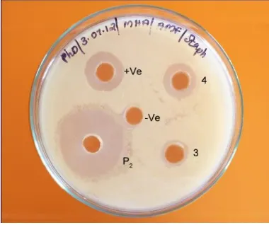
The isolated actinomycetes were screened against common pathogens by cross-streak method. Out of 6 actinomycetes isolated, four isolates namely P1, P2, 3, 4 showed promising antibacterial activities after screening against test organism (Table 1). Inability of test organisms to grow in the presence of actinomycete suggests antibiotic production by the actinomycete. Among them isolate P2 which possessed high broad spectrum activity against bacteria in comparison to other isolates was selected for further studies (Fig.2).
Table (1): Cross Streak Assay.
S. No. Bacterial
Pathogens
Antagonistic activity of Isolated Actinomycetes Strains
Isolate P1 Isolate P2 Isolate 3 Isolate 4
1. S. aureus + +++ ++ +++
2. Bacillus sp. ++ ++ - -
3. Enterococcus sp. - - - +
4. E. coli + +++ +++ ++
5. Klebsiella sp. + +++ +++ ++
6. P. aeruginosa - ++ - -
7. Salmonella sp. ++ +++ +++ -
8. S. marscescenes +++ +++ - +++
9. Candida sp. - + - +
10. Aeromonas sp. + +++ - +
[image:6.595.198.396.477.641.2]+++ Better inhibition, ++ Good inhibition, + Moderate inhibition, - No inhibition
Fig. 2: Cross Streak Assay Isolate P2
Secondary Screening
www.wjpr.net Vol 3, Issue 9, 2014. 1064 acetate extract of isolate (P2) showed a very broad spectrum of antimicrobial activity against both gram positive and gram negative bacteria used in the test; the result of the same can be seen in the Fig.3 below. The antibacterial activity of the extract was highest against S. aureus with a zone of inhibition of 27.7 ± 0.88 mm (Fig.4).
Fig. 3: Antibacterial activity of Extract against Clinical pathogens.
E.A.E- Ethyl acetate extract
Values are presented as the mean ± SEM (standard error of mean); n = 3 for all groups. ***P<0.001, *P<0.05, considered significant.
Fig. 4: Antibacterial Assay – S. aureus
Thin Layer Chromatography and Bioassay of Antibiotic
[image:7.595.202.392.458.617.2]www.wjpr.net Vol 3, Issue 9, 2014. 1065 Structural analysis of the antibiotic by using UV Spectroscopy
The structural elucidation of the compound was performed by using U.V. The λ max of antibacterial compound was found to be 316 (Fig.5).
0.29 0.3 0.31 0.32 0.33 0.34 0.35 0.36 0.37 0.38 0.39
304 308 312 316 320 324 328 332 336 340
A
b
so
rb
an
ce
…
Absorba…
Wavelength (nm)
Fig. 5: Determination of λ Max of Antibiotic.
Identification and Characterization of Actinomycetes
The actinomycetes were identified based on its morphological, cultural characters and cover slip culture techniques. Aerial mass color of the substrate mycelium was determined by observing the plates after 7 to 10 days. It was done only after observing the heavy spore mass surface. The isolate P2 showing maximum zone of inhibition was stained with gram’s staining procedure and was found to be gram positive rod in chain. The colonies were rough and elevated and the morphological observation in light microscope reveals chains of spores which is a specific characteristic of Streptomyces species.
www.wjpr.net Vol 3, Issue 9, 2014. 1066 Table-(2): Morphological and Cultural characteristics of Isolate P2.
Table (3): Biochemical Characteristics of Isolate P2.
S. No Name of the Test Properties
1. Indole -
2. Methyl Red +
3. Voges Proskauer -
4. Citrate Utilization + 5. Nitrate Reduction +
6. Urease +
7. Catalase +
8. Oxidase +
9. Starch Hydrolysis - 10. Gelatin Liquefaction + 11. Casein Hydrolysis + 12. Triple Sugar Iron Agar alk/alk + Positive, - Negative
DISCUSSION
In the present scenario microbial natural products appear as the most promising sources for developing future antibiotics. Actinomycetes are the most biotechnologically valuable prokaryotes responsible for the production of about half of the discovered bioactive secondary metabolites including antibiotics [5]. In the present study out of 6 actinomycetes isolated from earthworm gut, isolate P2 possessed high broad spectrum activity. Organic solvents with different polarities have been used by many researchers for the extraction of antimicrobial compounds from actinomycetes [23]. The antimicrobial compounds from the isolate P2 showed broad spectrum of activity against both Gram positive and Gram-negative organisms which supports the popular notion that actinomycetes can be sources of potent antibacterial agents that can be of value in the treatment of infections especially those caused by clinically resistant bacteria, such as Staphylococcus aureus and Klebsiella pneumoniae [2]. The antibacterial activity of the actinobacteria isolated from the molluscans samples was studied by [24], who reported that the tested ten actinobacterial isolates were active against gram positive and gram negative bacteria. The results of the primary and secondary screening
S. No Morphological & Cultural Characteristics Isolate P2
1. Growth on Culture medium Excellent
2. Aerial Mycelium Grayish White
3. Substrate Mycelium Pale Yellow (-)
4. Melanoid Pigmenation Negative
5. Type of Spore Long chain of spores
[image:9.595.145.418.234.431.2]www.wjpr.net Vol 3, Issue 9, 2014. 1067 reveals that Ethyl acetate extract of isolate (P2) showed a very broad spectrum of antimicrobial activity against both gram positive and gram negative bacteria used in the test, which were in parallel with the results of [25]. According to [26], the extracts from ethyl acetate showed maximum antimicrobial activity against bacteria and fungi, other solvent extracts showed moderate activity against test organism which is similar to the present study. The results of the present study clearly indicated that the antimicrobial activity of potential strain is due to the production of extracellular bioactive compounds. The published literature stated that most of the antibiotics from actinomycetes are extracellular in nature [27].
The earthworm gut is favorable for the development of actinomycetes due to neutral pH, optimal humidity and temperature. In addition, the increased organic carbon and nitrogen content in earthworm gut may also stimulate microbial activity [9]. The actinomycetes isolate P2 grew well on Starch casein nitrate agar medium and produced grayish white aerial mycelium and pale yellow coloured substrate mycelium. Starch casein agar was reported as a suitable medium for the isolation of actinobacteria from soil, water and less explored ecosystems such as mountain, forest, desert ecosystems [28]. The consistency of the isolate was powdery in nature. The mycelial color difference between different strains of Actinomycetes may be due to the synthesis of various secondary metabolites [29]. The colonies were rough and elevated and the morphological observation in light microscope reveals chains of spores which is a specific characteristic of Streptomyces species.
www.wjpr.net Vol 3, Issue 9, 2014. 1068 CONCLUSION
In order to meet the increasing demand of natural medicines in recent years, bioactive components with medicinal values from earthworms have already provoked increased interest. The results obtained from the present study indicated that earthworm gut is promising and could be a vital source of habitat possessing antimicrobial activity. The immense scope of actinomycetes for the exploration of therapeutically active biomolecules has been recognized recently. In the future, there is a possibility of finding new antibiotics from gut flora of earthworm. For proper identification of the antimicrobial compounds, it is essential to obtain in pure form which requires a series of purification procedures and different chemical analysis. Still a little effort was made in this approach.
REFERENCES
1. Farnet CM, Zazopoulos E. Improving drug discovery from microorganisms. In Natural products: Drug discovery and Therapeutic medicine. Zhang L, Demain AL (Eds.). Humana press. Inc, Totowa. NJ, 2005; 95.
2. Oskay M, Tamer AU, Azeri C. Antibacterial activity of some actinomycetes isolated from farming soils of Turkey. Afr J Biotechnol, 2004; 3(9): 441- 446.
3. Ho C, Lo C, Lai N, Cheah H, Wong N. ASEAN Rev. Biodiversity Environ Conser, 2002; 9: 1-7.
4. Okami Y, Hotta K. Search and discovery of new antibiotics. In: actinomycetes in biotechnology. Good fellow M, Williams ST and Mordarski M (Eds.). Academic Press. Inc, San Diego, California, 1988; 33-67.
5. Yuan L, Zhang Y, Yu L, Sun C, Wei Y, Liu H, Li W, Zhang Y. Actinopolymorpha Cephalotaxi sp. nov., a novel actinomycete isolated from rhizosphere soil of the plant Cephalotaxus fortune.Int J Syst Evol Microbiol,2010; 60: 51-54.
6. Olano C, Carmen Méndez, José A Salas. Antitumor Compounds from Marine Actinomycetes. Marine Drug, 2009; 7(2): 210-248.
7. Luzhetskyy A, Pelzer S, Bechthold A. The future of natural products as a source of new antibiotics. Curr Opin Invest Drugs, 2007; 8(8): 608-613.
www.wjpr.net Vol 3, Issue 9, 2014. 1069 9. Karsten GR, Drake HI. Comparative assessment of the aerobic and anaerobic microfloras
of earthworm guts and forest soils. Appl Environ Microbiology, 1995; 61: 1039 – 1044. 10.Chu TL, Szabo IM, Szabo I. Nocardioform gut actinomycetes of Glomeris- Hexasticha
Brandt (Diplopoda). Biol Fert Soils, 1987; 3: 113-116.
11.Hamaki T, Suzuki M, Fudou R, Jojima Y, Kajiura T, Tabuchi A, Sen K and Shibai H. Isolation of novel bacteria and actinomycetes using soil-extract agar medium. J Biosci Bioeng, 2005; 99(5): 485-492.
12.Lakshmanaperumalsamy P, Chandramohan D, Natarajan R. Antibacterial and antifungal activity of Streptomyces from PortoNovo coastal environment. Mar Biol, 1978; 11: 15-24. 13.Westley JW, Evans RH, Sello LH, Troupe N, Liu CM, Blount JF. J Antibiot,1979;32(2):
100-107.
14.Remya M, Vijayakumar R. Isolation and characterization of marine antagonistic actinomycetes from West Coast of India. Facta Universitatis Medicine and Biology, 2008; 15(1): 13-19.
15.Pandey B, Ghimire P, Agrawal VP. Studies on the antibacterial activity of actinomycetes isolated from Khumbu region of Mount Everest. J App Mirobiol, 2004; 20: 45-54.
16.Sahin N, Ugur A, Investigation of the Antimicrobial activity of some Streptomyces isolates. Turk J Biol, 2003; 27: 79-84.
17.Sathiyaseelan K, Stella D. Isolation, Identification and Antagonistic Activity of Marine Actinomycetes Isolated from the Muthupet Mangrove Environment. International Journal of Pharmaceutical and Biological Archives, 2011; 2(5): 1464-1468.
18.Benedict RG, Lindenfelser LA, Stodola FH, Traufler DH. Studies on Streptomyces griseocarneus and the production of hydroxystreptomycin. J Bacteriol,1951; 62(4): 487-497.
19.Kandasamy S, Muthusamy G, Thangaswamy S, Senthilkumar B. Screening and Identification of Antibiotic Producing Actinomycetes and their Antagonistic Activity against Common Pathogens. World Res J Antimicrob Agents, 2012; 1(1): 07-10.
20.Williams ST, Good fellow M, Alderson G. Genus Streptomyces Waksman and Henrici 1943.339AL. In: Bergey’s Manual of Systematic Bacteriology. Williams & Wilkins Company, Baltimore, 1989; 4: 2452-2492.
21.Bergey’s Manual of Determinative Bacteriology. 9th ed., Actinomycetales, 2000.
www.wjpr.net Vol 3, Issue 9, 2014. 1070 23.Selvameenal L, Radakrishnan M, Balagurunathan R. Antibiotic pigment from desert soil actinomycetes; biological activity, purification and chemical screening. Indian J Pharm Sci,2009; 71: 499-504.
24.Krishna SR, Sathish Kumar SR, Meenambekha L, Madhusudhan M. Antibacterial activity and characterisation of Actinobacteria isolated from Marine Bivalve Meretrix casta (Gmelin). Advances in Applied Science Research,2011; 2(4): 431-439. 25.Oskay M. Antifungal and antibacterial compounds from Streptomyces strains Afr J
Biotechnol, 2009; 8: 3007-3017.
26.Vijakumar R, Murugesan S, Panneerselvam A. Isolation, characterization and antimicrobial activity of actinobacteria from point calimere coastal region, east coast of India. Int Res J Pharm, 2010; 1: 358-365.
27.Valan arasu N, Duraipandiyan M, Agastian M, Ignacimuthu S. Characterization and phylogenetic analysis of novel polyene type antimicrobial metabolite producing actinomycetes from marine sediments: Bay of Bengal India. Med Mycol, 2008;18: 147-153.
28.Radhika S, Bharathi S, Radhakrishnan M, Balagurunathan R. J Pharm Res, 2011; 4(8): 2584-2586.
29.Dharmaraj S. Marine Streptomyces as a novel source of bioactive substances. World J Microb Biot, 2010; 26: 2123-2139.
30.Shirling EB, Gottileb D. Methods for characterization of Streptomyces species. Int J Syst Bacteriol, 1966; 16: 313-340.
31.Madigan M and Martinko J (eds.). Brock Biology of Microorganisms 11thed., Prentice Hall: 2005.
32.Bushell M. Prog Ind Microbiol, 1982; 17: 101-107.
33.Okazaki T, Okami Y. Studies on marine micorganisms II. Actinomycetes in Sagami Bay and their antibiotic substances. J Antibiot, 1972; 25: 461-466.