www.wjpr.net Vol 4, Issue 11, 2015.
71
APATITE NUCLEATION OF DENSE AND POROUS CHITIN
MEMBRANES
1
Leandro Medeiros Motta, *2,3Gláucio Diré Feliciano and 4Cristina Tristão de Andrade
1
Unit Pharmacy (UFAR), State University Center Foundation of the West Zone (UEZO), Rio
de Janeiro, Brazil.
2
Laboratory of Chemical and Biological Analysis (LAQB), State University Center
Foundation of the West Zone (UEZO), Rio de Janeiro, Brazil.
3
Estácio de Sá University (Estácio), Health Sciences Area, Rio de Janeiro, Brazil.
4
Federal University of Rio de Janeiro (UFRJ), Institute of Macromolecules Professor
Eloisa Mano, Rio de Janeiro, Brazil
ABSTRACT
The formation of apatite (CHA) from a synthetic body fluid was
induced by the presence of dense and porous chitin membranes.
Infrared spectra and thermograms obtained for chitin alone and for the
composite biomaterials were compared and indicated the deposition of
CHA. Scanning electron micrographs showed the formation of
spherical crystals of CHA on the surfaces of both membranes. Images
obtained for the cross- sections ceramic material with apparently lower
crystallinity was observed for dense membranes.
KEYWORDS: apatite nucleation, dense and porous membranes,
chitin, crystallinity.
INTRODUCTION
Polymeric membranes have been developed as matrices for artificial mechanical support of
osteogenic cells in bone tissue regeneration. When implemented, many of these materials are
encapsulated by fibrous tissue and remain as foreign bodies without adherence to the bone.
To overcome this problem, the use of apatite in polymeric composites have been
recommended, in order to obtain materials capable of mimicking the extracellular matrix of
bone tissue.[1]
Volume 4, Issue 11, 71-76. Research Article ISSN 2277– 7105
Article Received on 21 Aug 2015,
Revised on 13 Sep 2015, Accepted on 07 Oct 2015
*Correspondence for
Author
Leandro Medeiros
Motta
Unit Pharmacy (UFAR),
State University Center
Foundation of the West
www.wjpr.net Vol 4, Issue 11, 2015.
72 Calcification is a natural process that occurs during many situations in the human body, for
example, after a bone is fractured. The natural process of calcification can be reproduced and
investigated in laboratory biomaterials such as chitin. The calcification of biomaterials can be
called deposition "in situ". The deposition studies "in situ" are of great interest in many
scientific and technological fields. In the field of materials, they lead to the development of
more resistant materials and functional [Cassiano]. In the medical field, they help
understanding the natural formation of bones and teeth and allow obtaining osteogenic
implants and prostheses for orthopedic correction.[2] as well as help in developing new frameworks for tissue engineering,[3] Moreover, the prostheses generally do not fail due to calcification or pathological calcification. Detailed knowledge of biomineralization processes
help prevent the pathological calcification and, consequently, the failure of medical implants.
Hydroxyapatite (HA) of the formula Ca10 (PO4)6 (OH)2, consists of the main inorganic
component of hard bone tissue. However, the in vivo stability of the synthetic HA slows
absorption. In order to improve absorption, porous HAs, or nanostructured fibers have been
developed. On the other hand, the HA differs in its stoichiometry, composition and
crystallinity of other biological apatites, which have a higher composition carbonate and are
deficient in calcium. Synthetic Fluid (SBF), with ion concentrations similar to those of
human blood plasma were used for obtaining apatite of a single phase (CHA), neutral
aqueous medium, and were considered as the best source for obtaining apatite carbonate.[2,3]
In a study it was observed that the chemical characteristics indicate that the analyzed chitosan
has high solubility in moderately acidic solutions, that is relevant to its applications in various
areas.The analysis of the healing process under seen macroscopic point discloses that
chitosan plays an important role in recovery from acute cutaneous lesions in rats by
accelerating the healing process and providing reduction of the width of injury, which
enhances their potential for medical application.[5]
In this study, the nucleation of apatite in dense and porous membranes of chitin was
investigated.
MATERIALS
PA lithium chloride (LiCl), Vetec Fine Chemicals Ltd., Rio de Janeiro; N, N-dimethyl
acetamide (C4H9NO), Vetec Fine Chemicals Ltd., Rio de Janeiro, RJ; Chitin powder
www.wjpr.net Vol 4, Issue 11, 2015.
73 Trial
SchmittiPenaeus shrimp shells were washed in distilled water at room temperature. After
drying, the skins were milled and subjected to treatment with 0.25N HCl for 30 minutes.
After neutralization, the demineralized material was treated with 1N NaOH solution at room
temperature for 24 hours to deproteinization.
To obtain dense chitin membranes a LiCl solution of 7% was prepared in
N,N-dimethylacetamide (DMAc) and used for the dissolution of polysaccharide to 1% (w/w). The
solution was poured into Petri plates and conditioned under 88% relative humidity, the 37oC for 3 days. After this period, the membranes were washed with deionized water until
complete elimination of the organic solvent.
To obtain porous membranes of chitin, a 5% solution of LiCl in DMAc was prepared.
Calcium carbonate 2% chitin and 0.5% were added and the resulting solution was poured into
Petri plates and conditioned as described above. The chitin membrane Ca(CO3)2 were flooded
with 1N HCl solution for 1 hour and washed with deionized water.
The simulated physiological fluid (SBF) was prepared according to the literature,[4] by mixing aqueous solutions of NaCl, NaHCO3, KCl, Na2HPO4.2H2O, MgCl2.6H2O,
CaCl2.2H2O, Na2SO4, and (CH2)3CNH2 in Erlenmeyer flask and the volume completed to
700 mL with water. Prior to adding the sixth solution, a volume of 15 mL of 1N HClwas
added. The pH was adjusted to 7.4 with additions of 1N HCl and the volume made up to 1
liter.
The chitin membranes were kept in SBF for a period of 7 days, after which they were dried at
50oC to constant weight.
The membranes of chitin/CHA were analyzed by infrared absorption spectroscopy, thermal
analysis and scanning electron microscopy.
RESULTS AND DISCUSSION
Figure 1 shows the absorption spectra in the infrared chitin (a) and the powder obtained from
chitin dense membrane after immersion in SBF (b). In Figure 1a, three peaks can be observed
www.wjpr.net Vol 4, Issue 11, 2015.
74 carbonate group at 1460 cm-1 are not resolved, the wider bands in these regions and the -OH group stretching region and the band 870 cm-1 indicate the deposition of CHA on the membrane.
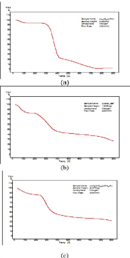
The thermal behavior of chitin and composite obtained by the deposition of CHA dense
membrane was evaluated up to 900 °C by thermogravimetric analysis. Adsorbed water loss
occurred by heating to 150 °C in both samples. Decomposition of chitin sample was
performed in two steps, with a significant loss of mass of between 330 and 400 °C and
between 400 and 750 °C. For the composite, the range studied, significant weight loss was in
the range 250 to 400 °C. From 400 °C, greater thermal stability was observed.
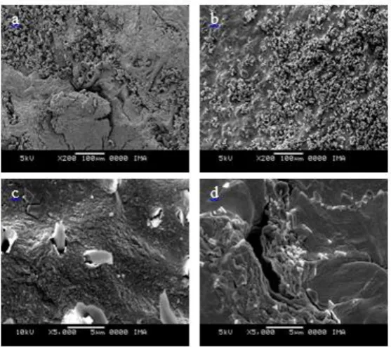
Figures 2 ad shows scanning electron microscopy images (SEM) of dense membranes (a, c)
and porous (b, d) after the nucleation of CHA in SBF. In Figures 1a and 1b, the morphology
of the surface with spherical CHA crystals can be observed. Figures 2c and 2d show
micrographs of cross sections, analyzed after immersing the membrane in liquid nitrogen. For
unclear reasons, CHA crystals can be seen in Figure 2d, whereas the dense membrane, the
CHA deposited appears as lower crystallinity material.
Figure 1 absorption spectra in the infrared chitin (a) and the composite chitin / CHA
www.wjpr.net Vol 4, Issue 11, 2015.
75 Figure 2: chitin alone thermograms of (a) the precipitate filtered FBS, after 7 days in
contact with the dense membrane chitin (b) and the dense membrane of chitin after 7
[image:5.595.171.437.86.610.2]www.wjpr.net Vol 4, Issue 11, 2015.
76 Figure 3: Scanning electron micrographs of the surfaces of dense membranes (a) and
porous (b) and cross sections of dense membranes (c) and porous (d), after immersion in
SBF for 7 days.
CONCLUSIONS
The results show that the chitin does not act as an inert material SBF and induces the
nucleation of CHA. CHA crystals were visualized by scanning electron microscopy on the
surface of dense and porous membranes and within porous membranes, after immersion in
SBF for 7 days.
ACKNOWLEDGEMENTS
The authors thank FAPERJ, FUJB and CNPq for financial support.
BIBLIOGRAPHIC REFERENCES
1. T. Kokubo; H.M. Kim; M. KawashitaBiomaterials., 2003; 24: 2161.
2. D. Bayaraktar; A.C. Tas J. Eur. Ceramic Soc., 1999; 19: 2573.
3. E. Landi; A. Tampieri; G. Celotti; R. Langenati; M. Sandri; S. SprioBiomaterials., 2005;
26: 2835.
4. R. Zhang; P.X. Ma; Macromolecular Bioscience., 2004; 4: 100.
5. R. M. Fráguas; D. A. Rocha; E. R. Queiroz;C. M. P. Abreu; R. V. Sousa; E.
[image:6.595.158.437.73.320.2]