PREVENTION OF RADIATION INDUCED HISTOPATHOLOGICAL
CHANGES IN ALBINO MICE BY
ALOE VERA
Priyanka Dadupanthi*
Biyani Girls College, Jaipur, Rajasthan, India.
ABSTRACT
The radio protection by Aloe leaf extract (1000mg/kg b.wt) was
studied in liver of Swiss albino mice before radiation exposure (3 Gy
gamma-radiation). Mice were autopsied at day 3 post irradiation and
liver was taken for histopathological studies. In this study mice, showing
distorted hepatic architecture, degranulated and vacouolated cytoplasm
in both contol and experimental set. But experimental set mice showed
mildly crenated and shrunken nuclei as compare to control. The result of
present study suggests that Aloe vera has a radioprotective effect due
to their antioxidant and radical scavenging activity.
KEYWORDS: Radiation, Aloe vera, Histopathology, Radioprotection, Albino mice
INTRODUCTION
In medical science radiation such as gamma as well as various radioisotopes is being used
considerably for both diagnostic as well as foe therapeutic purposes such as therapy of
cancer. Radiation destroys the biological molecules due to this it is very dangerous for living
systems. When individuals are exposed, the radiation energy is absorbed by the biological
systems, which causes radiolysis of tissue water and generates free radicals. The major free
radicals such as O2 2, H3O+ combine with each other and dissolved oxygen to
give a variety of potent oxidizing agents such as hydrogen peroxide, molecular oxygen and
perhydroxy radicals (Dragaric I.G. and Dragaric Z.D., 1971; Pradhan D.S., Nair C.K.K.,
Sreenivassan A., 1973; Dragaric I.G. and Scholes T.S., 1983).
The damage of tissue varies with dose of radiation exposure. It also depends on what kind of
radiation is given and on some other factors such as age, sex, species and nature of tissue.
Volume 6, Issue 5, 569-577. Research Article ISSN 2277–7105
Article Received on 26 Feb. 2017,
Revised on 18 March 2017, Accepted on 06 April 2017
DOI: 10.20959/wjpr20175-3361
*Corresponding Author’
Dr. Priyanka Dadupanthi
Biyani Girls College, Jaipur,
Chemical agents emerge to be a consequence of the anatomical position of liver and it plays
an important role in the metabolism (Plaa, 1988). Recent studies have recognized that
hepatotoxicity may be inflicting by thousands of synthetic chemicals, environmental
pollutants such as radiation and naturally occurring toxicants.
Latent liver damage, evoked in adult animals by preceding irradiation, manifests itself during
the course of liver regeneration after partial hepatectomy by various biochemical and
cytological changes, mainly by the inhibition of DNA-synthesis and mitotic activity and by
an increase in the occurrence of chromosomal aberration.
Several studies have shown histological and biological changes in liver after irradiation.
Dettmer et al. (1968) have noted that the condition of hepatocytes at the time of irradiation is
a determining factor for the subsequent damage. Grad and Stevens (1950) and Mehta et al.
(1975) have reported cytoplasmic degranulation, pyknosis and loss of architecture as a result
of irradiation. Low and moderate fractionated doses of X-rays lead to significant increase in
liver mass.
Therefore, interest has generated among scientists to develop the potential drugs of plant
origin for modification of radiation effects and hence the search for more effective, less
expensive, less toxic and easily available radioprotectors is still going on. To fulfill this need
various plant extracts and preparations are being investigated to evaluate their radioprotective
effects, because it is believed that a plant product has no or minimum side effects.
Aloe barbedensis (Mill.) commonly known as Aloe vera and belongs to family Liliaceae.
From several thousands of years Aloe has been used medicinally and polysaccharides which
are present in Aloe are always considered effective radioprotectors on radiation induced skin
damage (Wang et al.2004). In treatment of acute radiation dermatitis Aloe may be useful
(Wickline, 2004). Aloevera is widely used by patients with inflammatory bowel disease and
it has been claimed to have anti-inflammatory effects Langmead et al. 2004).
Liver is an important metabolic organ. It plays a key role in detoxification of pollutants, toxic
foods and drugs. Therefore, this study was undertaken to evaluate the value of nutritional
supplementation of Aloe vera against the radiation induced damage on the liver of Swiss
MATERIALS AND METHODS Animals
Swiss albino mice of 6-7 weeks old, weighing 24-26 gm were selected for this experimental
study. Animals were housed in polyvinyl chloride cages (290 × 320 × 390 mm) and
maintained under standard laboratory conditions. The animals had free access to food (mice
feed) and water. Tetracycline was also given along with drinking water to them once
fortnight as a preventive measure against infection. The maintenance and handling of the
animals were done according to the guidelines of the Committee for the Purpose of Control
and Supervision of Experimental Animals, animal ethics committee Ministry of Environment
and Forests, Government of India.
Source of Irradiation
Animals were irradiated at Cancer Treatment Center, S.M.S. Medical College and Hospital,
Jaipur by using Cobalt teletherapy unit (ATC-C9). Animals were kept properly in a
well-ventilated wooden box and distance between the animals in wooden box and radiation source
was 77.5 cm for exposure at the dose rate of 1.33 Gy / min. The dose rate was calibrated time
to time throughout the experimentation according to the decay table of Co60.
Aloe vera
Aloe barbadensis (Mill.) belongs to family Liliaceae and commonly known as Aloe vera.
Plant was collected from surrounding area, identified by the botanist, Department of Botany,
University of Rajasthan, Jaipur, allotted identification/voucher number RUBL-19886 and
same plant was placed in the departmental herbarium.
Experimental Design
For this study, selected adult male Swiss albino mice were divided into three groups (I, II and
III).
Group I: Animals of this group were given double distilled water (DDW) orally (volume equal to that used for Aloe administration in experimental mice) for 15 consecutive
days and called sham irradiated (normal) group.
Group II: Animals of this group were administered Aloe extract orally at the dose of 1000 mg /kg body weight (once in a day) for 15 consecutive days to study its toxic effects on
liver.
1000 mg /kg body weight (once in a day) for 15 consecutive days, whereas animals of control
set were given double distilled water (DDW) orally (volume equal to that used for Aloe
administration in experimental sets) for 15 consecutive days.
Just after 1 hour of last administration of extract and DDW, animals of group III, was
exposed to sublethal dose 3 Gy gamma radiation.
Histopathology
A minimum of 5 animals from group II and each set of control and experiment of group III
were sacrificed by cervical dislocation on day 3 of post irradiation and liver was taken for
histopathological observations.
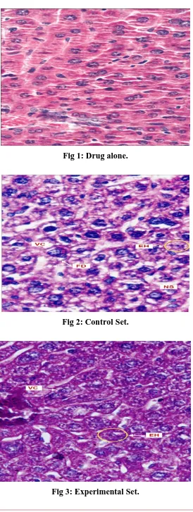
OBSERVATIONS
In present investigation animals of group I showed normal hepatic architecture of liver. There
were no changes in the cytoplasm and nuclei. A normal hepatic structure of liver is seen in
animals of group II which were supplemented Aloe alone for 15 consecutive days.
Several histopathological changes were found in the liver of Swiss albino after exposure to 3
Gy of gamma radiation individually as well as with pretreatment of Aloe vera. The changes
observed on day-3 after exposure was cytoplasmic degranulation, vacuolation, hyperaemia,
pycnotic and crenated nuclei. Many cells were lacking their nuclei. In the combined treatment
of radiation and Aloe vera similar changes were observed but they were more pronounced
showing synergistic effects. The liver of Aloe vera pretreated animals exhibited less severe
damage as compared to non-drug treated animals. An earlier and faster recovery was also
noticed in Aloe vera pretreated animals. (Figs.2).
Radiolesions like distorted hepatic architecture, few enucleated hepatocytes, degranulated
and vacuolated cytoplasm, mildly crenated and shrunken nuclei were observed in liver of
Aloe treated 3 Gy irradiated mice (experimental set) along with some normal hepatocytes at
day 3 post irradiation. However, severity of these histopathological changes was certainly
Fig 1: Drug alone.
[image:5.595.160.433.81.392.2]DISCUSSION
From the beginning, effects of radiation on liver were not clear and therefore due to its
radiosensitivity it remained as a controversial organ for a long time. Even the eminent
workers failed to observe any visible pathological change in hepatic tissue following
exposure to moderate or large doses of radiations (Hall and Whipple, 1919). Liver is
relatively resistant organ against radiation (Koletsky and Gustafson (1952); Kelly and Hirsch
(1955); and Gupta (1980).
Earlier workers such as Gupta (1972) and Bhatia et al. (1978) reported that mammalian liver
is a sensitive organ to internal irradiation at different post-natal ages (1 to 6 weeks).
In 3 Gy irradiated mice there were no radiation sickness and 30 days mortality was observed.
However, several workers such as Saharan (1977), Saini (1977), Maharwal (2002), Jagetia
and Baliga (2003) have reported various severe signs of radiation sickness and 30 days
mortality because of whole body irradiation of mice with high doses of gamma radiation.
Results of their study indicated that exposure of mice to 3 Gy did not because severe damage
in both bone marrow and gastrointestinal tract and therefore, signs of radiation sickness did
not appear.
Severity of hepatolesions increased and maximum damage was seen at day 3 in the form of
distorted hepatic architecture, karyorrhexis, chromatolysis, crenation, shrinkage and
fragmentation of nuclei and degranulation and vacuolization of cytoplasm. Similarly,
Bhartiya (1970), also reported maximum histopathological changes such as hyperaemia,
oedema, lymphocytic infiltration, pycnosis, cytoplasmic degranulation and vaculation at day
2 post irradiation in liver of gerbils exposed to 3 Gy gamma radiation and but resumption of
almost normal hepatic picture was reported after one week.
To provide protection against radiation induced deleterious effects cysteine was used for the
first by Patt et al. in 1949. Subsequently, several chemicals have been synthesized and tested
to evaluate their protective effects on different organ systems including liver, which is an
important metabolic organ and performs several functions.
Reports of Friedburg (1956) and Doul et al. (1961) also showed that herbicide, 3 amino1,
2-triazole provides slight protection to mouse liver against radiation induced damage. Similarly,
serotonin (Vittorio et al. 1963) provided protection to liver against radiation induced damage,
which is in agreement with present findings.
It has been observed that chemical radioprotectors, which generally provide maximum
protection, have to be given in high doses, which are toxic to animals. Therefore, various
natural products, herbal preparations and plant extracts have been tested by several workers
during last 30 years and reported that such protectors have various advantages over synthetic
protectors as they are nontoxic or less toxic, easily available, less expensive and easy to take
being a part of regular diet. Studies on such protectors are relatively a new area of research
and can be very promising for human beings.
Jain (2002) reported that oral administration of Amaranthus and Spinacia extracts singly
and/or in combination to Swiss albino mice reduced the 5.5 Gy induced damage in liver and
enhanced the recovery process. Similar findings have also been reported by Maharwal et al.
(2005) in liver of Rajgira extract treated 6, 8 and 10 Gy irradiated Swiss albino mice.
Thus, results of this study indicate that radioprotective effect of Aloe manifested in various
forms in liver. Treatment with Aloe prior to irradiation reduced the magnitude (severity) of
radiation induced various histopathological changes (distortion in hepatic architecture,
crenation, shrinkage and loss of liver cells nuclei, degranulation and vacuolization of
cytoplasm, lymphatic infiltration and dilation of sinusoids) and therefore, increase in kupffer
cell population was inhibited in mice liver. In this study, Aloe extract was tested for providing
protection to mice liver against radiation induced injury.
CONCLUSION
Results of this study suggest that pretreatment of mice with Aloe reduced the severity of
radiation induced various histopathogical changes in liver.
ACKNOWLEDGEMENTS
Author is gratefully acknowledging the necessary facilities provided by the Head Department
of Zoology University of Rajasthan, Jaipur, (India). I am also thankful to the Radiotherapy
Department, SMS Medical College & Hospital, Jaipur, India for providing the irradiation
REFERENCES
1. Dragaric IG, Dragaric ZD. The radiation chemistry of water. Academic Press. New York,
1971; 256.
2. Pradhan DS, Nair CKK, Sreenivassan A. Radiation injury repair and sensitization of
microorganisms. Proc. Ind. Nat. Sci. Acad, 1973; 39: 516.
3. Dragaric IG, Scholes TS. Health effects of exposure to low level of ionizing radiation.
Committee on the biological effects of ionizing radiation, 1983; 21.
4. Plaa. Toxic response of the liver. In “Toxicology the basic science of the poisons” New
York 1988; 206.
5. Dettmer, C.M., Kramer, S., Driscell, D.H. and Pante, G.E. A comparison of the chronic
effect of irradiation upon the normal, damaged and regenerating rat liver. Radiol, 1968;
21: 993.
6. Grad B, Stevens CE. Histological changes produced by a single large injection of
radioactive phosphorus (P32) in albino rats and in C3H mice. Cancer Res. 1950; 10: 289.
7. Mehta, S., Mehta, L. and Mongia, S. P. Effects of whole body X-irradiation on liver of
albino rats. Ind. J. Exptl. Biol, 1975; 13: 73.
8. Wang ZW, Zhou JM, Huang ZS, Yang AP, Liu ZC, Zeng YK. Aloe polysaccharides
mediated radio protective effect through the inhibition of apoptosis. J. Radiat. Res, 2004;
45: 447.
9. Wickline MM. Prevention and treatment of acute radiation dermatitis: a literature review.
Oncol. Nurs. Forum, 2004; 31: 237.
10.Langmead L, Feakins RM, Goldthorpe S, Holt H, Tsironi E, Desliva A, Jewell DP,
Rampton DS. Randomized double-blind, placebo-controlled trial of oral Aloe vera gel for
active ulcerative colitis. Aliment. Pharmacol. Ther, 2004; 19: 739.
11.Hall CC, Whipple GH. Roentgen intoxication: Disturbance in metabolism produced by
deep massive doses of hard roentgen rays. Am. J. Sci, 1919; 15: 453.
12.Kolletsky S, Gustafson G. Liver damage in rats from radioactive colloidal gold. Lab.
Invest, 1952; 1: 312.
13.Kelly, Hirsh. „‟Clinical Radiation Pathology” Rubbin and Casseret. (Eds) W.B.
Saunders Company, Philadelphia, 1955; 1: 263.
14.Gupta M.L. A comparative study on the radioresponse of liver in vertebrates. Ph.D.
thesis, University of Rajastan, Jaipur, India, 1980.
15.Gupta MS. Effect of radiophosphours on the prenatal and postnatal development of liver
16.Bhatia AL, Gupta ML, Singh RP. Radioresponse of mice liver to continuous β-irradiation
from tritiated water. J. Radiat. Res, 1978; 14: 194.
17.Saharan BR. MPG protection against radiation sickness and weight loss and its
correlation with mortality of mice after whole body gamma radiation Strahlenther, 1977;
157: 137.
18.Saini MR. The protective effect of 2-MPG (Thiola) against radiation induced changes in
hematopoietic tissues of Swiss albino mice after external iradiation. Ph.D. Thesis,
University of Rajasthna, Jaipur, India, 1977.
19.Maharwal J. Radioprotective effect of certain plant extract on liver and intestine of Swiss
albino mice. Ph. D. thesis, University of Rajasthan, Jaipur, India, 2002.
20.Jagetia GC, Baliga MS. Treatment of mice with a herbal preparation (mentant) protects
against radiation induced mortality. Phytother. Res, 2003; 17: 876.
21.Bhartiya, The efforts of radiation on the gastrointestinal tract and associated glands in the
India Desert Gerbils. Ph. D. thesis, University of Rajasthan, Indian, 1970.
22.Patt, H.M., Tyree, E.B., Straub, R.L. and smith, D.E. Cysteine protection against
X-irradiation. Curr. Sci, 1949; 110: 213.
23.Friedburg W. Effect of reduced liver and kidney catalase concentrations on lethality of
X-irradiation on rats. Proc. Soc. Exptl. Biol. Med, 1956; 93: 52.
24.Doul MM, VandeKerkhof PC, VanVlijmen IM, DeBakker ES, Zwiers F, DeJong EM.
The efficacy of a new topical treatment for psoriasis: Eur. J. Acad. Dermatol, 1961; 11:
13.
25.Chatterjee PD, Bose A. Determination of dry mass of rat liver cells to study the protective
mechanism of cysteamine against whole body X-irradiation. Exptl. Cell. Res, 1962; 27:
168.
26.Eldjarn L. Quoted by neuron K.F.C. “Current topics in radiation research” Elbert M.
and Howard, A. (Eds.), North Holland Publishing Co. Amsterdam, 1964; 54.
27.Vittorio PV, Wright E W, Sinott BE. A study of the protective effect of serotonin against
whole body X-irradiation in mice with the aid of Cr51. Cand. J. Biochem. Physiol, 1963;
41: 347.
28.Jain, M. Evaluation of antioxidative efficacy of certain plant extract: A study on mice
liver. Ph. D. thesis, University of Rajasthan, Jaipur, India, 2002.
29.Maharwal, J., Samarth, R.M. and Saini, M.R. Antioxidant effect of Rajgira leaf extract in