A Thesis Submitted for the Degree of PhD at the University of Warwick
http://go.warwick.ac.uk/wrap/72809
This thesis is made available online and is protected by original copyright. Please scroll down to view the document itself.
Developing new routes towards
precision and function in materials
synthesis and properties
Dafni Moatsou
submitted for the degree of Doctor of Philosophy
Department of Chemistry
Table of Contents... I List of Figures, Schemes, and Tables ...VII Figures ... VII
Schemes ... XIV
Tables ... XV
Acknowledgements ... XVII Declaration of Authorship ...XVIII List of Publications... XIX Summary of Thesis...XX Abbreviations... XXI
Chapter 1 - Introduction...1
1.1. Sequence in Nature... 2
1.2. Protein conjugates... 3
1.3. Enzyme mimics ... 4
1.3.1. Polymeric nanoreactors ... 5
1.3.1.1. Self-assembled polymers ... 6
1.3.1.2. Nano-sized particles... 7
1.4. Sequence-controlled polymerizations... 11
1.4.1. Reversible deactivation radical polymerizations ... 13
1.4.1.1. Nitroxide-mediated polymerization (NMP) ... 13
1.4.1.2. Transition metal-catalyzed polymerizations... 14
1.4.1.3. Reversible addition-fragmentation chain transfer (RAFT) polymerization... 15
1.4.2. “Living” polymerizations... 17
1.4.2.1. Ring-opening metathesis polymerization (ROMP) ... 17
1.5. Conclusions ... 20
2.1. Abstract... 31
2.2. Introduction ... 31
2.3. Results and discussion ... 37
2.3.1. Relative ratios ... 38
2.3.2. Multifunctional precision polymers... 45
2.3.3. Single monomer insertions using exo norbornenes... 56
2.4. Conclusions ... 64
2.5. Materials and methods... 65
2.5.1. Synthesis ... 66
2.5.1.1. Synthesis ofN-hexyl-endo-norbornene-5,6-dicarboximide (endoNbHex)... 66
2.5.1.2. Synthesis ofN-hexyl-exo-norbornene-5,6-dicarboximide (exoNbHex) ... 67
2.5.1.3. Synthesis of 7-coumarinyl-exo-5-norbornene-2-carboxylate (exoNbCoum)... 67
2.5.1.4. Synthesis of pentafluorophenylexo-5-norbornene-2-carboxylate (exoNbPFP) ... 68
2.5.1.5. Synthesis of (1-pyrenyl)methylexo-5-norbornene-2-carboxylate (exoNbPyr) ... 69
2.5.1.6. Synthesis of (trimethylsilanyl)methylexo-5-norbornene-2-carboxylate (exoNbTMS) ... 70
2.5.1.7. Homopolymerizations... 71
2.5.1.8. Copolymerization ofendoNbHex andexoNbCoum (P2.3-2.5) ... 71
2.5.1.9. Synthesis of multi-functional poly(norbornene) (P2.6)... 72
2.5.1.10. SingleexoNbCoum insertions into the ROMP ofendoNbHex ... 72
2.6. References ... 73
Chapter 3 - Single monomer additions in ring-opening metathesis polymerization...80
3.1. Abstract... 81
3.2. Introduction ... 81
3.2.1. End group functionalization... 82
3.2.2. Sacrificial copolymerization ... 83
3.2.3. Single monomer addition ... 86
3.2.4. Alternating copolymers ... 86
3.3.1.1. Homopolymerization of DxpPhe ... 90
3.3.1.2. Polymerization of dioxepin from an activated alkylidene ... 91
3.3.1.3. Addition to living poly(NbHex)... 101
3.3.2. Evaluation of the reactivity of functional dioxepins... 111
3.3.2.1. Reactivity ratios ... 111
3.3.2.2. Calculation of the reactivity ratios of dioxepins andexoNbHex ... 112
3.3.2.3. Calculation of the reactivity ratios of dioxepins andendoNbHex... 115
3.3.3. Sequential polymerization of norbornene ... 117
3.3.3.1. Polymerization ofendonorbornenes in the presence of DxpPhe ... 117
3.3.3.2. Polymerization ofexonorbornenes in the presence of DxpMe... 122
3.4. Conclusions ... 126
3.5. Materials and Methods ... 127
3.5.1. Synthesis ... 128
3.5.1.1. Synthesis of 2-phenyl-4,7-dihydro-2H-1,3-dioxepin (DxpPhe) ... 128
3.5.1.2. Synthesis of 2-methyl-4,7-dihydro-2H-1,3-dioxepin (DxpMe) ... 129
3.5.1.3. 2-(1-pyrenyl)-4,7-dihydro-2H-1,3-dioxepin (DxpPyr)... 129
3.5.1.4.N-(2-morpholinoethyl)-exo-norbornene-5,6-dicarboximide (exoNbMorph)... 130
3.5.1.5. Homopolymerization of DxpPhe ... 130
3.5.1.6. Chain extension of poly(NbHex) (P3.1) with DxpMe (P3.2) ... 130
3.5.1.7. Synthesis of poly(NbHex)-b-poly(DxpMe)-b-poly(NbPyr) (3.5) ... 131
3.5.1.8. Hydrolysis ofP3.5... 132
3.5.1.9. Addition of DxpMe to a living ROMP ofexoNbHex... 132
3.5.1.10. Addition of DxpPyr to a living ROMP ofendoNbHex (P3.8) ... 132
3.5.1.11. Determination of reactivity ratios ... 133
3.5.1.12. Sequential polymerization ofendonorbornenes ... 135
3.5.1.13. Hydrolysis ofP3.9,P3.10,P3.11, andP3.12... 135
3.5.1.14. Sequential polymerization ofexonorbornenes ... 136
3.5.1.15. Hydrolysis ofP3.19... 136
3.6. References ... 137
4.2. Introduction ... 141
4.3. Results and Discussion ... 146
4.3.1. GFP-dye conjugation ... 148
4.3.2. Polymerizations... 153
4.3.3. GFP-poly(OEGMA) bioconjugates... 158
4.3.4. Thermo-responsive properties... 163
4.3.5. Activity of the bioconjugates ... 172
4.4. Conclusions ... 174
4.5. Materials and Methods ... 175
4.5.1.1. Synthesis ofN-(6-(diethylamino)-9-(2-((prop-2-yn-1-yloxy) carbonyl)phenyl)-3H -xanthen-3-ylidene)-N-ethylethanaminium (Rhod-alk) ... 176
4.5.1.2. CuAAC reaction of azide-functional GFP andRhod-alk... 177
4.5.1.3. Synthesis of prop-2-yn-1-yl 2-phenyl-2-((phenylcarbonothioyl) thio)acetate (CTA1)... 178
4.5.1.4. RAFT polymerizations – Synthesis ofP4.1,P4.2, andP4.3... 179
4.5.1.5. CuAAC reaction of azide-functional GFP and polymersP4.1,P4.2, andP4.3... 180
4.6. References ... 182
Chapter 5 - Catalytic nanogels: study of the effect of structural properties... 188
5.1. Abstract... 189
5.2. Introduction ... 189
5.2.1. Aldolase mimics:L-Proline ... 189
5.2.1.1. Catalytic activity ... 189
5.2.1.2. Effect of water ... 191
5.2.1.3. Polymer-supportedL-proline... 193
5.2.2. Nanogels... 195
5.3. Results and discussion ... 198
5.3.1. Nanogel synthesis... 198
5.3.2.L-Proline-containing nanogels: Tuning the hydrophobicity ... 199
5.3.2.3. Tuning the cross-linking density ... 224
5.4. Conclusions ... 230
5.5. Materials and Methods ... 231
5.5.1.1. Synthesis ofO-methacryloyl-trans-4-hydroxy-L-proline hydrochloride (ProMA) ... 233
5.5.1.2. Hydrophobic nanogel synthesis ... 233
5.5.1.3. Representative catalytic aldol reaction... 234
5.5.1.4. Recycling of the catalytic nanogels... 235
5.5.1.5. Evaluation of the nanogel hydrophobicity with Nile Red ... 235
5.5.1.6. Synthesis of the core-shell nanogels (CS5.1)... 236
5.5.1.7. Synthesis of the core-gradient shell nanogels (GS5.2)... 236
5.5.1.8. Copper-stained nanogels for TEM imaging ... 237
5.5.1.9. Synthesis of the core-heavily cross-linked shellCS5.3nanogels... 237
5.5.1.10. Synthesis of the “impregnated” core-shell nanogels (ICS5.5) ... 238
5.5.1.11. Synthesis of hydrophobic MMA-based nanogels with different CLD ... 238
5.5.1.12. Synthesis of theCS5.6core-shell nanogels ... 239
5.6. References ... 239
Chapter 6 - Photo-induced cross-linking of thymine-functional nanogels ... 245
6.1. Abstract... 246
6.2. Introduction ... 246
6.2.1. Thymine dimerization ... 246
6.3. Results and discussion ... 248
6.3.1. Photo-cross-linked nanogels ... 248
6.3.1.1. Styrene-based nanogels for UV-induced cross-linking ... 249
6.3.1.2.N-isopropylacrylamide-based nanogels for UV-induced cross-linking... 260
6.4. Conclusions ... 276
6.5. Materials and Methods ... 277
6.5.1.1. Synthesis of 1-(vinylbenzyl)thymine (VBT)... 278
6.5.1.2. Synthesis of the thymine-containing styrene-based nanogels ... 279
Figures
Figure 1.1. Top-down and bottom-up strategies followed to obtain nanostructures mimicking protein and enzyme functions... 3 Figure 1.2. Schematic representation of various self-assembled structures formed by block copolymers in a block-selective solvent.44... 6 Figure 1.3. Schematic representation of the catalytic dispersed particles (A), catalytic hollow capsules (B-C), and the free catalyst-loaded polymer (D)... 8 Figure 1.4. Schematic representation of the three intervals of emulsion polymerization.42.... 9 Figure 1.5. DNA origami.105... 12 Figure 1.6. Common ROMP catalysts.149... 19 Figure 2.1. Microstrucures of the polymers and semilogarithmic plots from the copolymerization of maleimide-type monomers and styrene depending on the styrene conversion.86... 35 Figure 2.2. Schematic representation of (A) the steric interaction between the metallacyclobutane and theendonorbornene substituent and (B) the growing polymer chain and theendonorbornene substituent (adapted from the literature108)... 37 Figure 2.3. Chemical structures of the norbornenes used for the evaluation of the relative ROMP rates. ... 38 Figure 2.4. Monomer conversions for the ROMP ofendoNbHex and exoNbHex, calculated by1H NMR spectroscopy. ... 39 Figure 2.5. Assigned 1H NMR spectra from the homopolymerizations of exoNbHex (top) andendoNbHex (CDCl3, 400 MHz)... 40
Figure 2.6. Molecular weight distributions obtained by SEC in THF for polymer P2.1 and P2.2. ... 41 Figure 2.7. Schematic representation of the strategy followed for the synthesis of P2.3, P2.4, and P2.5. Arrows denote monomer addition. ... 43 Figure 2.8. Semilogarithmic plots of monomer conversion versus time for the copolymerization of endoNbHex (squares) and exoNbCoum (circles) in CDCl3 at
room temperature at threeendo monomer conversions: 25% (P2.3), 50% (P2.4) and 70% (P2.5). ... 44 Figure 2.9. 19F NMR spectra of exoNbPFP (top) and P2.6 (bottom) showing the characteristic fluorine signals of pentafluorophenyl (CDCl3, 282 MHz). ... 46
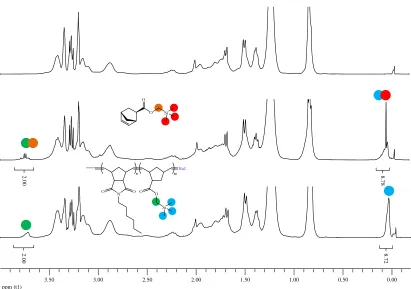
Figure 2.10. 1H NMR spectra of the polymerization ofendoNbHex before (top), 12 minutes after (middle) and 2 hours after (bottom) the addition of exoNbPFP in the reaction mixture (CDCl3, 400 MHz). ... 47
Figure 2.11. 1H NMR spectra of the polymerization mixture before (top), 15 minutes after (middle) and two hours after (bottom) the addition ofexoNbTMS (CDCl3, 400 MHz).
... 48 Figure 2.12. 1H NMR spectra of the polymerization mixture before (top), 15 minutes after (middle) and three hours after (bottom) the addition ofexoNbPyr (CDCl3, 400 MHz).
... 49 Figure 2.13. 1H NMR spectra of the polymerization mixture before (top), 12 minutes after (middle) and three hours after (bottom) the addition of exoNbCoum (CDCl3, 400
NbTMS) (CDCl3, 500 MHz). ... 51
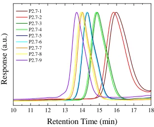
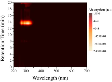
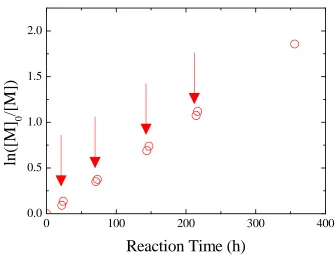
Figure 2.15. Semilogarithmic plots of monomer conversions versus time for the ROMP of endoNbHex (■) and the sequential addition of exoNbPFP (●), exoNbTMS (▲), exoNbPyr (▼), and exoNbCoum (♦) for the synthesis of the multifunctional copolymer P2.6. ... 52 Figure 2.16. Size exclusion chromatograms of the multifunctional copolymer P2.6 in THF using DRI detection as well as UV detection atλ= 309 nm andλ= 344 nm... 54 Figure 2.17. Overall DP as a function of polymerization time. ... 55 Figure 2.18. Schematic representation of the strategy followed for the single insertions of exoNbCoum in the ROMP ofendoNbCoum. ... 56 Figure 2.19. Size exclusion chromatograms from the polymerization of endoNbHex before (P2.7-1, P2.7-3, P2.7-5, and P2.7-7) and after (P2.7-2, P2.7-4, P2.7-6, and P2.7-8) addition of a single equivalent ofexoNbCoum, as well as the final copolymer P2.7-9, in THF... 57 Figure 2.20. 2D SEC/UV-vis spectrum for the final polymer P2.7-9 in THF. ... 59 Figure 2.21. Semilogarithmic plot of monomer conversion versus time for the ROMP of endoNbHex and the sequential addition of four single equivalents ofexoNbCoum. The arrows indicate the time points of theexoCoum addition... 60 Figure 2.22. Absorption spectra of the isolated copolymers from the ROMP of endoNbHex before (P2.7-1, P2.7-3, P2.7-5, and P2.7-7) and after (P2.7-2, P2.7-4, P2.7-6, and P2.7-8) addition of a single equivalent ofexoNbCoum, as well as the final copolymer P2.7-9, in CH2Cl2... 61
Figure 2.23. Amount of coumarin moieties per polymer chain (Nc) as calculated for each
polymer obtained from the single equivalent addition ofexoNbCoum into the ROMP ofendoNbHex. Arrows denote monomer addition times. ... 62 Figure 2.24. MALDI-ToF mass spectra of P2.7-1 and P2.7-2 after the addition of a single equivalent of exoNbCoum. Tables indicate the expected and measured masses in Da for the respective peaks. ... 63 Figure 3.1. Schematic representation of the strategy followed for the sacrificial polymerization of acid-labile monomers in order to afford polymers with a single functionalization.17... 84 Figure 3.2. 1H NMR spectra of the monomer DxpPhe (top) and an aliquot taken from the polymerization after 72 hours (bottom) (CDCl3, 400 MHz)... 91
Figure 3.3. Assigned 1H NMR spectra of the isolated polymers P3.1 before (top) and P3.2 after (bottom) addition of DxpMe. The insets show the alkoxy proton regions (3-5 ppm) of the respective spectra. (CDCl3, 300 MHz)... 93
poly(NbHex), S2 to poly(NbHex)-COH, and S3 to cyclized poly(NbHex). ... 104 Figure 3.11. Assigned 1H NMR spectrum of the P3.7 polymer and the acetal region expanded (CDCl3,300 MHz). ... 105
Figure 3.12. Chromatograms of polymer P3.8 and at different intervals after the addition of DxpPyr, detected by DRI (top) and UV (λ= 343 nm) (bottom) (SEC in THF). ... 107 Figure 3.13. MALDI-ToF mass spectra for P3.8 and after addition of DxpPyr at different time intervals. ... 108 Figure 3.14. Expanded region of the MALDI-ToF mass spectra for P3.8 and after addition of DxpPyr at different time intervals, as well as the predicted isotopic distributions for poly(NbHex) (S4), poly(NbHex)-DxpPyr (S5), and poly(NbHex)-COH (S6). Structures correspond to the simulated isotopic distributions. ... 109 Figure 3.15. Assigned 1H NMR spectrum of the P3.8-300 polymer and the 4.5-3.8 ppm region expanded. The signal marked with a star corresponds to the unfunctionalized P3.8 chain-end protons (CD2Cl2, 400 MHz)... 110
Figure 3.16. Polymer DxpMe content (F1) with respect to monomer DxpMe feed ratio (f1) in
the copolymerization withexoNbHex as determined by 1H NMR spectroscopy. Line shows ideal random copolymerization. ... 113 Figure 3.17. Polymer DxpPhe content (F1) with respect to monomer DxpPhe feed ratio (f1) in
the copolymerization with exoNbHex determined by 1H NMR spectroscopy. Line shows ideal random copolymerization. ... 114 Figure 3.18. Polymer DxpPhe content (F1) with respect to monomer DxpPhe feed ratio (f1) in
the copolymerization with endoNbHex determined by 1H NMR spectroscopy. Line shows ideal random copolymerization. ... 115 Figure 3.19. Plot of joint confidence intervals (95%) of the reactivity ratios for DxpPhe (r1)
andendoNbHex (r2)... 116
Figure 3.20. Molecular weight distributions of P3.9, P3.10, P3.11, and P3.12 obtained by SEC... 119 Figure 3.21. Number-average molecular weights and dispersities of P3.9, P3.10, P3.11, and P3.12 with respect to the DP of the poly(NbHex) blocks, determined by 1H NMR spectroscopy. Dashed line represents the linear fit of theMndatapoints... 120
Figure 3.22. SEC molecular weight distributions of the copolymers P3.9, P3.10, P3.11, and P3.12, and their hydrolyzed counterparts P3.13, P3.14, P3.15, and P3.16... 121 Figure 3.23. Chromatograms of polymers P3.17, P3.18, P3.19, P3.20, and P3.21... 123 Figure 3.24. Chemical structure and the assigned1H NMR spectrum of P3.18. (CDCl3, 300
Figure 4.6.Chromatogram obtained from the LC-MS ofG2-Rhod. ... 152 Figure 4.7. Mass spectra of G2-N3and G2-Rhod, and the corresponding spectra based on the
theoretical isotope distributions. Major peaks in each spectrum coincide with the theoretical peaks for each species and have been highlighted... 153 Figure 4.8. Molecular weight distributions of P4.1, P4.2, and P4.3 obtained by SEC in THF (2% Et3N)... 155
Figure 4.9. Comparison of the chromatograms obtained for P4.1, P4.2, and P4.3 using a DRI and a UV detector (SEC in THF, 2% Et3N)... 156
Figure 4.10. Assigned 1H NMR spectrum of polymer P4.1 showing the characteristic peaks of the end groups (CD3OD, 500 MHz). ... 157
Figure 4.11. Schematic representation of the nine bioconjugates resulting from conjugation of each of the three proteins G216-N3, G2.216-N3, and G2-N3 with each of the three
polymers P4.1, P4.2, and P4.3. ... 158 Figure 4.12. Chromatograms from the crude protein-polymer bioconjugates (SEC in Tris buffer). ... 159 Figure 4.13. PAGE gels of the proteins upon conjugation with P4.1 (A) lane 1: ladder, lane 2: P4.1, lane 3: G2-N3, lane 4: G2-P4.1, lane 5: G216-N3, lane 6: G216-P4.1, lane 7:
G2.216-N3, lane 8: G2.216-P4.1, upon conjugation with P4.2 (B) lane 1: ladder, lane
2: P4.2, lane 3: G2-N3, lane 4: G2-P4.2, lane 5: G216-N3, lane 6: G216-P4.2, lane 7:
G2.216-N3, lane 8: G2.216-P4.2, and upon conjugation with P4.3 (C) lane 1: ladder,
lane 2: P4.3, lane 3: G2-N3, lane 4: G2-P4.3, lane 5: G216-N3, lane 6: G216-P4.3, lane
7: G2.216-N3, lane 8: G2.216-P4.3... 160
Figure 4.14. PAGE gels of the proteins and densitometric analysis upon conjugation with P4.1 (A) lane 1: ladder, lane 2: P4.1, lane 3: G2-N3, lane 4: G2-P4.1, lane 5: G216-N3,
lane 6: G216-P4.1, lane 7: G2.216-N3, lane 8: G2.216-P4.1, upon conjugation with
P4.2 (B) lane 1: ladder, lane 2: P4.2, lane 3: G2-N3, lane 4: G2-P4.2, lane 5: G216-N3,
lane 6: G216-P4.2, lane 7: G2.216-N3, lane 8: G2.216-P4.2 ... 162
Figure 4.15. Photograph showing the observed pink precipitate and its corresponding assigned 1H NMR spectrum, showing the characteristic signals from the POEGMA homopolymer (CDCl3, 400 MHz) ... 163
Figure 4.16. PAGE gel and densitometric analysis of the G216-P4.2 bioconjugate upon purification by the thermal precipitation of excess polymer. ... 164 Figure 4.17. Cloud point curves for GFP (purple line), P4.1, P4.2, and P4.3 (black lines) and the corresponding bioconjugates after CuAAC with G2-N3(red lines), G216-N3(green
lines), and G2.216-N3(blue lines). All lines are averages of three measurements. ... 165
heating-cooling cycles. ... 173 Figure 5.1. Cryo-TEM image of poly(styrene)-co-poly(NIPAM) core-shell particles.82.... 198 Figure 5.2. Schematic representation of the synthesis of theL-proline functional hydrophobic nanogels. ... 199 Figure 5.3. Size distributions averaged by intensity for the hydrophobic nanogels in water at 25ºC... 200 Figure 5.4. Hydrodynamic diameters of the hydrophobic nanogels with respect to the co-monomer (MMA: squares, EMA: circles, BuMA: triangles, LMA: diamonds) and the degree of functionalization (DoF, wt%). Error bars show size dispersity. ... 201 Figure 5.5. Representative TEM images of the hydrophobic nanogels N5.M0.5-2 (A), N5.E0.5-2 (B), N5.B0.5-2 (C), and N5.L0.5-2 (D). Formvar-coated grid (Scale bar: 100 nm)... 202 Figure 5.6. Fluorescence emission spectra of the nanogels loaded with Nile Red (λex= 550
nm)... 205 Figure 5.7. Fluorescence emission spectra of Nile Red loaded in the N5.M0.5-2 and the N5.M0.5-15 nanogels (λex= 550 nm). ... 207
deviation as experiments were done in triplicate... 223 Figure 5.21. Size distributions of the hydrophobic methyl methacrylate-based nanogels with different CLD (0-50 wt%) and 2 wt% DoF, determined by DLS at 25 ºC... 225 Figure 5.22. Effect of cross-linking density on hydrodynamic size of the MMA-based hydrophobic nanogels in water... 226 Figure 5.23. Catalytic efficiency of the MMA-based hydrophobic nanogels as a function of their CLD. Conversions were determined by1H NMR spectroscopy after 24 hours. 228 Figure 5.24. Schematic representation of the synthesis of a hydrophobic shell to encapsulate surfaceL-proline moieties into a heavily cross-linked hydrophobic environment. ... 229 Figure 5.25. Size distributions of the N5.M50-2 and the CS5.6 hydrophobic nanogels at 25 ºC. ... 229 Figure 6.1. Schematic representation of the irradiation-induced dimerization of thymine, and thus cross-linking of the nanogels. ... 249 Figure 6.2. Size distributions for the VBT-containing styrene-based nanogels determined by DLS in water... 250 Figure 6.3. Hydrodynamic diameters of the VBT-functional styrene-based nanogels determined by DLS in water at 25 ºC. Error bars indicate size dispersity... 251 Figure 6.4. Representative TEM images of the N6.ST10 (left) and the N6.ST30 (right) nanogels (Scale bars: 200 nm). ... 251 Figure 6.5. Size histograms from the N6.ST10 and N6.ST30 particles measured by TEM. 252 Figure 6.6. DOSY NMR spectrum of N6.ST30 (500 MHz, C3D7NO). The polymer (blue
rectangle), the solvent (yellow rectangle), and the surfactant (green rectangle) are highlighted, as well as the signals that correspond solely to the VBT moiety (red arrows). ... 253 Figure 6.7. Normalized absorption spectra of N6.ST0 poly(styrene) nanogels and the VBT-containing nanogels N6.ST2, N6.ST10, N6.ST20, and N6.ST30 in water. ... 254 Figure 6.8. Absorption spectra for the styrene-based nanogels with different VBT loadings (2,10, 20, and 30 wt%) upon irradiation. ... 255 Figure 6.9. Digital photograph of the N6.ST2 (left) and the N6.ST2-15 (right) nanogel dispersions, showing the discoloration after prolonged irradiation. ... 256 Figure 6.10. Hydrodynamic diameters of the styrene-based VBT-loaded nanogels at different irradiation times, determined by DLS in water at 25 ºC. Error bars correspond to size distributions. ... 256 Figure 6.11. Size distributions for the styrene-based nanogels with different VBT loadings, before and after irradiation, determined by DLS in water at 25 ºC. ... 257 Figure 6.12. TEM images of the N6.ST10-18 (left) and the N6.ST30-18 (right) nanogels showing the morphology of the nanogel after irradiation. (Scale bars: 200 nm)... 258 Figure 6.13.1H NMR spectra of the N6.ST30 nanogels at different irradiation times, dissolved in C3D7NO containing TMS-PSA (400 MHz). Highlighted regions show the
Scheme 1.1. Schematic representation of the equilibrium in NMP. ... 13
Scheme 1.2. Chemical structures of commonly used nitroxides in NMP... 14
Scheme 1.3. Reaction mechanism of ATRP. ... 15
Scheme 1.4. Proposed mechanism for RAFT polymerization... 16
Scheme 1.5. Mechanism of ROMP... 17
Scheme 2.1. Schematic representation of the polymerization of NbHex using G1 as the catalyst and EVE as the quenching agent. ... 38
Scheme 2.2. Schematic representation of the strategy followed for the copolymerization of endoNbHex and exoNbCoum, resulting in three copolymers with different compositions... 43
Scheme 2.3. Schematic representation of the strategy followed for the synthesis of the multifunctional P2.6. ... 46
Scheme 3.1. Schematic representation of the approach followed by Hillmyer et al. for the synthesis of a sequence-defined polymer by ROMP.27... 86
Scheme 3.2. Proposed mechanisms for ROIMP28(left) and ALTMET29(right). ... 87
Scheme 3.3. Schematic representation of the homopolymerization of DxpPhe using G1 as the catalyst. ... 90
Scheme 3.4. Generic reaction scheme for the chain extension of poly(NbHex) with functional dioxepins... 92
Scheme 3.5. Schematic representation of the synthetic pathway for the synthesis of polymers P3.3, P3.4, P3.5, and the hydrolysis of P3.5... 97
Scheme 3.6. Strategy for the sequential polymerization of endoNbHex in the presence of DxpPhe. ... 118
Scheme 3.7. Schematic representation of the procedure followed for the synthesis of P3.17, P3.18, P3.19, P3.20, and P3.21viasequential additions followed by hydrolysis. .... 122
Scheme 4.1. Schematic representation of the three GFP analogues with the modified residues highlighted, and the chemical structure corresponding to the residues. ... 147
Scheme 4.2. Schematic representation of the CuAAC reaction using the three GFP derivatives G216-N3, G2.216-N3, and G2-N3 and Rhod-alk to form G216-Rhod, G2.216-Rhod, and G2-Rhod respectively... 148
Scheme 4.3. Schematic representation of the strategy followed for the synthesis of the temperature-responsive protein-polymer bioconjugates... 154
Scheme 5.1. Generally accepted mechanism for the catalytic cycle of L-proline in the aldol reaction between a ketone and an aldehyde... 192
Scheme 5.2. Schematic representation of the benchmark aldol reaction catalyzed by L -proline... 203
Table 2.1. Molecular weights and dispersities for the polymers synthesized by the ROMP of exoNbHex (P2.1) and endoNbHex (P2.2) (determined by SEC in THF against poly(styrene) standards)... 42 Table 2.2. Molecular weights and dispersities for the polymerization of endoNbHex with single equivalents of exoNbCoum being added at t = 22, 71, 144, and 214 hours (determined by SEC in THF against poly(styrene) standards). ... 58 Table 3.1. Molecular weights and dispersities for the polymers before (P3.1) and after (P3.2) the addition of DxpMe... 94 Table 3.2. Molecular weights and dispersities for polymers P3.3, 3.4, 3.5, and the hydrolysis product P3.5h... 99 Table 3.3. Molecular weights and molecular weight distributions for P3.8 and the samples after addition of DxpPyr. ... 108 Table 3.4. Molecular weights and dispersities of P3.9, P3.10, P3.11, and P3.12. ... 119 Table 3.5. Molecular weight and molecular weight distributions determined by SEC of polymers P3.17, P3.18, P3.19, P3.20 and P3.21... 124 Table 3.6. Quantities of reagents used for the copolymerizations of DxpMe and exoNbHex towards the calculation of their reactivity ratios... 133 Table 3.7. Quantities of reagents used for the copolymerizations of DxpPhe and exoNbHex towards the calculation of their reactivity ratios... 134 Table 3.8. Quantities of reagents used for the copolymerizations of DxpPhe andendoNbHex towards the calculation of their reactivity ratios... 134 Table 4.1. Molecular weights and dispersities for P4.1, P4.2, and P4.3 (determined by SEC in THF against poly(methyl methacrylate) standards)... 155 Table 4.2. Calculated quantities for the CuAAC reaction of the azide functional proteins and the alkyne functional dye Rhod-alk. ... 178 Table 4.3. Reagent quantities and conditions used for the synthesis of the alkyne-functional polymers ... 180 Table 4.4. Quantities calculated for the CuAAC of P4.1, P4.2, and P4.3 to the three different proteins G2-N3, G216-N3, and G2.216-N3... 181
This PhD has not been easy, but I have had a lot of support in the past three (and a
bit) years and, now, I feel like time flew.
I want to thank all of the people whom I have collaborated with around the world:
Prof. Michael Jewett, Dr. Arnaz Ranji, and Dr. Jian Li from NU; Dr. Kei Saito and
Dr. Gagan Kaur from Monash University; Prof. Andreas Kilbinger and Dr. Amit
Nagarkar from the University of Fribourg; Dr. Annhelen Lu from the University of
Warwick. I would also like to acknowledge EPSRC for funding.
I cannot thank enough Rachel (Professor O’Reilly to you) for bringing me here in the
first place, helping me through some rough patches, and always being the diligent
guide she is. I sometimes cannot believe how many exciting things have happened
because of her.
If this thesis makes sense to anyone, that is a result of the kind help from Mat, Lewis,
Becky, Zan, Rob, Guillaume, Alice, Anaïs, Helen, Anne, and James that were
volunteered to proof-read. I also want to thank everyone in the Do’Reilly
(O’Dovelly?) groups, including Annie, for making life as a lab rat more social, and
for always leaving snacks at the tea point. I couldn’t leave out of this the O’Reilly
try-athletes, and their successors, who introduced me to a lifestyle that allows me to
go for a run to relax. Special thanks to the people that didn’t give up on me even on
my grumpiest days; well, I don’t need to explain why, it’s my thesis, so “thanks”.
I know my family still has no idea what my PhD thesis is about, but they have
influenced a great deal its outcome. Thank you for supporting me though all of these
years in education, which are now coming to an end. Probably… Αλέξανδρε, I hope
This thesis is submitted to the University of Warwick in support of my application
for the degree of Doctor of Philosophy. It has been composed by myself and has not
been submitted in any previous application for any degree. The work presented
(including data generated and data analysis) was carried out by the author except in
the case of Chapter 4, which was carried out in collaboration with Arnaz Ranji and
Jian Li from Professor Michael Jewett’s group at Northwestern University, USA,
Chapter 5, which was carried out in collaboration with Annhelen Lu, from Professor
Rachel O’Reilly’s group at the University of Warwick, UK, and in the case of
Chapter 6, that was carried out with the help of Dominic Gray from Professor Rachel
O’Reilly’s group at the University of Warwick, UK. Any materials synthesized or
data analyses carried out by persons other than the author are clearly labelled in the
1. “Tuning the Catalytic Activity of L-Proline Functionalized Hydrophobic Nanogel
Particles in Water”, A. Lu,‡D. Moatsou,‡D. A. Longbottom, R. K. O’Reilly,Chem.
Sci., 2013,4, 965-96 (Chapter 5)
2. “Studying the activity of the McMillan catalyst embedded within hydrophobic
crosslinked polymeric nanostructures”, B. Moore, D. Moatsou, A. Lu, R. K.
O’Reilly,Polym. Chem., 2014,5, 3487-3494
3. “Precision Polymers: A Kinetic Approach for Functional Poly(norbornenes)”, D.
Moatsou, C. F. Hansell, R. K. O’Reilly,Chem. Sci., 2014,5, 2246-2250 (Chapter 2)
4. “The effect of polymer nanostructure on diffusion of small molecules using
Tryptophan as a FRET probe”, B. L. Moore, A. Lu, D. Moatsou, R. K. O’Reilly,
Eur. Polym. J., 2015,62, 380-3853
5. “Recyclable L-Proline Functional Nanoreactors with Temperature-Tuned Activity
Based on Core-Shell-Corona Nanogels”, A. Lu,‡D. Moatsou,‡I. Hands-Portman, D.
A. Longbottom, R. K. O’Reilly,ACS Macro Lett., 2014,3, 1235–1239 (Chapter 5)
6. “Biofunctionalizable flexible bucky paper by combination of multi-walled carbon
nanotubes and polynorbornene-pyrene. Application to the bioelectrocatalytic
reduction of oxygen”, S. Cosnier, R. Haddad, D. Moatsou, R. K. O’Reilly, Carbon,
accepted
7. “Self-assembly of temperature-responsive protein-polymer bioconjugates”, D.
Moatsou,‡ J. Li,‡ A. Ranji, A. Pitto-Barry, I. Ntai, M. C. Jewett, R. K. O’Reilly,
This thesis explores three methods for the synthesis of materials with precise
sequence and functions.
Chapter 1 gives a brief introduction to the main concepts that underpin the methods
used throughout the thesis.
Chapter 2 examines the use of norbornenes for the synthesis of precision polymers
via ring-opening metathesis polymerization by taking advantage of the different
reactivity of different isomers of the monomers.
Chapter 3 suggests the use of dioxepins for single monomer insertion in the
ring-opening metathesis polymerization of norbornenes.
Chapter 4 describes the synthesis of a temperature-responsive protein-polymer
bioconjugate and the study of its properties.
Chapter 5 evaluates the synthetic parameters of L-proline-containing nanogels as a
function of their ability to catalyze organic reactions.
Chapter 6 discusses the attempt to modulate the cross-linking of nanogels by a
3
J proton coupling constant (commonly written as3JHH)
ADMET acyclic diene metathesis
AIBN 2,2′-azobis(2-methylpropionitrile)
ALTMET alternating diene metathesis
ATRP atom transfer radical polymerization
BuMA n-butyl methacrylate
CLD cross-linking density
CRP controlled radical polymerization
CTA chain transfer agent
CuAAC copper-catalyzed azide-alkyne cycloaddition
d doublet
DCM dichloromethane
Dh hydrodynamic diameter
DLS dynamic light scattering
ĐM molecular weight distribution (Mw/Mn)
DMAP 4-(dimethylamino) pyridine
DMF N,N-dimethylformamide
DMSO dimethylsulfoxide
DNA deoxyribonucleic acid
DoF degree of functionalization
DOSY diffusion-ordered spectroscopy
DP degree of polymerization
ee enantiomeric excess
EGDMA ethylene glycol dimethacrylate
EMA ethyl methacrylate
endoNbHex N-hexyl-endo-norbornene-5,6-dicarboximide
eq. equivalents
ESI electrospray ionization
EVE ethyl vinyl ether
exoNbCoum 7-coumarinyl-exo-5-norbornene-2-carboxylate
exoNbHex N-hexyl-exo-norbornene-5,6-dicarboximide
exoNbPFP pentafluorophenylexo-5-norbornene-2-carboxylate
exoNbPyr (1-pyrenyl)methyl-exo-5-norbornene-2-carboxylate
exoNbTMS trimethylsilylexo-5-norbornene-2-carboxylate
FDA food and drug administration
G1 Grubbs Catalyst, 1st Generation
(bis(tricyclohexylphosphine)benzylidine ruthenium(IV)
dichloride)
GO graphene oxide
HPLC High performance liquid chromatography
HRMS high resolution mass spectrometry
ki initiation rate constant
kp propagation rate constant
LC liquid chromatography
MALDI matrix-assisted laser desorption ionization
MMA methyl methacrylate
Mn number-average molecular weight distribution
MS mass spectrometry
Mw weight-average molecular weight distribution
MW molecular weight
MWCO molecular weight cut-off
NIPAM N-isopropylacrylamide
NMBA N-N’-methylenebis(acrylamide)
NMP nitroxide-mediated polymerization
NMR nuclear magnetic resonance
PAGE poly(acrylamide) gel electrophoresis
PEG poly(ethylene glycol)
PEGMA poly[oligo(ethylene glycol) methyl ether methacrylate]
PEO poly(ethylene oxide)
ProMA O-methacryloyl-trans-4-hydroxy-L-proline hydrochloride
RAFT reversible addition-fragmentation chain-transfer
RDRP reversible-deactivation radical polymerization
Rf retardation factor
RNA ribonucleic acid
ROIMP ring-opening-insertion-metathesis polymerization
ROMP ring-opening metathesis polymerization
t triplet
TEA triethylamine
TEM transmission electron microscopy
Tg glass transition temperature
THF tetrahydrofuran
TLC thin layer chromatography
TMS-PSA 3-(trimethylsilyl)propanesulfonic acid
ToF time of flight
UV ultraviolet
UV-vis ultraviolet-visible
VBT 1-(vinylbenzyl)thymine
wt% weight percent
δ chemical shift
1.1. Sequence in Nature
Nature has always outperformed synthetic chemists when it comes to controlling the
sequence of macromolecules. With an arsenal consisting of just over two dozen building
blocks, a remarkable repertoire of functional natural polymers such as DNA and proteins
is now known to be the basis of life. Decades before Watson and Crick solved the
double-helical structure of DNA in 1953,1 Albrecht Kossel had isolated the five
nucleobases that are to a great extent responsible for the remarkable properties of DNA
and RNA.2 The fact that intricate functions are dictated by such a small number of
building blocks highlights the importance of the sequence in which these building
blocks are connected. The importance is emphasized when genetic disorders are
considered, whereby the sequence of the genome is compromised potentially having
damaging effects on the host organism. The implications of sequence-ordered
macromolecules are equally prominent in proteins where the primary structure, that is
the sequence of the amino acid residues, is to a large extent responsible for the
three-dimensional tertiary structure, and thus the function of the protein. Small variations in
the sequence of the protein result in significantly different properties, ensuing
tremendous variation in naturally occurring proteins. As each protein is synthesized in
order for a specific function to be accomplished, multiple biological processes ensure
that the sequence of a synthesized protein is correct. However, often proteins with
dissimilar sequences have similar functions as a result of homologous three-dimensional
microstructures in the way the sequences fold. While there is still a lot of ground to
cover in our understanding of how sequences correlate to specific functions, a large
amount of work is dedicated to mimicking these functions by combining materials with
synthetic macromolecules in order to evaluate their properties (bottom-up approach)
(Figure 1.1).3
Figure 1.1.Top-down and bottom-up strategies followed to obtain nanostructures mimicking protein and enzyme functions.
1.2. Protein conjugates
Proteins that are conjugated with synthetic non-peptide molecules have drawn the
attention of the scientific community as they offer the potential of enhancing the
properties of the biomolecule. This has become more tangible with the development of
methods that allow incorporation of non-natural amino acids into proteins.4, 5
Non-natural amino acids can bear functionalities that are not commonly found in Non-natural
systems, and that can directly alter the protein properties, such as fluorescence,6-8
are non-natural amino acids that allow further orthogonal reactions, post-synthetically;
for example amino acids bearing groups capable of undergoing “Click” reactions.
“Click” chemistry represents a range of reactions that, as described by Sharplesset al.,
and later specified for macromolecules, should be equimolar, easy to purify, rapid, high
yielding, but perhaps more importantly they should be chemoselective.13, 14The ability
to perform a reaction between two groups in the presence of other commonly reactive
moieties renders “Click” reactions an invaluable tool in protein conjugation. Non-natural
amino acids that have been previously incorporated into proteins and that bear
functionalities that can undergo such reactions bear alkyne,15-17azide,18-20maleimide,21
halide,22aminothiol,23allyl,24tetrazole,25keto,26and norbornene18, 27moieties. While
these functionalities significantly broaden the range of molecules that can be conjugated
onto the host protein, there is an increasing interest in the conjugation of proteins with
synthetic polymers. This concept will be extensively discussed in Chapter 4. Other
substrates for protein conjugation leading to potentially interesting applications include
surfaces,28 quantum dots,29 chemotherapeutic agents,30 and nanoparticles.31 The
prevailing motivation for all these conjugates is to combine the protein properties with
those of the conjugated substrate, potentially enhancing them, therefore making such
systems more attractive for applications such as in industrial,32 biomedical,33, 34 and
biotechnological fields.35, 36
1.3. Enzyme mimics
An example of sequence-control leading to function is enzymes which are proteins often
referred to as “Nature’s catalysts” because of their ability to accommodate complex
reactions.37Synthetic approaches towards the synthesis of proteins such as enzymes are
importance to healthcare and industrial applications.38Nonetheless, a large body of work
has been dedicated to the alteration of the properties of such biomolecules in an attempt
to enhance and expand their functions. As an alternative approach, polymeric
nanoreactors have emerged as reproducible and scalable mimics of enzymes.39
1.3.1. Polymeric nanoreactors
While enzymes are highly efficient catalysts, a substantial effort has been made to
replicate their potential using synthetic materials, in an attempt to understand their
catalytic mechanisms and enhance the modern catalytic methods used in industry.
Polymer chemistry is one of the fields that have significantly contributed, as synthetic
macromolecules offer the potential to replicate the properties of enzymes that promote
those catalytic functions; one of those properties being the presence of a hydrophobic
compartment while the enzyme is dispersed in water.
An important aspect of enzyme catalytic activity is the sequence of the amino acid
residues that form active catalytic sites. These sites are often only accessible by
substrates with specific size, shape, hydrophilicity and hydrogen bonding ability.40As
such, a recent development in polymeric enzyme mimics is the use of molecularly
imprinted polymers. These are highly cross-linked polymers synthesized in the presence
of a template molecule that, upon removal, produces a cavity containing specific
functional groups at well-defined positions and orientations.41With regards to systems
dispersed in water, polymer chemistry also offers the potential to mimic the
nanoenvironment of the enzyme catalytic sites based on nanostructures such as micelles,
1.3.1.1. Self-assembled polymers
Ever since the introduction of controlled polymerizations, the range of copolymer
architectures that can be synthesized has significantly widened with perhaps the most
commonly studied being block copolymers. These are often pursued given the ability of
two blocks to segregate when exposed to selective media. As such, by combining a
hydrophobic and a hydrophilic block, an amphiphilic copolymer is formed which, when
in water, self-assembles into high order structures (Figure 1.2), with the hydrophobic
block being shielded from the water phase by the hydrophilic block.44This effectively
forms a hydrophobic pocket, resembling that often found surrounding the catalytic
active site of enzymes, thus encouraging their use as bio-mimetic materials.45As such, a
polymeric nanoreactor can be produced when a catalytic moiety that is inefficient in
aqueous environments, is either tethered onto the polymer, or encapsulated within the
hydrophobic compartment of the aggregate.46
Figure 1.2.Schematic representation of various self-assembled structures formed by block copolymers in a block-selective solvent.44
There are a few examples that have demonstrated the formation of catalytic nanoreactors
by incorporating the catalytic functionality onto a copolymer which then was
concentration of the catalyst results in accelerated catalysis, as opposed to the results
obtained from either the free catalyst in water or polymer micelles in the absence of the
catalytic moiety.47, 48
Although a large number of enzyme-mimicking nanostructures rely on well-defined
copolymers, random copolymers have also proven popular catalytic systems as they are
easy to obtain lowering their commercial cost.49 Based on the formation of “local
micelles”, the copolymers exhibit superior catalytic activity in water (Figure 1.3).50-53
1.3.1.2. Nano-sized particles
The catalytic nanoreactors described so far rely on the self-assembly of copolymers in
water, however hydrophobic pockets in aqueous media can also be obtained by
dispersions/solutions of dendritic polymers,54orviaheterogeneous polymerizations that
result in the formation of particles dispersed in the medium. These particles can be
readily loaded with the catalytic moiety and used as catalytic nanoreactors in water
(Figure 1.3A).55-58Other examples involve the use of such particles as templates for the
synthesis of polymeric hollow capsules that bear the catalytic moiety (Figure 1.3B),59
while in some cases the polymer was synthesized around the dispersed catalyst in order
to modulate its properties (Figure 1.3C).60, 61One other example is the synthesis of a
core-shell particle in water, whereby the shell was cross-linked and the hydrophobic
Figure 1.3.Schematic representation of the catalytic dispersed particles (A), catalytic hollow capsules (B-C), and the free catalyst-loaded polymer (D).
Such dispersed particles do not only serve as excellent nanoreactors, but they have also
been studied in a variety of applications,63such as rheological modifiers,64wastewater
treatments,65drug delivery,66, 67photonic crystals,68and sensors.69, 70Perhaps the main
reason why dispersed particles find so many applications in such diverse fields is the
ease of obtaining large quantities of particlesviasimple reactions (with the exception of
dendritic particles). Generally, these can be distinguished into four categories: emulsion,
suspension, and precipitation polymerization, and microfluidics, while other synthetic
procedures based on these processes have also been reported, such as miniemulsion and
inverse emulsion polymerization.71-75In all cases the final polymer is insoluble in the
reaction medium; however the solubility of the reaction components is different. One
prominent difference is the use of a water-soluble initiator in emulsion and precipitation
polymerization, as opposed to an insoluble initiator in the case of suspension
polymerization. Additionally, while the monomer is insoluble in the reaction medium in
emulsion and suspension polymerization, it is soluble in the case of dispersion
polymerization.
1.3.1.2.1. Emulsion polymerization
Emulsion polymerization has become popular since 1947 when Harkins proposed the
polymerization requires the presence of an emulsifier (i.e. surfactant) that allows
dispersion of the hydrophobic components of the reaction in the aqueous medium.
Common emulsifiers are either charged such as sodium dodecyl sulfate, or nonionic
such as Triton X. With the concentration of the surfactant being higher than its critical
micelle concentration (CMC), small surfactant micelles form (Figure 1.4). These
encapsulate small amounts of the hydrophobic monomer, however the majority of the
monomer is dispersed in large droplets whose size depends on the stirring rate, while a
small fraction of the monomer is dissolved in the aqueous phase (depending on its water
solubility). The initiator is also dissolved in water. Upon initiation of the polymerization
the water soluble initiating radicals migrate into the micelles where the polymerization
propagates in a manner similar to bulk polymerization. The reaction proceeds with
diffusion of the water-dissolved monomer into the micelles, while the monomer droplets
reduce in size as monomer dissolves into the water. It should be noted that the rate of the
polymerization is different at three distinct intervals of the reaction (Figure 1.4).
Initially, the reaction rate increases until the amount of micelles is constant, then, the
rate is constant and dependent on the diffusion of monomer into the polymer-containing
micelles. In the third stage, the rate constantly drops as the monomer droplets have been
consumed and all monomer is contained in the micelles.78The size distribution of the
resulting particles is usually narrow and can range from the nanometer to the micron
scale.79 Often the polymers synthesized by heterogeneous polymerizations are
cross-linked in order to retain their shape at different conditions. As such, the resulting
particles are essentially nano-sized polymer gels (i.e. nanogels) dispersed in the
medium.
1.3.1.2.2. Responsive particles
Dispersed particles, and more specifically cross-linked particles such as nanogels, have
characteristic soft particle-particle interactions with the ability to form highly packed
suspensions, as a result of their elastic composition.80, 81The balance between osmotic
pressure and polymer elasticity determines the size of the particles, which can be
fine-tunedviathe amount of crosslinker and the polymer-solvent interactions. This can be
further adjusted using systems that selectively respond to external stimuli thus allowing
the particles to selectively swell and de-swell, while degradation and dissolution can
also be achieved.82A range of responsive systems have been reported in the literature,
with the most common examples being responsive to changes in temperature,83pH,84
irradiation wavelength,85 and current.86 Upon exposure to such changes, the
physicochemical properties of the polymer chains are altered, resulting in change of
their solvation, and thus swelling or de-swelling of the particles. Such changes, for
example are the coil-to-globule transition of temperature-responsive polymers, resulting
electrostatic repulsion forces that cause pH-responsive polymer particles to reversibly
swell upon increase of the concentration of ions.88
1.4. Sequence-controlled polymerizations
Since the seminal work on synthetic polymers by Staudinger and Carothers,89-91 the
range of macromolecules that can be obtained is diverse not only in terms of
composition, but also in their architecture. Intricate structures such as block,
alternating/periodic, gradient, cyclic, star, graft, (hyper)branched, and dendrimer
(co)polymers can be achieved, as greater precision in the way monomers are
polymerized is achieved. With the development of controlled and “living”
polymerizations, macromolecules with predictable compositions and properties are
synthesized, while a wealth of strategies allow materials with more precise sequence
distributions to be obtained.
Although generally perceived as poorly-controlled reactions in terms of the resulting
polymer characteristics, step-growth polymerizations can be exploited for the synthesis
of sequence-defined polymers. Based on the orthogonal reaction between monomers,
solid-supported synthesis of such polymers has been proven highly efficient and has
been extensively used for the synthesis of peptides.92Although complete control over
monomer sequence is achieved, this method is limited by incomplete coupling reactions
with the use of protecting groups often being necessary, and non-native folding of the
products, therefore allowing the synthesis of only small polymers (up to 50 repeat
units).93 Nonetheless, a wealth of reactions and monomers has been reported in the
literature regarding the iterative addition-activation process that results in
Another elegant approach involves the use of Nature’s toolbox for the synthesis of
sequence-controlled materials. Mimicking the natural paths, such as complementary
nucleobases, on-demand sequences of DNA strands can be obtained, thus leading to
higher order materials, such as DNA origami (Figure 1.5).101-104
Figure 1.5.DNA origami.105
As Nature creates sequences based on templates, another promising approach for the
synthesis of well-defined sequences involves the use of specific interactions between
building blocks and a template. As such, conventional monomers can be introduced in a
synthetic pathway that dictates their polymerization with high precision.106-111
Although far from the examples shown with DNA origami, intricate polymer structures
can also be obtained by “living” and reversible-deactivation radical polymerizations
(RDRP). Additionally, recent reports have explored the ability to kinetically control
these polymerizations in order to achieve higher order copolymer structures and achieve
1.4.1. Reversible deactivation radical polymerizations
Reversible deactivation radical polymerizations (RDRP)113 are a family of
polymerizations that combine the advantages of traditional free radical polymerization
and living polymerizations. As such, a range of vinyl monomers can be polymerized
under conditions less demanding than classical anionic polymerization. Most common
such processes are nitroxide-mediated polymerization (NMP), reversible
addition-fragmentation chain transfer (RAFT) polymerization, and transition metal-catalyzed
radical polymerizations – including atom transfer radical polymerization (ATRP) and
Cu0-mediated polymerizations. These techniques emerged within the past thirty years
and have been extensively used for the synthesis of polymers with well-defined
molecular weights, dispersities (typicallyĐM< 1.5), and architectures.114, 115
1.4.1.1. Nitroxide-mediated polymerization (NMP)
NMP is historically the first of RDRPs to be developed and it relies on the equilibrium
between an active species that involves active radical chain ends and a dormant species
whereby the polymer end is capped by combination with nitroxide radicals (Scheme
1.1).116-120
The alkoxyamine initiators introduced in the reaction are commonly
2,2,6,6-tetramethyl-1-piperidinyloxy (TEMPO) and 2,2,5-trimethyl-4-phenyl-3-azahexane-N-oxyl (TIPNO)
(Scheme 1.2).
Scheme 1.2.Chemical structures of commonly used nitroxides in NMP.
Although efficient and compatible with a range of monomer functionalities, NMP
suffers from low polymerization rates that require high temperatures (commonly above
100oC), while polymerization of methacrylates is challenging.121
1.4.1.2. Transition metal-catalyzed polymerizations
Matyjaszewskiet al.and Sawamotoet al.separately described the polymerization of
styrene and methyl methacrylate, respectively, in the presence of transition metal
complexes.122, 123Widely known as atom transfer radical polymerization (ATRP), this
method relies on the equilibrium provided by the metal catalyst between the propagating
polymer and its dormant species. Since the dormant form of the polymer is dominant,
side reactions are suppressed, providing the desired control over the polymer
characteristics. Upon transfer of the (pseudo)halogen from the polymer to the transition
metal complex, the propagating radical is formed while the transition metal adopts a
Scheme 1.3.Reaction mechanism of ATRP.
ATRP is a versatile polymerization method with the ability to polymerize a wide range
of monomers under mild conditions and in a variety of solvents (as well as in bulk), thus
making it attractive in many applications. An interesting development has also been the
use of zerovalent metals as the catalytic species in polar solvents (DMSO and water).125
While the mechanism (and the nomenclature) is still under debate,114it has proven to be
a powerful tool in the synthesis of well-defined polymers.
1.4.1.3. Reversible addition-fragmentation chain transfer (RAFT) polymerization
RAFT polymerization emerged in 1998 as a type of RDRP that allows the synthesis of
complex architectures using a thiocarbonylthio-based chain transfer agent (CTA) with
very low dispersities.126 Although some aspects had been under debate,127, 128 the
mechanism of the polymerization is similar to free radical polymerization with two
added equilibria steps; chain transfer and equilibration.129These are depicted in Scheme
Scheme 1.4.Proposed mechanism for RAFT polymerization.
The key aspect of the RAFT mechanism the rapid equilibrium between dormant and
propagating species that provides equal probability for all the chains to grow. Thus,
control of the polymer molecular weight is achieved via the monomer and CTA
stoichiometry. Termination reactions are minimal as a result of the low radical
concentration. It is generally accepted that different CTAs can polymerize different
monomers, with the determining parameter being the selection of the R and the Z
groups.129, 130 The R group governs the pre-equilibrium and thus should be stable for
fragmentation to be favored, while also being able to re-initiate. On the other hand, the Z
group determines the stability of the thiocarbonylthio bond and therefore the retention of
a radical in the main equilibrium. Additionally, both the R and the Z group can be
accordingly modified in order to provide the final polymer with functional end groups.
Nonetheless, polymers synthesized by RAFT can readily undergo post-polymerization
1.4.2. “Living” polymerizations
Polymers that have lost their capacity to further grow were characterized as “dead”,
therefore when styrene under anionic polymerization conditions was observed to be able
to further react upon addition of monomer, the term “living” polymer was coined.132
Since then, living polymerizations are identified as those that “retain their ability to
propagate for a long time and grow to a desired maximum size while their degree of
termination or chain transfer is still negligible”.132Most common such polymerizations
are anionic,133 cationic,134 group-transfer,135 ring-opening,136 and ring-opening
metathesis polymerizations.137
1.4.2.1. Ring-opening metathesis polymerization (ROMP)
Amongst the mechanisms that govern living polymerizations, the knowledge of the
mechanism of olefin metathesis is perhaps the youngest.138-141 Based on the generic
olefin metathesis mechanism, ROMP was explained by the mechanism proposed by
Chauvin (Scheme 1.5).142
back biting
Scheme 1.5.Mechanism of ROMP.
The metal carbene initiator (Figure 1.6) initially coordinates with a cyclic olefin to form
a metallacyclobutane intermediate via a [2+2] cycloaddition, followed by its
same reactivity as the metal carbene and thus in the presence of excess olefin propagates
the reaction in a similar manner. Termination can occurviathe addition of a quenching
agent that removes and deactivates the metal from the end of the polymer chainviacross
metathesis. Some concerns over the living nature of ROMP stem from two key
characteristics: metathesis reactions are reversible, and backbiting is possibleviaolefin
metathesis. Nevertheless, owing to the fact that the driving force in ROMP is relief of
the ring strain of the monomer, a variety of cyclic monomers can by polymerized, with
the exception of six-membered rings that are not likely to undergo ring-opening.143Most
commonly used monomers are norbornenes, cyclooctenes, cyclopentenes, and their
derivatives; however a constantly increasing range of reactive monomers is reported.
While both concerns over the living nature of ROMP are considered, the efficiency of
the polymerization is ultimately determined by the catalyst and the monomer.
1.4.2.1.1. ROMP catalysts
Initial catalysts used in ROMP involved transition metal chlorides, however the
reactions were heterogeneous and the polymerizations were uncontrolled. 144 Initial
examples of catalysts able to efficiently mediate ROMP were based on titanium and
tantalum. Schrocket al.introduced the first tungsten catalysts that were able to catalyze
ROMP in a controlled manner.145, 146 The main difference with previously used catalysts
was the metal-carbene complex, a motif that was later repeated for the synthesis of
Figure 1.6.Common ROMP catalysts.149
The importance of the introduction of ruthenium-based catalysts (mainly by Grubbset
al.) is highlighted from its greater tolerance towards functional groups.137 This was
initially observed for catalyst G1’ (Figure 1.6) only to be surpassed by catalyst G1,
nowadays known as Grubbs 1stgeneration catalyst.150-154Later developed catalysts were
also proven powerful, however suffered from uncontrolled polymerizations or
demanding syntheses.155
Based on these catalysts, several reports have emerged whereby a functional group is
introduced onto the metal alkylidene in order to obtain α-end-functional polymers, as a
direct result of the living nature of the polymerization. As such, fluorescent moieties,156
pre-synthesized polymers,157 and ATRP initiating groups158 are amongst the
functionalities obtained on the polymer chain-end in this manner.
1.4.2.1.2. Quenching agents
As previously mentioned, ROMP is considered a living polymerization, and as such it
requires quenching in order for termination to take place (Scheme 1.5). This is often
exploited for the ω-chain-end functionalization of the polymer. Early reports where
successful chain end functionalization with commercially available aldehydes.159-161
However, with the rise of Ru-based catalysts, such reagents can no longer be employed,
as their quenching efficiency is limited. On the other hand, polymerizations catalyzed by
ruthenium can be quenched with the use of vinyl ethers.162 This strategy has been
followed to introduce biotin,163 ATRP-initiating groups,164 hydrogen-bonding
functionalities,165 hydroxyl groups,166 and aldehydes.167 Commercially available
acrylates are also effective ROMP quenchers, however metatheses of acrylates are only
partially efficient leading to mixture of products.168Most of the aforementioned
end-group functionalization methods rely on the fast metathesis with the quenching agent;
however this is often not the case, thus large excess of the quencher is required for
efficient termination.
1.5. Conclusions
Based on our understanding of how some complex processes are efficiently carried out
by natural macromolecules, such as enzymes, the wealth of literature dedicated to
replicate them can be assigned to two categories: top-down and bottom-up. In the first
case, materials provided by Nature are used and altered in order to enhance their
potential while in other cases the components responsible for the desired functionality
are placed in a system that will allow their improved performance. As such, the use of
protein conjugates was seen often to provide the biomacromolecule with new attributes,
while polymeric nanoreactors have demonstrated excellent potential as catalytic enzyme
mimics. In the second case, the macromolecules are synthesized from small building
blocks, attempting to replicate the precision in the sequence of model natural polymers,
such as enzymes. This is the result of the newly introduced controlled polymerizations
1.6. References
1. J. D. Watson and F. H. C. Crick,Nature, 1953,171, 737-738.
2. M. E. Jones,Yale J. Biol. Med., 1953,26, 80-97.
3. Z. Dong, Q. Luo and J. Liu,Chem. Soc. Rev., 2012,41, 7890-7908.
4. C. J. Noren, S. J. Anthony-Cahill, M. C. Griffith and P. G. Schultz, Science,
1989,244, 182-188.
5. A. Dumas, L. Lercher, C. D. Spicer and B. G. Davis,Chem. Sci., 2015,6, 50-69.
6. D. Summerer, S. Chen, N. Wu, A. Deiters, J. W. Chin and P. G. Schultz,Proc.
Natl. Acad. Sci. U. S. A., 2006,103, 9785-9789.
7. A. T. Krueger and B. Imperiali,ChemBioChem, 2013,14, 788-799.
8. M. Amaro, J. Brezovský, S. Kováčová, J. Sýkora, D. Bednář, V. Němec, V.
Lišková, N. P. Kurumbang, K. Beerens, R. Chaloupková, K. Paruch, M. Hof and
J. Damborský,J. Am. Chem. Soc., 2015,137, 4988-4992.
9. M. J. Schmidt, J. Borbas, M. Drescher and D. Summerer,J. Am. Chem. Soc.,
2014,136, 1238-1241.
10. N. E. Fahmi, L. Dedkova, B. Wang, S. Golovine and S. M. Hecht,J. Am. Chem.
Soc., 2007,129, 3586-3597.
11. D. Mendel, J. A. Ellman and P. G. Schultz,J. Am. Chem. Soc., 1991,113,
2758-2760.
12. A. Gautier, D. P. Nguyen, H. Lusic, W. An, A. Deiters and J. W. Chin,J. Am.
Chem. Soc., 2010,132, 4086-4088.
13. H. C. Kolb, M. G. Finn and K. B. Sharpless,Angew. Chem. Int. Ed., 2001,40,
2004-2021.
14. C. Barner-Kowollik, F. E. Du Prez, P. Espeel, C. J. Hawker, T. Junkers, H.
Schlaad and W. Van Camp,Angew. Chem. Int. Ed., 2011,50, 60-62.
15. A. Deiters, T. A. Cropp, M. Mukherji, J. W. Chin, J. C. Anderson and P. G.
Schultz,J. Am. Chem. Soc., 2003,125, 11782-11783.
16. D. P. Nguyen, H. Lusic, H. Neumann, P. B. Kapadnis, A. Deiters and J. W.
Chin,J. Am. Chem. Soc., 2009,131, 8720-8721.
17. M. Pott, M. J. Schmidt and D. Summerer,ACS Chem. Biol., 2014,9, 2815-2822.
18. J. W. Chin, S. W. Santoro, A. B. Martin, D. S. King, L. Wang and P. G. Schultz,
19. N. J. Agard, J. A. Prescher and C. R. Bertozzi,J. Am. Chem. Soc., 2004,126,
15046-15047.
20. H. Neumann, K. Wang, L. Davis, M. Garcia-Alai and J. W. Chin,Nature, 2010,
464, 441-444.
21. S. Marburg, A. C. Neckers and P. R. Griffin,Bioconjugate Chem., 1996,7,
612-616.
22. J. W. Chin, T. A. Cropp, J. C. Anderson, M. Mukherji, Z. Zhang and P. G.
Schultz,Science, 2003,301, 964-967.
23. D. P. Nguyen, T. Elliott, M. Holt, T. W. Muir and J. W. Chin,J. Am. Chem.
Soc., 2011,133, 11418-11421.
24. Z. Zhang, L. Wang, A. Brock and P. G. Schultz,Angew. Chem. Int. Ed., 2002,
41, 2840-2842.
25. J. Wang, W. Zhang, W. Song, Y. Wang, Z. Yu, J. Li, M. Wu, L. Wang, J. Zang
and Q. Lin,J. Am. Chem. Soc., 2010,132, 14812-14818.
26. L. Wang, Z. Zhang, A. Brock and P. G. Schultz,Proc. Natl. Acad. Sci. U. S. A.,
2003,100, 56-61.
27. K. Lang, L. Davis, J. Torres-Kolbus, C. Chou, A. Deiters and J. W. Chin,Nature
Chem., 2012,4, 298-304.
28. I. Willner and E. Katz,Angew. Chem. Int. Ed., 2000,39, 1180-1218.
29. J. C. Claussen, A. Malanoski, J. C. Breger, E. Oh, S. A. Walper, K. Susumu, R.
Goswami, J. R. Deschamps and I. L. Medintz, J. Phys. Chem. C, 2015,119,
2208-2221.
30. J. M. Lambert,Curr. Opin. Pharmacol., 2005,5, 543-549.
31. E. Katz and I. Willner,Angew. Chem. Int. Ed., 2004,43, 6042-6108.
32. J.-M. Choi, S.-S. Han and H.-S. Kim, Biotechnol. Adv., 2015,
doi:10.1016/j.biotechadv.2015.1002.1014.
33. B. R. Knudsen, M. L. Jepsen and Y.-P. Ho,Expert Rev. Mol. Diagn., 2013,13,
367-375.
34. E. V. Suprun, V. V. Shumyantseva and A. I. Archakov,Electrochim. Acta, 2014,
140, 72-82.
35. I. Willner, B. Willner and E. Katz,Rev. Mol. Biotechnol., 2002,82, 325-355.
36. I. Willner, B. Basnar and B. Willner,FEBS J., 2007,274, 302-309.
38. C. Chiarabelli, P. Stano and P. L. Luisi,Curr. Opin. Biotechnol., 2009,20,
492-497.
39. R. J. R. W. Peters, I. Louzao and J. C. M. van Hest,Chem. Sci., 2012,3,
335-342.
40. R. Breslow, S. Bandyopadhyay, M. Levine and W. Zhou,ChemBioChem, 2006,
7, 1491-1496.
41. G. Wulff,Chem. Rev., 2002,102, 1-28.
42. M. J. Monteiro,Macromolecules, 2010,43, 1159-1168.
43. A. Lu and R. K. O’Reilly,Curr. Opin. Biotechnol., 2013,24, 639-645.
44. A. Blanazs, S. P. Armes and A. J. Ryan,Macromol. Rapid Commun., 2009,30,
267-277.
45. Y. Zhao, F. Sakai, L. Su, Y. Liu, K. Wei, G. Chen and M. Jiang,Adv. Mater.,
2013,25, 5215-5256.
46. A. Khaskel, P. Barman and U. Jana,R. Soc. Chem. Adv., 2015,5, 13366-13373.
47. L. Liu, M. Rozenman and R. Breslow, J. Am. Chem. Soc., 2002, 124,
12660-12661.
48. Y. Yin, L. Wang, H. Jin, C. Lv, S. Yu, X. Huang, Q. Luo, J. Xu and J. Liu,Soft
Matter, 2011,7, 2521-2529.
49. Y. Yin, Z. Dong, Q. Luo and J. Liu,Prog. Polym. Sci., 2012,37, 1476-1509.
50. R. Svensson, V. Pamedytyt, J. Juodaityt, R. Makuška and R. Morgenstern,
Toxicology, 2001,168, 251-258.
51. W. B. Motherwell, C. E. Atkinson, A. E. Aliev, S. Y. F. Wong and B. H.
Warrington,Angew. Chem. Int. Ed., 2004,43, 1225-1228.
52. R. Skouta, S. Wei and R. Breslow,J. Am. Chem. Soc., 2009,131, 15604-15605. 53. E. Huerta, P. J. M. Stals, E. W. Meijer and A. R. A. Palmans,Angew. Chem. Int.
Ed., 2013,52, 2906-2910.
54. A.-M. Caminade, A. Ouali, M. Keller and J.-P. Majoral,Chem. Soc. Rev., 2012,
41, 4113-4125.
55. B.-B. Jang, K.-P. Lee, D.-H. Min and J. Suh, J. Am. Chem. Soc., 1998, 120,
12008-12016.
56. L. J. Twyman, A. S. H. King and I. K. Martin,Chem. Soc. Rev., 2002,31, 69-82.