Margaret Anne Whelan 1,2 Sadek K. Hilal1 Richard Palmer Gold1 Martin G. Luken3 W. Jost Michelson3
This article appears in the September/ Octo-ber 1982 issue of AJNR and the December 1982 issue of AJR.
Received October 14, 1980; accepted after revision April 20, 1982.
Presented at the annual meeting of the
Ameri-can Society of Neuroradiology, Los Angeles, March 1980.
'Department of Radiology, Columbia-Presb
y-terian Medical Center, New York, NY 10032.
2Present address: Department of Radiology, New York University Medical Center, 560 First
Ave., New York, NY 10016. Address reprint r e-quests to M. A. Whelan.
'Department of Neurosurgery, Columbia-Pres-byterian Medical Center, New York, NY 10032.
AJNR 3:555-559, September/October 1982 0195-6108/82/0305-0555 $00.00 © American Roentgen Ray Society
Computed Tomography of
the Sacrum:
2. Pathology
555
Fifteen cases of primary sacral pathology were analyzed. High-resolution computed tomography was found to be the most accurate means of studying these cases. Certain anatomic changes involving the central canal and sacral foramina were found to be helpful in determining the type of pathology. Although conventional plain films, radio-nuclide bone scans, and myelography were useful in certain cases, computed tomog-raphy was found to be the procedure of choice in the workup of sacral problems
Materials and Methods
The computed tomographic (CT) scans of 15 patients with sacral abnormalities were analyzed. All scans were obtained in the supine position with no angulation of the table or gantry. Thirteen of the 15 patients were examined using the Pfizer model 450 scanner with a 512 x 512 matrix, a scan time of 5 or 10 sec, and a slice thickness of 2-10 mm. Of the 15 patients studied, nine had congenital lesions (three adults, six children) and six had neoplastic disease (four adults, two children).
Results
Congenital Sacral Lesions
Congenital lesions were diagnosed as meningocele (case 1), lipoma (cases 2-4), dermoid (cases 5 and 9), lipomeningocele (case 6), low conus, tethered cord (case 7), and lipoma, tethered cord (case 8). All but one of the six children in this group was strongly suspected of having a congenital lesion either because of a superficial dermal marker (hair tuft, hemangioma) or associated anomalies such as anal atresia. Of the adults, two of the three had histories strongly suggestive
of a congenital abnormality (cases 2 and 3), while one patient (case 1) was
thought to have a neoplasm when first examined. All the patients with congenital lesions had varying degrees of spinal dysraphism on plain films. In two cases,
the defect was minimal and not appreciated on the initial films, while in one case
of adult meningocele the sacral deformity was believed to represent neoplasm.
Bone scanning was not performed in any of the patients with congenital lesions.
Myelography was performed in six patients, two with metrizamide and four with Pantopaque. A low conus with tethered cord was demonstrated in five patients; an associated lipoma was present in four and a dermoid in one. However, the
most informative study of the sacrum in all these patients was the high-resolution CT scan.
All the congenital lesions demonstrated dorsal dysraphism with widening of
the central sacral canal and sparing of the sacral foramina. The four lipomas
556 WHELAN ET AL. AJNR:3, September IOctober 1982
A
B
Fig. 1.-Sacral lipoma. Widened sacral canal conlains large fat density (arrow).
Fig. 2.- Sacral meningocele. A, Metrizamide -filled meningocele is seen widening central canal and protruding into buttock through dorsal bony
defect. B, Lower section. Smaller ventral extrusion (arrow).
A
B
c
D
identify a smaller ventral extrusion (fig. 2B). Only one patient
received intravenous contrast material, because it was not
believed to be helpful in delineating congenital lesions of the
sacrum. The one patient who had an intravenous contrast
study (case 9) had meningitis and demonstrated minimal dural enhancement.
Neoplastic Sacral Lesions
Neoplastic lesions were diagnosed as neurofibroma
(cases 10 and 11), chordoma (cases 12 and 13), Ewing
Fig. 3.- Ewing sarcoma. A, Bowel gas and feces obscure right sacral lesion on plain films. B, Tomogram clearly demonstrates bone lesion in right upper sacrum. C, CT of upper sacrum con-firms bony lesion (arrow). D, Slightly higher sec -tion reveals soft-tissue component both within spinal canal (arrow) and in paraspinal area.
sarcoma (case 14), and histiocytic lymphoma (case 15).
Although all patients with tumors had abnormal plain films, the lesions were not initially appreciated in three patients
(one child and two adults). The diagnosis was clarified in
the child because of a strong clinical suspicion leading to a
tomogram, which clearly visualized the lesion in the right
sacral ala (case 14) (fig. 3). Both the adults (cases 10 and
13) were thought to have disk disease. In fact, one patient
(case 13) was operated on for a herniated disk, at which
time his primary problem, chordoma, was discovered.
AJNR:3, September/October 1982 CT OF SACRAL PATHOLOGY 557
Fig. 4.- Sacral neurofibroma. A, AP view of sacrum clearly demonstrates well circumscribed left sacral lesion. B, eT shows uniform widening of first sacral foramina on left with sparing of sacral canal.
A
A
B
B
c
Fig. 5.-Neurofibromatosis involving sacrum. eT demonstrates widening of first (Al and second (B) sacral foramina
on right with extension of neurofibroma into pelvis (e) (arrows). Myelogram (D) shows extradural defect on right secondary to large neurofibroma as well as a number of smaller neurofibromas in lower lumbar region.
o
and all were abnormal. Four patients had bone scans (cases 10, 12, 14, and 16). The chordoma (case 12) and the sarcomas (cases 14 and 16) showed abnormal uptake, while the neurofibroma (case 10) showed a focal photon-deficient area. High-resolution CT proved the most diagnos-tic study in all three cases.
Both neurofibromas showed selective uniform expansion
[image:3.615.56.559.76.634.2]-558 WHELAN ET AL. AJNR:3, September/October 1982
A
B
A
B
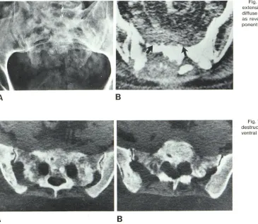
ponent of these lesions. The Ewing sarcoma revealed a discrete abnormality of the bony matrix as well as a soft-tissue component of the lesion that was not appreciated on tomography (fig. 3D). The lymphosarcoma showed bony
destruction of the lumbosacral as well as iliac regions bilat -erally. CT also demonstrated a large ventral soft-tissue
component (fig. 7). By demonstrating both bony and
soft-tissue components. CT was the best means of delineating
the true extent of these neoplasms.
Discussion
It is difficult to evaluate the sacrum using conventional
radiography because of its dorsal curvature and normal overlying bowel gas and fecal material. To compensate for this dorsal curvature. it has been recommended that patients
with suspected sacral problems have an anteroposterior (AP) sacral view with the x-ray tube angled 15°-20°
ceph-alad. This view will avoid foreshortening of the sacrum (fig. 4). To overcome the problem of gas and feces. especially in children. an AP tomogram is necessary when the abnor-mality cannot be definitely established by the plain film (fig.
3). To further complicate the plain film evaluation of the
sacrum. a variant on the lateral projection has been de-scribed that appears as a sharply demarcated bony defect
overlying the first or second sacral vertebra [1]. This variant
represents the fossa cribrosa. the site of penetration of blood vessels into the bone. and can be confused with
Fig. 5.- Chordoma. A. Plain film demonstrates extensive destruction of sacrum. B. CT confirms diffuse destructive pattern of bony sacrum as well
as revealing ventral and dorsal soft-tissue co m-ponent (arrows).
Fig. 7 .- Lymphosarcoma. CT of sacrum shows destruction of sacrum and iliac bone (A) with large
ventral soft-tissue components bilaterally (B).
neoplasms or intrasacral meningoceles. Lateral tomograms
should be helpful in this instance [1].
Myelography and bone scanning may further delineate a
problematic area in the sacrum. although not all sacral
lesions will cause a myelographic defect or pick up radio-nuclide agents. The advent of high-resolution CT. however. has resolved these problems by providing a noninvasive means of accurately evaluating the sacrum [2-5]. Therefore. once a sacral lesion has been established or strongly sug-gested by either clinical. plain film. or tomographic criteria. a high-resolution CT scan is the next procedure of choice.
Sacral lesions include congenital. neoplastic. metabolic. inflammatory. and traumatic processes. In evaluating the CT
scan of the sacrum. it can be divided into four areas. sacral canal. sacral foramina. bony matrix. and perisacrai soft
tissues. Analysis of the alteration of these various features
enabled us to separate the congenital from neoplastic
le-sions in our series.
The congenital lesions showed varying degrees of spinal dysraphism with primary widening of the midline sacral
canal. Determination of the CT numbers allowed distinction
of fat. calcium. and cerebrospinal fluid densities. In deline-ating the position of the conus. however. CT alone was not sufficient. Here. myelography with either metrizamide or
Pantopaque was necessary. In the congenital lesions. me
[image:4.612.60.425.83.401.2]nec-AJNR:3, September/October 1982 CT OF SACRAL PATHOLOGY 559
essary. Although complications have been reported with
metrizamide, the more serious complications, namely,
tran-sient visual disturbance and paresis, are unusual. Further
-more, children and young adults seem less affected by metrizamide [6-8].
It must be mentioned that a study has been reported in the literature [9] stating that water-soluble contrast agents
such as metrizamide decrease the ability to recognize
var-ious intracanalicular tissues such as fat, fibrocartilage, and
bone, and therefore, air myelography should be coupled with CT in evaluating lumbosacral problems. Using the fourth-generation Pfizer 0450 scanner, it has not been difficult to distinguish fat or bone from metrizamide. Fur-thermore, it is often difficult to manipulate subarachnoid air into the exact area of interest.
With regard to neoplastic lesions of the sacrum, the
efficacy of CT has previously been reported [10-1 2]. De
-pending on the results of the sacral scan, a decision as to the necessity of myelography can be made. Certain CT
characteristics were believed to add specificity to the
diag-nosis. Both neurofibromas demonstrated primary expansion of the sacral foramina sparing the sacral canal. Since the
sacral nerve roots exit through the foramina, this CT finding
was considered diagnostic of a neoplastic nerve lesion such as neurofibroma, ganglioneuroma, or schwannoma (figs. 4 and 5). Both chordomas revealed diffuse bone destruction with obliteration of the sacral canal and foramina (fig. 6). In
addition, both revealed large soft-tissue components both
ventrally and dorsally on CT. The CT scan helped in the
surgical approach as both cases were operated on in two
stages, one using a ventral and one a dorsal approach, thus
allowing maximal removal of the tumor. In the child with
Ewing sarcoma, CT showed a bony abnormality in the superior part of the right sacrum with a small ventral soft-tissue component, as well as an intracanalicular extension
at L5-S1 on the right (fig. 3). If there remains a question as
to the extent of tumor involvement in the lower lumbar region, myelography can be performed. All the neoplasms in this series demonstrated extradural defects in the lower lumbar region while one patient with neurofibromatosis showed on myelography small neurofibromas in the lumbar and cervical region (fig. 50).
After the extent of the neoplasm has been documented
with CT and myelography, follow-up CT provided a nonin
-vasive method of evaluating residual tumor after surgery or the response of malignant lesions to radiation and/or chemotherapy.
From this experience we believe high-resolution tom
og-raphy is not only the most accurate evaluation of the sacrum,
but analysis of the alteration of the various sacral comp
o-nents, canal, foramina, bony matrix, and soft tissues adds
specificity to the diagnosis. All congenital lesions expand
the central sacral canal, while neoplasms involving an
indi-vidual nerve root will expand the sacral foramina. Malignant
neoplasms such as chordoma and sarcoma alter the bony
matrix to varying degrees and frequently demonstrate
as-sociated soft-tissue masses.
Plain films of the sacrum, even with tomography, can be difficult to evaluate and may not fully clarify the etiology of
the clinical problem. We believe that high-resolution CT
should be performed in all patients with suspected sacral
lesions where more conventional plain film examinations do not provide the anatomic information necessary for definitive patient management.
REFERENCES
1. Kreyenbuhl W, Hessler C. A variation of the sacrum on the
lateral view. Radiology 1973;109:49-52
2. Baleriaux-Waha, Osteux M, Terwinghu G, deMeeus A,
Jean-nart L. The management of anterior sacral meningocele with
computed tomography. Neuroradiology 1977; 14: 45-46
3. Crellin RQ, Jones ER. Sacral extradural cysts-a rare cause
of low backache and sciatica. J Bone Joint Surg [Br]
1973;55: 20-31
4. Gilula LA, Murphy WA, Tailor CC, Patel RB. Computed t
omog-raphy of the osseous pelvis. Radiology 1979; 132: 1 07 -114
5. Janecki CJ, Nelson CL, Dohn OF. Intrasacral cyst-report of
a case and review of the literature. J Bone Joint Surg [Am] 1972;54: 423-428
6. Barry JF, Harwood-Nash DC, Fitz CR, Byrd SE, Boldt OW.
Metrizamide in pediatric myelography. Radiology 1977;
124:409-418
7. Drayer BP, Rosenbaum AE, Reigel DB, Bank WO, Deeb ZL.
Metrizamide computed tomography cisternography: pediatric
application. Radiology 1977; 124: 349-357
8. Hugosson C, Hindmarsh T, Bergstrom K. Myelography with
metrizamide in infants and children. Acta Radiol [Suppf]
(Stock h) 1977;355: 1 93-1 99
9. Scatliff JH, Bidgood WD, Killebrew K, Staab EV. Computed
tomography and spinal dysraphism: clinical and phantom
stud-ies. Neuroradiology 1979;17: 71-75
10. Berger PE, Kuhn JP. Computed tomography of tumors of the
musculoskeletal system in children. Radiology 1978; 127:
171-175
11. deSantos LA, Bernardino ME, Murray JA. Computed tom
og-raphy in the evaluation of osteosarcoma. Experience with 25
cases. AJR 1979; 132: 535-540