Donald L. Kreipke 1 Jack J. Moss1 James M. Franco1 Michael D. Maves2
David J. Smith3
This article appears in the March/April 1984 issue of AJNR and the May 1984 issue of AJR.
Received June 3, 1983; accepted after revision September 26, 1983.
'Department of Radiology, Indiana University Medical Center, Indianapolis, IN 46202. Address reprint requests to D. L. Kreipke, Department of Radiology, Wishard Memorial Hospital, 1001 W. 10th St., Indianapolis, IN 46202.
2Department of Otolaryngology, Indiana Univer-sity Medical Center, Indianapolis, IN 46223.
3Department of Plastic Surgery, Indiana Univer-sity Medical Center, Indianapolis, IN 46223.
AJNR 5:185-189, March/April 1984 0195-6108/84:0502-0185 $00.00 © American Roentgen Ray Society
185
Computed Tomography and
Thin-Section Tomography in
Facial Trauma
The efficacy of radiographic methods in detecting and classifying facial fractures was assessed. Thirty-one patients with maxillofacial trauma were studied with plain radiog-raphy, coronal and lateral pluridirectional tomography (PT), and axial and direct coronal computed tomography (CT). PT and CT were compared to assess how many fractures each method could demonstrate. In addition, plain films were used in combination with each special study to see how efficacious each combination was at classifying fractures into types, such as blow-out, tripod, etc. To reflect the fact that it is sometimes impossible to obtain lateral PT or direct coronal CT scans at this institution, the same analysis was done using just coronal PT and axial CT. With two projections, CT was better than PT at demonstrating fractured surfaces (168 vs. 156) and in classifying fractures in combina-tion with plain films (48 vs. 43). However, when only one projeccombina-tion from each special study was used, PT surpassed CT in showing fractures (137 vs. 124) and in classifying fractures (42 vs. 40). Failures with each method occurred when the plane of section was parallel or oblique to the plane of the structure being examined, that is, axial CT failed to show the floor of the orbit well and coronal PT failed to show the anterior maxillary sinus wall well. Imaging in two planes, including the coronal plane, is desirable for greatest accuracy in fracture detection, whether by CT, PT, or both. CT is generally better for the display of soft-tissue abnormalities.
The initial radiographic evaluation of facial trauma is made with plain films. Although it is possible to assess some facial fractures with physical examination and plain films alone, suspect findings or complex injuries warrant further investi-gation [1]. This may be with either computed tomography (CT) or pluridirectional tomography (PT). Of these two, PT offers better spatial resolution, while CT has superior contrast resolution. Also, the two modalities differ in that PT is performed with coronal and sagittal sections, while CT is done with coronal and axial sections. Thus, these methods differ in both resolution and projections available. Several recent communications have indicated a preference for CT over PT [1-3]. We prospectively compared PT and CT studies in a series of facial trauma patients to investigate their relative efficacy as they are applied in our institution.
Subjects and Methods
Thirty-one patients with facial trauma were studied with plain films, PT, and CT. All patients had blunt trauma, usually acquired in motor vehicle accidents or personal altercations. No missile injuries were included. CT and PT were performed to look for fractures suspected but not definitely seen on plain film or to investigate further complex fractures identified on plain films.
PT studies were performed on a Philips Poly tome employing hypocycloidal motion with a 48° arc. Exposure time was 6 sec. Coronal cuts at 3 mm intervals were obtained in all patients. After review of the coronal cuts, lateral sections were obtained as needed at 3 or 5 mm intervals. Twenty-two patients received lateral views.
186 KREIPKE ET AL. AJNR:5, Mar/Apr 1984
TABLE 1: Number of Fractures Identified by Axial/Coronal CT
and Coronal/Lateral Pluridirectional Tomography
No. of Fractures Identified
Fractured Surface Totals Exclusively
CT PT CT PT
Anterior maxillary wall . .. ... . 21 14 8 1
Lateral maxillary wall 21 21 1 1
Medial maxillary wall . 9 9 0 0
Orbital floor . . . . ... 11 19 1 9
Inferior orbital rim .. 12 16 2 6
Frontozygomatic suture 11 14 1 4
Lateral orbital wall . . . . .
.
17 11 7 1pterygoid plate . . . 6 8 0 2
Nasion . . . . . . . 11 11 0 0 Nasal septum . . . . . . .
.
. . . 4 4 0 0Zygomatic arch . . . . . . . .
.
17 7 10 0Anterior frontal wall 8 8 1 1
Posterior frontal wall 1 1 0 0
Medial orbital wall . . . . .... 7 2 5 0
Orbital roof ... . 3 2 1 0
Cribriform plate 1 1 0 0
Sphenoid bone ... 2 2 0 0
Superior orbital rim. 3 3 0 0
Zygoma . . . . . . . . .
.
1 1 0 0Medial orbital rim 2 2 0 0
Totals 168 156 37 25
Note.- PT ~ pluridirectional tomography.
sections were then obtained if the patient could tolerate the
hyper-extended position. Two patients had supine coronals, and 25 had
prone coronals. Slice thickness was 6 mm, and table index was 5
mm.
Plain films and CT and PT images from each patient were reviewed,
and a single collective impression was made by the reviewers. The. studies were evaluated in two ways: First, the CT or PT image was
interpreted by all reviewers together without the benefit of plain films,
and the number of definite fractures was totaled. Fractures were
called definite if both sides of the bony wall and a distinct interruption
of the cortex were seen. Single or multiple fractures through a bony
wall counted as one fractured surface. Next, CT and PT were
evaluated in conjunction with the plain films, and the types of fractures
were classified (e.g., Le Fort, blow-out).
Not all patients had lateral PT or coronal CT, usually because other
injuries or significant pain precluded their lying in any position other
than supine. Therefore, only axial CT or coronal PT was obtained in
these patients. We suspected that some data might be lost in doing
just one-projection CT or PT. Therefore, to examine this limitation,
the entire set of studies was reviewed in the same format using only
axial CT and coronal PT findings.
Results
CT identified a total of 168 fractured surfaces, and PT showed 156 (table 1). The difference in total fractures identi-fied is less meaningful than the differences for certain specific fractures. When the fractures were classified into types, CT again identified more types of facial injuries than PT: 48 vs. 43 (table 2).
Contrasting results were obtained when only axial CT and
TABLE 2: Types of Fractures Identified by Axial/Coronal CT or
Coronal/Lateral Pluridirectional Tomography When Used with Plain Films
No. of Fr
ac-Fracture Type tures Identified CT PT
Maxillary blow-out ... _ ... . 3 5
Ethmoid blow-out ... . 3 1
Tripod . . . . . . . . . . . . . ... . 9 6
Le Fort I 6 8
Le Fort II . 5 3
Le Fort III 4 2
Maxillary bone . . . . . . _ . . 7 7
Frontal sinus .... .... _ ... . 6 6
Orbital roof 2 2
Cribriform plate .... _ ... _ ... _ .. . 1 1
Sphenoid . . . ... . . . ... . 1 1
Orbital wall . . . . . . _ . . . .. 1 1
Totals 48 43
Note.-PT ~ pluridirectional tomography .
TABLE 3: Numbers of Fractures Identified by Axial CT and
Coronal Pluridirectional Tomography
Fractured Surface
Anterior maxillary .
Lateral maxillary ... ... .
Medial maxillary
Orbital floor . . . . . . . . . . . . . . . . Inferior orbital rim
Frontozygomatic suture
Lateral orbital wall .
Pterygoid plate ... _ .. .
Nasion . . . ... .
Nasal septum ... ... .
Zygomatic arch ... .
Anterior frontal wall ... _ . . . Posterior frontal wall
Medial orbital wall ... _ ..
Orbital roof . . . . . . . . . . _ .. Cribriform plate . . . . . . . . _ . . Sphenoid bone . . . . . . . . .
Superior orbital rim ... .
Zygoma bone
Medial orbital rim ... _ . _ .
Totals
Note.-PT ~ pluridirectional tomography.
No. of Fractures Identified
Totals
CT PT
21 3
21 21
6 9
o
198 16
1 13
17 12
4 7
3 11
2 4
17 3
8 5
1 0
6 3
3 2
1 1
2 2
3 3
o
1o
2124 137
Exclusively
CT 19 1
o
o
1o
5o
o
o
14 3 1 3 1o
o
o
o
o
48 PT 1 1 3 19 9 12o
3 8 2o
o
o
o
o
o
o
o
1 2 61coronal PT were used. PT found 137 fractured surfaces, while CT showed 124 (table 3). The differences for individual frac-tures are even more striking, for example, anterior maxillary and floor of orbit. In correlation with plain films, PT was able to classify 42 fractures, while CT classified 40 (table 4).
[image:2.612.54.296.111.352.2] [image:2.612.314.556.347.593.2]AJNR:5, Mar/Apr 1984 FACIAL TRAUMA 187
TABLE 4: Types of Fractures Identified by Axial CT or Coronal Pluridirectional Tomography When Used with Plain Films
Fracture Type
Maxillary blow-out ... . Ethmoid blow-out.
Tripod . . ... . Le Fort I ... .
Le Fort II . Le Fort III Maxillary bone Frontal Orbital roof Cribriform plate Sphenoid .. Orbital wall
Totals
Note.-PT = pluridirectional tomography.
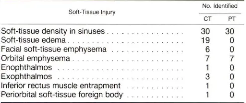
TABLE 5: Types of Soft-Tissue Injuries
Soft· Tissue Injury
Soft-tissue density in sinuses ... . Soft-tissue edema.
Facial soft-tissue emphysema ... . Orbital emphysema.
Enophthalmos
Exophthalmos . . . . Inferior rectus muscle entrapment
Periorbital soft-tissue foreign body .
Note. -PT = pluridirectional tomography.
Discussion
No. Identified
CT PT
0 3
2 1
7 6
5 8
5 3
3 2
5 7
6 5
2 2
1 1
1 1
3 3
40 42
No. Identified
CT PT
30 30
19 0
6 0
7 7
1 0
3 0
1 0
1 0
Tabulation of fractured surfaces without assistance of plain films provides some measure of differential sensitivity be-tween the two methods. CT identified 12 more fractures than did PT; however, CT and PT did not reveal the same 156 fractures. Each method demonstrated several fractures not seen by the other: 37 fractures were seen by CT but not PT, while PT exclusively identified 25 fractures not seen by CT. These exclusively identified fractures and their locations are shown in table 1 .
PT was better than CT at defining inferior orbital rim and orbital floor fractures. This disagreement occurred in two types of patients: (1) those in whom only axial CT cuts were obtained and (2) those in whom small floor and rim fractures showed very little displacement (figs. 1 and 2). In the former instance, the plane of the fractured surface is parallel to the CT cut. In the latter example, the small fracture presumably could not be well seen because of the partial volume of normal bone, making it difficult to see a definite interruption of the surface. The superior spatial resolution of PT allowed these fractures to be seen more easily.
CT exclusively defined lateral and medial orbital wall
frac-tures many more times than did PT. The lateral wall of the orbit is oriented obliquely to the beam in both coronal and lateral projections in PT. This spatial arrangement has been shown by Littleton [4] to produce a poor image on PT. CT has the advantages of having a different beam/target orien-tation from PT and of showing the entire length of the lateral orbital wall on the axial cuts (figs. 3 and 4).
The medial orbital wall fractures were nearly always accom-panied by hemorrhage or soft-tissue swelling in the ethmoid sinuses. The improved contrast resolution of CT allows for good differentiation between ethmoid wall and blood. With much soft-tissue density in the ethmoid sinuses, the thin lamina papyracea often was nearly invisible on PT (fig. 5).
Maxillary sinus anterior wall fractures were demonstrated exclusively by CT eight times. In seven of these cases, the corresponding PT study had coronal projections only. Here, the anterior sinus wall is oriented parallel to the PT cuts, thus making the anterior wall fracture hard to demonstrate. In two cases where both axial/coronal CT and coronal/lateral PT were performed, one had an anterior maxillary fracture exclu-sively identified by CT, while the other had exclusive demon-stration by PT. Thus, when CT and PT have two projections each, their abilities to show maxillary sinus anterior wall fractures are equal.
Zygomatic arch fracture identification was very discrepant: 10 were seen exclusively by CT and none by PT. In eight of these instances, the zygomatic arches were not included on the lateral PT cuts (usually, these were of patients in whom arch fractures were already demonstrated on the plain films, and it was thought to be redundant to visualize these fractures by taking lateral PT cuts out to the arches). By subtracting these eight cases, the CT score becomes two arch fractures exclusively demonstrated, nearly equal to PT.
We also analyzed the data by using CT and PT in conjunc-tion with plain films and then classifying the fractures. Table
2 shows types and numbers of fractures demonstrated when
plain films are added; CT also surpassed PT in this compari-son. Increased CT ability to identify Le Fort and tripod frac-tures reflects CT's better ability to demonstrate fractures through the medial and lateral orbital walls. PT was more efficacious than CT at demonstrating maxillary blow-out frac-tures, a reflection of its greater ability to show small fractures in the orbital floor.
When the same analyses were undertaken using one pro-jection for each method, PT surpassed CT in total number of fractures identified (table 3). Actually, the total number of fractures demonstrated decreased for both CT and PT, but the greatest drop was caused by axial CT's inability to dem-onstrate orbital floor and inferior rim fractures. Of note, how-ever, is that coronal PT missed a posterior wall frontal sinus fracture that was seen on axial CT. In classifying fractures with the additional assistance of plain films, PT again sur-passed CT (table 4). These results imply that the coronal plane is important in visualizing maxillofacial fractures.
[image:3.612.54.299.108.262.2] [image:3.612.54.299.328.431.2]188 KREIPKE ET AL. AJNR:5, Mar/Apr 1984
A
B
c
Fig. 1.-Right inferior orbital rim fracture. A, PT image shows minimally displaced fracture (arrow). B, Corresponding CT image does not demonstrate rim
fracture (arrow marks fracture site). C, Axial CT image fails to show right rim fracture (straight arrow indicates fracture site). Left inferior orbital rim and arch
fractures are visible on this cut (curved arrows).
A
B
B
Fig. 2.-Left orbital floor fracture. A, Coronal PT
shows left maxillary blow-out fracture (arrow). B, Coronal CT does not show fracture (arrow denotes fracture site).
Fig. 3.- Left lateral orbital wall fracture. A, Axial
AJNR:5, Mar/Apr 1984 FACIAL TRAUMA 189
Fig. 4.- Left lateral orbital wall fracture. A, Co-ronal CT shows fracture in inferior part of lateral orbital wall (arrow). e, Same fracture is not dem-onstrated definitively on corresponding coronal PT
(arrow).
Fig. 5.-Right medial orbital wall fracture. A,
Coronal CT shows definite interruption of medial wall (arrow). e, Coronal PT shows little contrast between ethmoidal blood and medial orbital wall. Fracture is difficult to visualize (arrow denotes frac-ture area).
A
A
mm were used. In our study, the 6 mm sections that over-lapped by 1 mm gave coarse reformatted images that were difficult to interpret. Therefore, reformatted coronal and sag-ittal images were not incorporated into our survey.
An additional advantage of CT is its ability to demonstrate soft-tissue injury. In two studies, about one-third of the pa-tients had intracranial injury demonstrated by CT ("I, 2). No cases in our series had intracranial abnormalities on CT. Of the soft-tissue findings in our series (table 5), PT matched CT only in demonstrating soft-tissue density in the sinuses (such as blood) and orbital emphysema. PT was unable to show the more clinically significant information on the status of the globe and intraocular muscles. In addition, PT was unable to demonstrate a periorbital foreign body.
On the basis of this study we make the following recom-mendations:
1. To identify the maximum number of fractures, two pro-jections (axial/coronal for CT or coronal/lateral for PT) should be obtained.
2. If two-projection CT or PT can be obtained, then CT is the better procedure, except in injuries to the inferior obital rim or orbital floor.
3. In investigating bony injury to the orbital inferior rim or
B
B
floor, PT is the better procedure. However, the ability of CT to demonstrate intraorbital contents would make it a better
choice if specific information about these structures is desired. 4. Coronal imaging is important in enumerating fractures. If only axial CT can be done, and reformatting is not helpful,
then coronal PT should be obtained as well.
REFERENCES
1. Noyek AM, Kassel EE, Gruss JS, Wortzman G, Holgate RC, Cooper PW. Sophisticated CT in complex maxillofacial trauma.
Laryngoscope 1982;92:[Suppl 27)1-17
2. Rowe LD, Miller E, Brant-Zawadzki M. Computed tomography in maxillofacial trauma. Laryngoscope 1981;91 :745-757 3. Zilkha A. Computed tomography in facial trauma. Radiology
1982; 144: 545-548
4. Littleton JT. Physical principles. In: Littleton JT, ed. Tomography:
physical principles and clinical applications. Baltimore: Williams & Wilkins, 1976:33-94
5. Littleton JT, Shaffer KA, Callahan WP, Durizch ML. Temporal bone: comparison of pluridirectional tomography and high reso -lution computed tomography. AJR 1981;137:835-845
6. Brant-Zawadzki MN, Minagi H, Federle MP, Rowe LD. High resolution CT with image reformation in maxillofacial pathology.
[image:5.612.112.555.80.462.2]