Scanning Electron Microscopy, and Phase-Contrast Microscopy
Kiyo Nakabayashi, Makoto Negoro, Takashi Handa, Hiroomi Keino, Masaya Takahashi, and Kenichiro Sugita
PURPOSE: To analyze the properties and embolic effect of microfibrillar collagen (MFC), Gelfoam powder, and polyvinyl alcohol (PVA) materials that are used in embolization procedures in the head and neck. METHODS: The shape and surface of these embolic agents were examined with scanning electron microscopy and phase-contrast microscopy. The mean number of areas of T2-weighted high signal intensity was measured on MR images in a rat embolization model to estimate the embolic effect. RESULTS: By scanning electron microscopy and phase-contrast microscopy, MFC appears fibriform and has various sizes and an irregular surface. Gelfoam is of uniform size and has a smooth surface. PVA materials are granulated and have a rough surface. MFC is somewhat suspendable and its shape changes moderately after suspension. Gelfoam is very suspendable and its shape changes rapidly. PVA showed only mild swelling. The embolic effect of MFC was the lowest of the materials examined. Large PVA particles (250 to 500mm) showed a lesser embolic effect than Gelfoam or small PVA particles (50 to 150mm) or medium-sized PVA particles (150 to 250mm). No significant differences were observed among the embolic effects of Gelfoam, small PVA particles (50 to 150mm), and medium PVA particles (150 to 250 mm). CONCLUSIONS: MFC and large PVA particles (250 to 500mm) should be used for embo-lization of vascular anatomy involving potentially dangerous anastomoses. Gelfoam, PVA particles of 150- to 250-mm diameter, and PVA particles of 50- to 150-mm diameter are adequate for embolization involving homogeneous and peripheral anatomy.
Index terms: Interventional materials, embolic agents; Interventional materials, polyvinyl alcohol foam
AJNR Am J Neuroradiol18:485–491, March 1997
Many embolic particulate agents have been used in head and neck embolization. The use of microfibrillar collagen (MFC), Gelfoam powder, and polyvinyl alcohol (PVA) is well docu-mented.
MFC (Avitene, Avicon, Fort Worth, Tex) is a topical hemostatic agent prepared from purified bovine collagen that has been used to control capillary bleeding in various surgical applica-tions (1). MFC was described as an embolic
agent in 1978 (2); however, recanalization oc-curs, and it is primarily used for preoperative embolization (3). Because of the presence of bovine collagen, MFC is potentially antigenic; however, animal and human studies have not demonstrated any significant antigenic effects (4).
Gelfoam (Upjohn; Kalamazoo, Mich) was used to control hemorrhage during surgical pro-cedures in 1945 (5). Speakman (6) reported the intravascular use of Gelfoam in 1964 (6). It is available as a powder (with particles ranging from 40 to 60mm in size) that can be placed in contrast material and injected through small-lumen catheters. In general, caution is advised when using Gelfoam in the external carotid ar-tery because of its small size and the multiple anastomoses between the external carotid ar-tery and the intracerebral circulation (7). It can deeply penetrate the tumor bed, causing tu-moral hemorrhage (8).
Received May 17, 1995; accepted after revision September 12, 1996. From the Department of Neurosurgery, Nagoya (Japan) University School of Medicine (K.N., M.N., T.H., K.S.); the Department of Perinatol-ogy, Institute of Developmental Research, Aichi, Japan (H.K.); and the Department of Research, Nihon, Schering KK, Osaka, Japan (M.T.).
Address reprint requests to Kiyo Nakabayashi, MD, Department of Neurosurgery, Yokkaichi City Hospital, 2-2-37 Shibata, Yokkaichi City, Mie 510, Japan.
AJNR 18:485–491, Mar 1997 0195-6108/97/1803–0485
©American Society of Neuroradiology
PVA is converted into a spongelike material by foaming agents and is hardened with form-aldehyde (9). PVA particles have proved to be a useful embolic material, particularly when long-term occlusions are desirable (10). To obtain a more uniform size, we divided blended PVA par-ticles into three size categories: 50 to 150 mm, 150 to 250mm, and 250 to 500mm. PVA has a high coefficient of friction that sometimes makes smooth injection difficult. Various man-uevers have been suggested to overcome this problem (11–13). The clinical efficiency of these embolic materials in therapeutic neurora-diology has been well described; however, the objective estimations of the embolic effect have been limited. We examined the shape and sur-face of the embolic materials by scanning elec-tron microscopy and used phase-contrast mi-croscopy to study the changes in the shape of these embolic agents in suspension. We also examined their embolic effect in rats by using magnetic resonance (MR) imaging.
Materials and Methods
The examined materials were MFC (,1000mm), Gel-foam powder (40 to 60mm) and PVA. A blended mixture of PVA was divided into three size fractions: small (50 to 150mm), medium (150 to 250mm), and large (250 to 500 mm). Each particulate fraction was purified by using an exact sieve, and the size range of each fraction was con-firmed with scanning electron microscopy.
Scanning Electron Microscopy and Phase-Contrast Microscopy
MFC, Gelfoam, and medium-sized PVA particles were examined. The dried specimens were coated with gold in a vacuum evaporator and subsequently examined with scanning electron microscopy (330,3300, and31500) (14).
Each material (10 mg) was suspended in 5 mL of contrast medium (Iopamiron-300, Nihon Schering KK; Osaka, Japan) and 5 mL of normal saline. The changes in each material that occurred during suspension were eval-uated by using phase-contrast microscopy (310 and 325).
Rat Embolization Model
Adult male Sprague-Dawley rats weighing 350 to 450 g were anesthetized with ketamine (40 mg/kg). The neck area was shaved and cleaned. Preoperatively, a suspen-sion of 10 mg of embolic material in 5 mL of contrast medium (Iopamiron 300) and 5 mL of normal saline was prepared.
Under a surgical microscope, we exposed the right common carotid artery, the internal carotid artery, and the external carotid artery via an anterior cervical incision. Each artery was clamped with microclips. A modified mi-crocatheter (distal internal diameter of 0.6 mm) was in-serted into the common carotid artery distal to the clamp. After the microclip of the internal carotid artery was re-moved, a 0.5-mL volume of the embolic suspension was injected slowly into the right common carotid artery of the experimental animals, and a 0.5-mL mixture of Iopamidol and normal saline was infused into the right common artery of five control rats. Any major defect that was present after the arteriotomy was closed with a 10-0 mi-crosuture. The proximal carotid clamp was released to achieve a first-pass effect exclusively into the internal ca-rotid artery (15). The clamp (microclip) on the external carotid artery was removed, and the skin was closed with 3-0 nonabsorbable sutures.
Embolization was performed with each of the following five materials: microfibrillar collagen (,1000mm) in five rats, Gelfoam powder (40 to 60mm) in six rats, small PVA particles (50 to 150mm) in nine rats, medium PVA parti-cles (150 to 250mm) in seven rats, and large PVA particles (250 to 500mm) in seven rats.
MR Examination
One day after injection, the rats were examined on a 4.7 T MR system. The animals, under pentobarbital anesthesia (40 mg/kg), were placed prone and T2-weighted (1500/50 [repetition time/echo time]) coronal images of the brain were obtained with a 2.5-mm section thickness at four anatomic levels using a 2563 128 matrix. The T2 high-signal-intensity area was measured to evaluate in-farction and ischemic brain edema in both hemispheres. The high-intensity area was measured by means of man-ual planimetry and was expressed as a percentage of the total brain area at that level. Data were expressed as the mean plus or minus standard deviation. Differences in the mean high-intensity area between the groups were determined by Scheff’s test after one-way analysis of variance. A P value of less than .05 was considered significant.
Histologic Examination
Results
Scanning Electron Microscopy and Phase-Contrast Microscopy (Fig 1)
By scanning electron microscopy, MFC ap-pears fibrinous and irregular in size and has a rough surface. Gelfoam powder is of intermedi-ate texture, and its surface is smooth. PVA is granulated and has a rough surface. By phase-contrast microscopy, MFC is suspendable and it enlarges moderately in suspension. Gelfoam powder is very suspendable, and the edges dis-solve in normal saline and Iopamidol. Extreme changes in its shape were noted. Suspensions of MFC and Gelfoam resemble a colloidal solution. PVA is less suspendable and expands mildly in suspension.
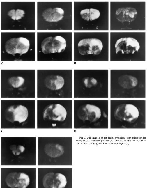
MR Imaging (Fig 2)
No significant differences were found among the groups of rats for any of the physiological variables measured, including arterial oxygen
pressure, partial pressure of carbon dioxide, hy-drogenion concentration, blood pressure, heart rate, temperature, and serum glucose. Table 1 shows the rate of occurrence of high-intensity areas in the rat brain after injections of various embolic agents. No area of high signal intensity was observed in the five control animals.
Areas of high signal intensity were observed in the ipsilateral cerebral cortex in all five rats in the MFC embolization group. In the Gelfoam embolization group, diffuse areas of high inten-sity were seen throughout the entire hemisphere on the side operated on in three of the six rats, and the remaining three rats showed high-inten-sity areas in the cerebral hemispheres bilater-ally. In the group in which embolization was done with small PVA particles, diffuse areas of high signal intensity were detected throughout the cerebral hemisphere on the embolized side in seven of the nine rats, and scattered areas of high intensity were seen in the cortex and/or basal ganglia on the embolized side in the re-maining two rats. Diffuse areas of high signal Fig 1.A–C, Scanning electron photomicrographs of microfibrillar collagen (A), Gelfoam powder (B), and PVA particles (150 to 250 mm) (C) (A,3300; B,31500; C,3300).
[image:3.587.48.549.84.377.2]intensity were detected in the entire cerebral hemisphere on the embolized side in two of the seven rats in which large PVA particles were used, and scattered areas of high intensity were seen in the cortex and/or basal ganglia in the remaining five rats.
Histologic Examination
Histologic examination of the brain sections resected from the levels corresponding to MR images showed infarction and ischemic edema. Table 2 presents a summary of the vessels oc-cluded after injection of the various embolic agents. MFC and large PVA particles occluded only the internal carotid artery on the embolic side (Fig 3). Gelfoam emboli were observed in the leptomeningeal arteries in all six rats in this
embolization group. Leptomeningeal arterial occlusion was also observed in seven rats in which small PVA particles were used and in two rats in which medium PVA particles were used (Fig 4). There was good correlation between the histologic areas of infarction and areas of high signal intensity (Fig 5).
The embolic effect of each material was eval-uated as the percentage of high-signal areas seen on four sections of brain per rat (Fig 6). The mean percentage of areas of high signal intensity for each embolic agent was 4.72% 6
4.06%, 37.25% 6 14.67%, 41.54% 6 14.27%,
[image:5.587.49.546.98.191.2]27.7% 611.59%, and 19.4% 611.8% for MFC, Gelfoam, small PVA particles, medium PVA particles, and large PVA particles, respectively. The embolic effect of MFC was less than that of Fig 3. Photomicrograph shows PVA particles (250 to 500mm) in the internal carotid artery (hematoxylin-eosin, original magnification330).
Fig 4. Photomicrograph of small lep-tomeningeal vessel shows PVA particles (50 to 150mm) (hematoxylin-eosin, orig-inal magnification330).
TABLE 1: Rate of occurrence of T2-weighted high-signal-intensity areas in the rat brain after embolization
Embolic Material No. of Animals
No. of Animals with Scattered Areas of High Signal Intensity in the Cortex
and/or Basal Ganglia
No. of Animals with Diffuse Areas of High Signal Intensity in the Whole
Hemisphere (Bilateral)
Microfibrillar collagen 5 5 0
Gelfoam powder 6 0 6 (3)
PVA (50–150mm) 9 2 7
PVA (150–250mm) 7 5 2
PVA (250–500mm) 7 5 2
TABLE 2: Vessels occluded after injection of embolic materials
Embolic Material No. of Animals No. of Animals with Occluded Internal Carotid Artery
No. of Animals with Occluded Distal Middle Cerebral Artery and/or
Anterior Cerebral Artery
Microfibrillar collagen 5 5 0
Gelfoam powder 6 6 6
PVA (50–150mm) 9 9 7
PVA (150–250mm) 7 5 2
[image:5.587.53.449.217.503.2]Gelfoam and small PVA particles (P,.01), and the effect of large PVA particles was less than that of small PVA particles (P, .05).
Discussion
MFC, Gelfoam, and PVA are commonly used embolic agents in procedures involving the head and neck. The choice of embolic agents depends on the objectives of the procedure, the selectivity desired, and the vascular anatomy of the pathologic territory. Particulate emboliza-tionrefers to a mechanical blockage of vascular territory done with precut particles that may be
of uniform or variable size and shape. Their capacity to occlude is related to their size, shape, and coefficient of friction (7). We exam-ined the surfaces of three embolic materials by using scanning electron microscopy and ob-served the changes in their shape during sus-pension by using phase-contrast microscopy. We also determined the embolic effect of these embolic agents in animal models.
Our observations with scanning electron mi-croscopy suggest that MFC apparently acts as a large particle because of its rough surface and irregular size. Gelfoam is small and has a smooth surface, allowing it to penetrate deeply and occlude the capillary or precapillary ves-sels. Because PVA particles have a rough sur-face, the potential for penetration is decreased by their external irregularities. Furthermore, the results of phase-contrast microscopy indicate that MFC acts as a large particle, retaining its irregular shape after being soaked and becom-ing swollen. Gelfoam powder can penetrate deeply even in suspension, but PVA has little potential for penetration, retaining its rough sur-face even after soaking and swelling.
MFC has the least embolic effect cerebrally, followed by large particles of PVA. Previous re-ports indicate that MFC can penetrate arterioles as small as 100mm in diameter (3). However, in our experiment, the concentration of the solu-tion used was lower than that, and the injected volume was smaller. The suspension concen-tration may influence the embolic effect of each agent. Ischemic edema of the ipsilateral cortex developed in the rats in which MFC was used owing to the thrombotic effect or watershed type of occlusion from the proximally occluded internal carotid artery. The embolic effect of large particles of PVA seems to be due to the thrombotic effect from the proximally occluded vessels, because large PVA particles are too big to penetrate and occlude capillary or precapil-Fig 5. Coronal section of rat brain
[image:6.587.52.538.80.475.2]em-bolized with microfibrillar collagen (A) and Gelfoam powder (B) (hematoxylin-eosin original magnification32.5).
[image:6.587.49.291.100.471.2]lary vessels. Further experiments are needed to explain the difference in embolic effects be-tween MFC and large PVA particles.
The cerebral embolic effects of Gelfoam and small PVA particles were greater than those of MFC. In particular, the animals in whom Gel-foam was used showed diffuse leptomeningeal arterial occlusion, bilateral cerebral infarction, and ischemic edema. These findings indicate very deep penetration of Gelfoam. The embolic effect of Gelfoam is related to its small size, smooth surface, and good affinity for the sol-vent. The embolic effects of small and medium PVA particles were similar to that of Gelfoam despite their rough surface and swollen shape after solution. The potential of PVA particles to promote thrombosis may increase the area of infarction and ischemic edema (16). The em-bolic effect of PVA is strongly affected by its particulate size range. Our results suggest that for particles smaller than 250mm, size has more influence on embolic effect than does external texture of the particles; however, for particles larger than 250 mm, texture has the greater influence.
Our observations of the shape and surface of various particulate embolic agents with scan-ning electron microscopy and phase-contrast microscopy are effective in explaining the em-bolic effect as assessed with MR imaging in rat embolization models. MFC and large PVA par-ticles are suitable for use when there is a risk of distant embolus, dangerous anastomosis, and a large-vessel tumoral bed. Gelfoam and small and medium PVA particles are better for embo-lization involving homogeneous and peripheral anatomy.
Acknowledgements
We are grateful to Masafumi Ito, Ikuo Takahashi, Kazu-hiro Fukui, and Kouzi Tuzi for their help with this work.
References
1. Battista OA, Erdi NZ, Ferraro CF. Novel microcrystals of poly-mers.J Appl Polymer Sci1967;11:481– 498
2. Kaufman SL, Strandberg JD, Barth KH, White RI. Transcatheter embolization with microfibrillar collagen in swine.Invest Radiol
1978;13:200 –204
3. Kumar AJ, Kaufman SL, Patt J, Posey JB, Maxwell DD, White RI. Preoperative embolization of hypervascular head and neck neo-plasms using microfibrillar collagen. AJNR Am J Neuroradiol
1982;3:163–168
4. Lee DH, Wriedt CH, Kaufmann JC, Pelz DM, Fox AJ, Vinuela F. Evaluation of three embolic agents in pig rete.AJNR Am J Neu-roradiol1989;10:773–776
5. Light RV, Prentice HR. Surgical investigation of a new absorbable sponge derived from gelatin for use in hemostasis.J Neurosurg
1945;2:435– 455
6. Speakman TJ. Internal occlusion of carotid-cavernous fistula.
J Neurosurg1964;21:303–305
7. Lasjaunias P, Berenstein A.Surgical Neuroangiography 2: Endo-vascular Treatment of Craniofacial Lesions. Berlin, Germany: Springer-Verlag; 1987:19 –29
8. Motozaki T, Otuka S, Sato S, et al. Preoperative embolization with Gelfoam powder for intracranial meningioma caused an unusual peritumoral hemorrhage.Neurol Surg1987;15:95–101
9. Tadavarthy SM, Moller JH, Amplatz K. Polyvinyl alcohol (Ivalon): a new embolic material.AJR Am J Roentgenol1975;125:609 – 616
10. Castaneda-Zuniga WR, Sanchez R, Amplatz K. Experimental ob-servations on short- and long-term effects of arterial occlusion with Ivalon.Radiology1978;126:783–785
11. Kerber CW, Bank WO, Horton JA. Polyvinyl alcohol foam: pre-packing emboli for therapeutic embolization.AJR Am J Roentge-nol1978;130:1193–1194
12. Horton JA, Marano GD, Kerber CW, et al. Polyvinyl alcohol foam: Gelfoam for therapeutic embolization: a synergetic mixture.AJNR Am J Neuroradiol1983;4:143
13. Herrera M, Rysavy J, Kotula F. Ivalon shaving: technical consid-erations of a new embolic agent. Radiology 1982;144:638 – 640
14. Choen AL, Marlow DP, Garner GE. A rapid critical point method using fluorocarbons (freon) as intermediate and transitional fluids.
J Microsc1968;7:331–342
15. Razack N, Soloniuk DS, Perkins E, Chandrasoma PT. Cerebro-vascular histopathology after intracarotid infusion of Gelfoam in the rat.Neurosurgery1993;33:116 –119