Nervous System: Comparison with Histopathologic Features
Blake A. Johnson, Evan K. Fram, Peter C. Johnson, and Ronald Jacobowitz
PURPOSE: To describe the MR features of primary central nervous system (CNS) lymphoma and to determine whether there is a correlation with histopathologic findings. METHODS: The MR images, pathologic specimens, and clinical records of 23 patients with primary CNS lymphoma were reviewed. The imaging and pathologic characteristics were tabulated and compared by using the standard tests for association in a two-dimensional contingency table. RESULTS: A total of 61 lesions were present in 23 patients; 12 patients (52%) had multiple lesions. All lesions were isointense or hypointense on T1-weighted images, and 53% were isointense or hypointense on T2-weighted images. Twenty patients received intravenous contrast material, and 43 (91%) of 47 lesions enhanced. The three patients who had nonenhancing lesions received steroids before the initial MR studies. Enhancement patterns differed between the immunocompetent and the immu-nocompromised hosts, with the latter group harboring a higher percentage of rim-enhancing lesions. Twenty-seven (44%) of the lesions were centered in a cerebral hemisphere and 14 (23%) were centered in the central gray matter. There was a statistically significant correlation between a higher degree of necrosis histologically and hyperintensity on T2-weighted MR images. The degree of necrosis also showed a positive correlation with rim enhancement. CONCLUSIONS: Primary CNS lymphoma has a variable MR appearance that correlates with the severity of intratumoral necrosis. These imaging characteristics, as well as lesion location, mean lesion size, and proclivity to harbor necrosis, are altered in the immunocompromised host.
Index terms: Brain neoplasms, magnetic resonance; Lymphoma
AJNR Am J Neuroradiol18:563–572, March 1997
Primary central nervous system (CNS) lym-phoma is an aggressive neoplasm with an inde-terminate pathogenesis. It is increasing in fre-quency in both immunocompetent and
im-munocompromised patients, and currently rep-resents 0.2% to 2.0% of primary malignant le-sions of the CNS (1–9). Primary CNS lym-phoma is virtually always the non-Hodgkin type, and the vast majority are B-cell lympho-mas. Primary T-cell lymphomas in the CNS have been reported, but they are rare (11, 12). Primary CNS lymphoma is histologically iden-tical to systemic extranodal non-Hodgkin lym-phoma, and may thus be categorized according to the working formulation for the classification of non-Hodgkin lymphomas (13). Previous investigators have evaluated the computed tomographic (CT) characteristics of primary CNS lymphoma (6, 14 –16) and their correla-tion with the histopathologic features (4, 17). The magnetic resonance (MR) imaging features have also been reported (7, 8, 14, 18). The purpose of this article is to describe the MR features of primary CNS lymphoma and to de-Received August 22, 1995; accepted after revision October 2, 1996.
The views expressed in this material are those of the authors and do not reflect the official policy or position of the US Government, the Department of Defense, or the Department of the Air Force.
Presented at the annual meeting of the American Society of Neurora-diology, Vancouver, Canada, May 1993.
From the Department of Radiology, David Grant Medical Center, Travis AFB, Calif (B.A.J., E.K.F.); the Divisions of Neuroradiology (B.A.J.) and Neuropathology (P.C.J.), Barrow Neurological Institute, St Joseph’s Hos-pital and Medical Center, Phoenix, Ariz; and the Department of Mathemat-ics, Arizona State University, Tempe (R.J.).
Address reprint requests to Blake A. Johnson, MD, Neuroradiology Section/SGSX, David Grant Medical Center, 101 Bodin Circle, Travis AFB, CA 94535.
AJNR 18:563–572, Mar 1997 0195-6108/97/1803–0563 ©American Society of Neuroradiology
termine any correlation with the histopathologic findings.
Materials and Methods
The MR images, histologic specimens, and clinical records of 23 patients with pathologically proved primary CNS lymphoma were reviewed. All specimens were ob-tained via open or stereotactic needle biopsy. Spin-echo MR imaging was performed on a 1.5-T unit. T1-weighted spin-echo axial and sagittal, 600/20/1 (repetition time [TR]/echo time [TE]/excitations), and long-TR dual-echo axial sequences (2500/30,90/1) were obtained. Other pa-rameters included a 21-cm field of view, a 256 3 192 matrix, and 5-mm-thick sections. Contrast-enhanced T1-weighted axial images were obtained after intravenous administration of 0.1 mmol/kg gadopentetate dimeglu-mine.
The imaging studies were evaluated for the number, location, and size of the lesions by two neuroradiologists who were blinded to the patients’ clinical history and his-tologic findings. In addition, MR signal characteristics of each lesion were evaluated on T1- and T2-weighted im-ages and rated as hyperintense, isointense, or hypointense relative to gray matter. The pattern of enhancement was categorized as solid, rim(ring)-like, or irregular. The de-gree of mass effect and perifocal white matter signal changes (edema) were graded as low, moderate, or high. The contour of the lesion was also noted (smooth, lobular, or irregular). Finally, the location of each lesion was doc-umented and assessed for contiguity with an ependymal or meningeal surface.
The pathologic slides were reviewed by a neuropathol-ogist using light microscopy. The tumors were classified according to the working formulation of malignant lym-phomas (13). The specimens were also evaluated for the degree of tumor cellularity and necrosis. Some of the bi-opsy specimens included adjacent brain parenchyma. These were evaluated for the presence of perifocal edema and glial response. The relative degree or severity of each of these features was graded as low, moderate, or high. A comparison of all specimens in this trial and extensive experience with CNS neoplasms provided the basis for the semiquantitative analysis performed by the neuropatholo-gist. Because no universally accepted criteria for quanti-fying the degree of necrosis, edema, or cellularity are available, the relative severity of these parameters was used for analysis.
The imaging and pathologic characteristics were tabu-lated and compared by using the standard tests for asso-ciation in a two-dimensional contingency table. Each of the MR characteristics (signal intensity, enhancement pattern, white matter change, mass effect, and lesion con-tour) was compared with each of the pathologic features (histologic subtype, cellularity, necrosis, edema, and glial re-sponse).
Results
Patients
The patients consisted of 14 men and nine women, 20 to 80 years old (mean, 57 years). Five of the men were positive for the human immunodeficiency virus and had acquired im-munodeficiency syndrome (AIDS) (age range, 20 to 36 years; mean, 30 years). Nine men and nine women comprised the immunocompetent cohort; their ages ranged from 40 to 80 years (mean, 65 years).
Imaging
immunocompetent group was 2.13 cm (range, 0.9 to 5.0 cm). This was larger than that seen in the AIDS cohort (mean, 1.64 cm; range, 0.8 to 3.0 cm).
Pathologic Findings
The histologic subtypes encountered were, in decreasing order of frequency, diffuse large cell (n513), immunoblastic (n54), small cleaved cell (n 5 2), small lymphocytic (n 5 2), and diffuse mixed cell (n 5 2). The AIDS patients had only diffuse large cell or immunoblastic subtypes. Other histologic features are summa-rized in Table 2. The degree of perifocal edema and gliotic response could not be assessed in seven specimens because there was no brain parenchyma adjacent to the tumor as sampled. All patients with a moderate to high degree of necrosis histologically had intermediate-grade (diffuse large cell) (n 55) or diffuse mixed cell (n 51) or high-grade (immunoblastic) (n54) lymphoma.
Radiologic-Pathologic Correlation
Pearson’s x2test for association between the MR features and the histologic findings revealed a statistically significant correlation between a high degree of necrosis and hyperintensity on T2-weighted MR images (Table 3). Tumors with no or low necrosis were most commonly isoin-tense to hypoinisoin-tense on T2-weighted images (Fig 2), and those with moderate to severe ne-crosis were more often hyperintense (Fig 3). The degree of necrosis also correlated with the pattern of enhancement (Table 4). Tumors with no or mild necrosis on light microscopy showed a statistically significant propensity to enhance in a solid pattern (Fig 4), whereas those with moderate to severe necrosis generally showed an irregular or rim-enhancing pattern (Fig 5). All AIDS patients had lesions that contained either moderate or severe necrosis, and rim-enhancing lesions were much more common in this group (Table 5). We found no statistically significant correlation between the MR features and degree of cellularity, edema, or adjacent glial response on the corresponding histologic specimen.
Discussion
[image:3.587.48.381.82.293.2]The mean age of 57 years in our cohort is comparable to that of other reported series of patients with primary CNS lymphoma (4, 5, 7, 19). As expected, the immunocompromised patients had a significantly lower mean age (32 years) than that of the immunocompetent co-hort (65 years). The earlier age at presentation Fig 1. Case 13: 61-year-old immuno-competent woman with diffuse large cell lymphoma.
A, Contrast-enhanced axial T1-weighted image shows an enhancing le-sion adjacent to the posterior aspect of the left lateral ventricle. Motion artifact causes some image compromise.
B, Axial T2-weighted image shows T2 prolongation in the distribution of the en-hancing lesion. Additional hyperintense lesions are present in the contralateral hemisphere (arrows) spanning the genu of the corpus callosum.
TABLE 1: Lesion location
Location Immunocompetent Patients (%)
AIDS Patients
(%) Total (%)
Hemisphere 16 (35) 11 (69) 27 (44)
Central gray 11 (24) 3 (27) 14 (23)
Corpus callosum 3 (7) 0 3 (5)
Intraventricular 3 (7) 1 (6) 4 (7)
Cerebellum 8 (18) 0 8 (13)
[image:3.587.50.288.328.429.2]for immunocompromised patients harboring CNS lymphoma manifests in a lower mean age reported in other series of AIDS patients with CNS lymphoma (8, 17). A male predominance in cases of primary CNS lymphoma is generally recognized (1– 4, 7, 9, 19, 20). Our series also had a male preponderance; all the AIDS pa-tients were males. However, the male-to-female ratio was equal in the immunocompetent group. The CT features of primary CNS lymphoma are well documented (4 – 6, 16, 17). The MR features have also been described (7, 8, 14, 18). Early observations indicated that primary CNS lymphoma has signal characteristics sim-ilar to those of other intracranial neoplasms: T1 and T2 prolongation resulting in a hypointense appearance on T1-weighted images and a hy-perintense appearance on T2-weighted images. Others have described primary CNS lymphoma as intracranial masses that are isointense with gray matter on long-TR images (7, 8). A hy-pointense appearance (relative to gray matter)
has also been described (14). We also noted the absence of T2 prolongation in a significant number of lesions. In fact, a majority of the lesions (53%) were either isointense (33%) or hypointense (20%) relative to gray matter on T2-weighted images. This feature distinguishes primary CNS lymphoma from most brain tu-mors, which are generally hyperintense unless
hemorrhage, calcification, or melanin is
present.
Most lesions (91%) identified on long-TR im-ages enhanced after administration of contrast material. Because the patients with nonenhanc-ing lesions had received steroid therapy, a spu-rious reduction of the percentage of lesions that enhance is likely. A high prevalence of en-hancement is consistent with other series, which range from 92% to 100% (3, 5, 7).
[image:4.587.51.546.97.428.2]The pattern of enhancement seen in our se-ries is also comparable to reported data, with 74% of lesions in the immunocompetent cohort showing homogeneous enhancement (7, 17).
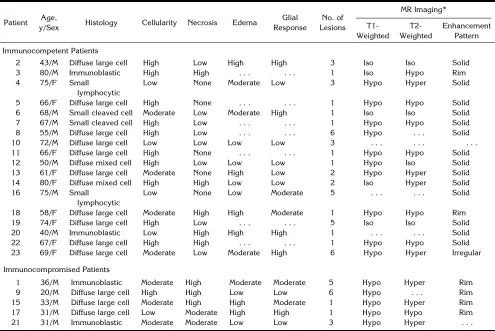
TABLE 2: Histologic and imaging findings
Patient Age,
y/Sex Histology Cellularity Necrosis Edema
Glial Response
No. of Lesions
MR Imaging*
T1-Weighted
T2-Weighted
Enhancement Pattern
Immunocompetent Patients
2 43/M Diffuse large cell High Low High High 3 Iso Iso Solid 3 80/M Immunoblastic High High . . . 1 Iso Hypo Rim 4 75/F Small
lymphocytic
Low None Moderate Low 3 Hypo Hyper Solid
5 66/F Diffuse large cell High None . . . 1 Hypo Hypo Solid 6 68/M Small cleaved cell Moderate Low Moderate High 1 Iso Iso Solid 7 67/M Small cleaved cell High Low . . . 1 Hypo Hypo Solid 8 55/M Diffuse large cell High Low . . . 6 Hypo . . . Solid 10 72/M Diffuse large cell Low Low Low Low 3 . . . . 11 66/F Diffuse large cell High None . . . 1 Hypo Hypo Solid 12 50/M Diffuse mixed cell High Low Low Low 1 Hypo Iso Solid 13 61/F Diffuse large cell Moderate None High Low 2 Hypo Hyper Solid 14 80/F Diffuse mixed cell High High Low Low 2 Iso Hyper Solid 16 75/M Small
lymphocytic
Low None Low Moderate 5 . . . Solid
18 58/F Diffuse large cell Moderate High High Moderate 1 Hypo Hypo Rim 19 74/F Diffuse large cell High Low . . . 5 Iso Iso Solid 20 40/M Immunoblastic Low High High High 1 . . . Solid 22 67/F Diffuse large cell High High . . . 1 Hypo Hypo Solid 23 69/F Diffuse large cell Moderate Low Moderate High 6 Hypo Hyper Irregular
Immunocompromised Patients
1 36/M Immunoblastic Moderate High Moderate Moderate 5 Hypo Hyper Rim 9 20/M Diffuse large cell High High Low Low 6 Hypo . . . Rim 15 33/M Diffuse large cell Moderate High High Moderate 1 Hypo Hyper Rim 17 31/M Diffuse large cell Low Moderate High High 1 Hypo Hypo Rim 21 31/M Immunoblastic Moderate Moderate Low Low 3 Hypo Hyper . . .
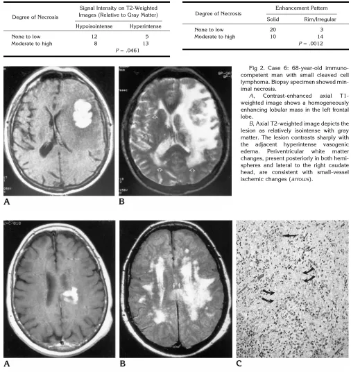
Fig 2. Case 6: 68-year-old immuno-competent man with small cleaved cell lymphoma. Biopsy specimen showed min-imal necrosis.
A, Contrast-enhanced axial T1-weighted image shows a homogeneously enhancing lobular mass in the left frontal lobe.
B, Axial T2-weighted image depicts the lesion as relatively isointense with gray matter. The lesion contrasts sharply with the adjacent hyperintense vasogenic edema. Periventricular white matter changes, present posteriorly in both hemi-spheres and lateral to the right caudate head, are consistent with small-vessel ischemic changes (arrows).
Fig 3. Case 14: 80-year-old immunocompetent woman with diffuse mixed cell lymphoma.
A, Contrast-enhanced axial T1-weighted image shows the irregular contour of the enhancing mass adjacent to the left lateral ventricle. The mass extends into the splenium of the corpus callosum.
B, Axial intermediate-weighted image depicts the lesion as hyperintense relative to gray matter (arrows). This patient also has significant small-vessel ischemic disease involving the periventricular white matter.
C, Pathologic specimen shows a heterogeneous cell population with variable cellular size and shape. The large nuclei have a vesicular chromatin pattern and contain marginated nucleoli (straight arrow). There are areas of extensive necrosis (curved arrows) and densely cellular components (open arrows) within the tumor. The adjacent brain parenchyma (not shown) displayed no significant edema or gliotic response.
TABLE 3: Signal intensity on T2-weighted images versus degree of histologic necrosis
Degree of Necrosis
Signal Intensity on T2-Weighted Images (Relative to Gray Matter)
Hypoisointense Hyperintense
None to low 12 5
Moderate to high 8 13
[image:5.587.316.547.108.177.2]P5.0461
TABLE 4: MR enhancement pattern versus degree of histologic necrosis
Degree of Necrosis
Enhancement Pattern
Solid Rim/Irregular
None to low 20 3
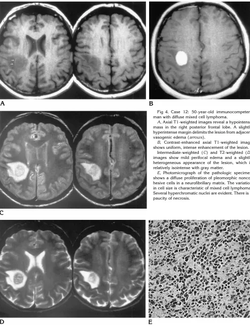
Fig 4. Case 12: 50-year-old immunocompetent man with diffuse mixed cell lymphoma.
A, Axial T1-weighted images reveal a hypointense mass in the right posterior frontal lobe. A slightly hyperintense margin delimits the lesion from adjacent vasogenic edema (arrows).
B, Contrast-enhanced axial T1-weighted image shows uniform, intense enhancement of the lesion.
Intermediate-weighted (C) and T2-weighted (D) images show mild perifocal edema and a slightly heterogeneous appearance of the lesion, which is relatively isointense with gray matter.
The frequency of rim enhancement in such pa-tients before treatment is much lower (17), and was seen in only 12% of lesions in our series. In immunocompromised patients, rim enhance-ment was much more common (Fig 6). This pattern of enhancement was seen in approxi-mately half the lesions in our immunocompro-mised patients, which is similar to the experi-ence of other investigators (6, 15–17). Although
characteristic, rim enhancement is not a distin-guishing feature for lesions in either population. The primary differential considerations based on enhancement pattern include other neo-plasms and infection.
In our series, lesion size (in largest diameter) tended to be smaller in AIDS patients (mean diameter, 1.6 cm; range, 0.8 to 3.0 cm) than in the immunocompetent group (mean diameter, 2.1 cm; range, 0.9 to 5.0 cm). Although some workers have noted this trend toward smaller lesions in immunocompromised patients (8), others have noted a tendency toward larger le-sions in AIDS patients with primary CNS lym-phoma (4, 18).
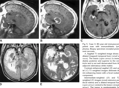
[image:7.587.50.542.85.451.2]Overall, 52% of patients in our series had multiple lesions. In the immunocompetent group, 50% had more than one lesion. This is at the high end of reported values for multiplicity, which range from 11% to 50% (2–5, 7, 9, 19, Fig 5. Case 3: 80-year-old immunocom-petent man with immunoblastic lym-phoma. Biopsy specimen revealed promi-nent necrosis.
A, Sagittal T1-weighted image shows a dilated (trapped) temporal horn (small ar-rows). The lesion (open arrow) is imme-diately posterior and superior to the ven-tricle and is not well demarcated from the adjacent edematous white matter.
Contrast-enhanced sagittal (B) and ax-ial (C) T1-weighted images delineate the rim-enhancing lesion with a focal nodular component.
Intermediate-weighted (D) and T2-weighted (E) images reveal extensive per-ifocal edema and a dilated temporal horn (open arrow) ventral to the lesion (straight arrow). The tumor is predominately hy-pointense, with a hyperintense focus anteriorly (curved arrow). Histologically, this lesion had areas of high cellularity and a high degree of necrosis (Table 1), which may explain the hypointense and hyperintense foci on MR images, respectively.
TABLE 5: Imaging findings: enhancement patterns
Pattern of Enhancement
Immunocompetent (%)
AIDS Patients
(%)
Total (%)
Solid 25 (74) 6 (46) 31 (66)
Rim 1 (3) 6 (46) 7 (15)
Irregular 4 (12) 1 (8) 5 (11)
None 4 (12) 0 4 (9)
[image:7.587.51.288.504.596.2]20). The higher rate of multiplicity displayed in the AIDS cohort (60%) is congruent with the results of other researchers, who reported mul-tiplicity rates ranging from 41% to 81% (14 –16). Almost half (45%) the lesions in this study were located peripherally within the hemi-spheres. Several authors report this as the most frequent location, with 46% to 50% of lesions so located (4, 5, 16, 19). The central gray matter is a characteristic location for primary CNS lym-phoma, albeit not the most frequent. Only 23% of lesions in our series were located in the deep gray matter structures. This is only slightly higher than the 18% prevalence reported by Jellinger et al (19) and lower than that reported by Jack et al in two series, in which there was a 30% (5) and a 33% (4) prevalence of lesions in the central gray matter. Intraventricular and posterior fossa lesions were less common.
Contact with an ependymal or meningeal sur-face is another putative characteristic feature of primary CNS lymphoma, reported in as many
as 75% of lesions (4). Roman-Goldstein et al (7) reported 58% abutting the ventricular system. This feature was less common in our series; only 28% of lesions were contiguous with an ependymal surface, and 8% with the meninges. Contiguity with an ependymal surface was more common in AIDS patients (38%) than in immu-nocompetent patients (24%) in our trial.
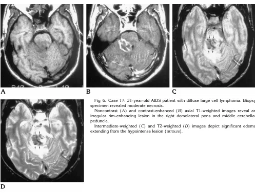
[image:8.587.48.545.77.458.2]We used the working formulation for lympho-mas to classify cases in our study and found low-, intermediate-, and high-grade subtypes. All of the AIDS patients with primary CNS lym-phoma reviewed by Cordoliani et al (18) were found to have intermediate- to high-grade sub-types. This was our experience as well. None of the AIDS patients in our series had low-grade lymphomas according to the working formula-tion, and only four (22%) of the 18 immuno-competent patients did. Diffuse large cell lym-phoma was the most frequent histologic subtype in our series (Table 2). Previous studies cite a similar experience (4, 7, 9, 17), while Fig 6. Case 17: 31-year-old AIDS patient with diffuse large cell lymphoma. Biopsy specimen revealed moderate necrosis.
Noncontrast (A) and contrast-enhanced (B) axial T1-weighted images reveal an irregular rim-enhancing lesion in the right dorsolateral pons and middle cerebellar peduncle.
others show immunoblastic lymphoma as the most common (3, 20) subtype. Immunoblastic lymphoma was the subtype in all the patients with primary CNS lymphoma reported by Loureiro et al (21). In our AIDS cohort, the immunoblastic subtype was present in 40% of the patients, while 60% were diffuse large cell lymphomas.
Lesions harbored by patients in the AIDS co-hort tended to show a higher degree of necrosis. The four immunocompetent patients in our study with lesions that showed a high degree of necrosis had immunoblastic (n 5 2), diffuse mixed cell (n 51), and large cell (n 51) sub-types. The other patients had lesions that showed no or mild necrosis. Conversely, all the AIDS patients had lesions that showed moder-ate or severe necrosis. This is similar to the experience of Lee and coworkers (17), who noted a paucity of necrosis in the lesions they examined, except for those in the AIDS patients. Similar to the experience of Jack et al (5), we found no imaging correlation with the patho-logic subtype of primary CNS lymphoma. Our analysis of the other histologic characteristics for correlation with imaging features revealed a significant association between the degree of necrosis and two of the imaging parameters: the T2 signal intensity and the pattern of enhance-ment on contrast-enhanced T1-weighted im-ages.
High signal intensity is expected with necro-sis. In the absence of a significant degree of necrosis, primary CNS lymphoma tends to be isointense to hypointense relative to brain on long-TR sequences. This may be due to the high nuclear-to-cytoplasmic ratio and dense cellu-larity evidenced by these tumors. The MR fea-tures produced by such a lesion are obviously altered when necrosis is present, which then dominates MR contrast.
Likewise, the more characteristic homoge-neous enhancement pattern would predictably be disrupted when necrotic debris replaces tu-mor centrally. In the setting of central necrosis, enhancement of the peripheral viable tumor surrounding central nonenhancing necrotic de-bris produces a ring-enhancing pattern.
Because this was a retrospective study, the biopsy method was not standardized. Some of the patients underwent open biopsy (n58), but a majority had needle biopsy (n515). In addi-tion, while all lesions were evaluated on the imaging studies, not all lesions were subjected
to biopsy. Although most lesions in a given patient displayed similar imaging characteris-tics, the biopsy specimen obtained may not fully reflect the histologic features either of that lesion or of all lesions in that patient. Because all the lesions identified on MR images were included in the statistical analysis, it is impor-tant to consider that not all were confirmed pathologically. In addition, the pathologic and radiologic features were graded in a relative fashion, as no universally accepted scale for quantification is available. This introduces sub-jectivity into the assessment of the various pa-rameters. However, our goal was not to provide quantification of the various features we evalu-ated but to look for correlations. A relative grad-ing of the severity of each feature was thus deemed acceptable.
Neuroimagers should be aware of the vari-able MR appearance and distribution of primary CNS lymphomas within the brain. These tumors may be hyperintense, isointense, or hypoin-tense relative to brain on T2-weighted images. The enhancement pattern is variable. Lesions are commonly multiple, hemispheric in loca-tion, and tend to enhance homogeneously in the immunocompetent host. In AIDS patients, pri-mary CNS lymphoma is even more likely to present with multiple lesions. Rim enhancement is much more prevalent in these patients, a feature that is most likely due to the high degree of necrosis. The severity of necrosis on the his-tologic specimen had a significant correlation with the MR signal characteristics on T2-weighted images and with the pattern of en-hancement. Familiarity with these features should provoke consideration of primary CNS lymphoma when such lesions are encountered. This may prompt early biopsy rather than con-servative management in the appropriate clini-cal setting.
References
1. Grant JW, Isaacson PG. Primary central nervous system lym-phoma.Brain Pathol1992;2:97–109
2. Grote TH, Grosh WW, List AF, Wiley R, Cousar JB, Johnson DH. Primary lymphoma of the central nervous system. Am J Clin Oncol1989;12:93–100
3. Hochberg FH, Miller DC. Primary central nervous system lym-phoma.J Neurosurg1988;6:835– 853
4. Jack CR, O’Neill BP, Banks PM, Reese DF. Central nervous sys-tem lymphoma: histologic types and CT appearance.Radiology 1988;167:211–215
cases of primary CNS lymphoma.AJR Am J Roentgenol1986; 146:271–276
6. Poon T, Matoso I, Tchertkoff V, Weitzner I, Gada M. CT features of primary cerebral lymphoma in AIDS and non-AIDS patients. J Comput Assist Tomogr1989;13:6 –9
7. Roman-Goldstein SM, Goldman DL, Howieson J, Blekin R, Neu-welt EA. MRI of primary CNS lymphoma in immunologically nor-mal patients.AJNR Am J Neuroradiol1992;13:1207–1213 8. Schwaighofer BW, Hesselink JR, Press GA, Wolf RL, Healy ME,
Berthoty DP. Primary intracranial CNS lymphoma: MR manifesta-tions.AJNR Am J Neuroradiol1989;10:725–729
9. Socie G, Piprot-Chauffat C, Schlienger M, et al. Primary lym-phoma of the central nervous system.Cancer1990;65:322–326 10. Eby NL, Grufferman S, Jlannelly CM, Schold SCJ, Vogel FS,
Bruger PC. Increasing incidence of primary brain lymphoma in the United States.Cancer1988;62:2461–2465
11. Bednar MM, Salerni A, Flanagan ME, Pendlebury WW. Primary central nervous system T-cell lymphoma.J Neurosurg1991;74: 668 – 672
12. McCue MP, Sandrock AW, Lee JM, Harris NL, Hedley-Whyte ET. Primary T-cell lymphoma of the brainstem.Neurology1993;43: 377–381
13. The Non-Hodgkin’s Lymphoma Pathologic Classification Project. National Cancer Institute sponsored study of classifications of
non-Hodgkin’s lymphomas: summary and description of a work-ing formulation for clinical usage.Cancer1982;49:2112–2135 14. Ciricillo SF, Rosenblum ML. Use of CT and MR imaging to
distin-guish intracranial lesions and to define the need for biopsy in AIDS patients.J Neurosurg1990;73:
15. Dina TS. Primary central nervous system lymphoma versus tox-oplasmosis in AIDS.Radiology1991;179:823– 828
16. Goldstein JD, Zeifer B, Chao C, et al. CT appearance of primary CNS lymphoma in patients with acquired immunodeficiency syn-drome.J Comput Assist Tomogr1991;15:
17. Lee Y-Y, Bruner JM, Tassel PV, Libshitz HI. Primary nervous system lymphoma: CT and pathologic correlation. AJR Am J Roentgenol1986;147:747–752
18. Cordoliani Y-S, Derosier C, Pharaboz C, Jeanbourquin D, Schill H, Cosnard G. Primary cerebral lymphoma in patients with AIDS: MR findings in 17 cases.AJR Am J Roentgenol1992;159:841– 847 19. Jellinger K, Radaskiewicz TH, Slowik F. Primary malignant
lym-phomas of the central nervous system in man.Acta Neuropathol (Berl)1975;Suppl VI:95–102
20. Helle TL, Britt RH, Colby TV. Primary lymphoma of the central nervous system.J Neurosurg1984;60:94 –103