With 8 text-figures Printed tn Great Britain
CURRENT-VOLTAGE RELATIONS
IN THE ISOLATED GIANT AXON OF THE COCKROACH
UNDER VOLTAGE-CLAMP CONDITIONS*
BY Y. PICHON AND J. BOISTEL
Laboratoire de Physiologie Animale, Faculte des Sciences, Rermes, France (Received 5 May 1967)
INTRODUCTION
Our understanding of the ionic exchanges taking place at the nerve membrane during electrical activity has been greatly increased by the application of the voltage-clamp technique. This experimental method consists of the application of a stepwise change in potential to a given area of membrane and the simultaneous measurement of the current flow produced by this given potential gradient. The voltage-clamp technique was first introduced by Cole (1949) and Marmont (1949) and was subse-quently developed by Hodgkin & Huxley (1952) and Hodgkin, Huxley & Katz (1952) using the squid giant axon. It has also been successfully applied to other excitable structures, including, for example, the isolated frog nerve fibre (Dodge & Franken-haeuser, 1958, 1959) and the isolated lobster giant axon (Julian, Moore & Goldman, 19626). There has, however, been no application of this technique to the relatively small insect axons, and previous work has been confined to the measurement of the changes in membrane potential induced by alteration of the ionic composition of the bathing medium. Thus, Boistel & Coraboeuf (1958) have shown that partial replace-ment of sodium chloride in the Ringer bathing the nerve cord of the cockroach, Periplaneta americana, by choline chloride results in a small increase in resting potential and an appreciable decrease in the height of the action potential. Similar experiments were also carried out by Yamasaki & Narahashi (1959a) who observed that a 8emilogarithmic plot of the overshoot of the action potential against sodium concentration resulted in a straight line, which only diverged slightly from the theo-retical relation predicted by the Nernst equation. Similarly, the resting potential was shown to be exponentially related to the external potassium concentration, for values higher than 20 mM/1. (Yamasaki & Narahashi, 1959a), although in this species, and also in Carausius morosus (Treherne & Maddrell, 1967), the line departed appreciably from the 58 mV. slope predicted by the Nernst equation. This departure was postu-lated to result from the involvement of other ions than potassium in determining the resting potential.
The above results were interpreted as strong evidence for the validity of the sodium theory in cockroach giant axons. More recently, this evidence has been strengthened by the observation of Narahashi (1965) that the puffer fish poison, tetrodotoxin, blocked the excitability of cockroach axons without significantly changing the resting
344 Y- PlCHON AND J. BOISTEL
potential, for this substance is known to inhibit the sodium activation mechanisms in frog muscle fibres (Narahashi, Deguchi, Urakawa & Ohkubo, i960) and in lobster axons (Narahashi, Moore & Scott, 1964) for example. Despite the above evidence, some recent findings have demonstrated the dependence of excitability of cockroach giant axons upon divalent cations (Narahashi & Yamasaki, i960; Narahashi, 1966). These findings raise the question of the contribution of calcium ions to the inward ionic current during the rising phase of the action potential.
Although the intracellular recording of action and resting potentials has yielded valuable insight into the ionic properties of the insect nerve membrane, this method cannot by itself give a detailed analysis of the different mechanisms which govern excitability. There is, for example, no direct method available to estimate sodium inactivation using only membrane potential measurements. The application of the voltage-damp technique to insect axons is obviously an ideal method of elucidating the mechanisms involved in membrane excitability. In particular, it would be desirable to apply the classical equations of Hodgkin & Huxley (1952) to insect axons.
The results presented in this paper represent the first attempt to apply the voltage-clamp technique to insect axons. A short report of this work has been given previously (Pichon, 1967).
METHODS
Extensive studies on insect axon have been carried out in recent decades using intra-cellular microelectrodes (Boistel & Coraboeuf, 1954, 1958; Yamasaki & Narahashi, 1957, 1959a, b; Boistel, i960; Narahashi & Yamasaki, i960; and others). This method has also been successfully employed with the voltage-clamp technique for a variety of nervous structures including the supramedullary nerve cell of the puffer (Hagiwara & Saito, 1959 a), the nerve cell membrane of Onchidium verruculatum (Hagiwara & Saito, 1959 J), the giant soma of Aplysia (Tauc & Frank, 1962) and the spinal motoneurone of the cat (Frank, Fuortes & Nelson, 1959; Nelson & Frank, 1964). Unfortunately, the application of the voltage-clamp technique to the insect axon is complicated by the difficulties encountered in controlling the membrane potential throughout the length of these structures. On the other hand, the small diameter of the fibres (40-50 ju. in Periplaneta) necessitates the use of very fine-tipped microelectrodes, the high resistance of which limits the intensity of the polarizing current to a rather low value.
eventually followed by a blockage (Narahashi, 1966). For these reasons, the 'sucrose-gap' method was discarded. Recently, Tomita & Wright (1965) have shown that replacement of sucrose, after a few minutes, by mineral oil maintained the repetitive responses of crustacean axons for many hours, whereas these axons became non-repetitive or even totally inexcitable after only 15-30 min. of immersion in isotonic sucrose. A similar technique has been used by Chapman (1966) for crab axons. An even more simple method would consist in using only oil to form the insulating sheath around the single fibre. This has been successfully done in recent experiments on the cockroach axon (Pichon & Boistel, 1966).
The method of Koppenhofer & Weymann (1965) involving a single feedback amplifier has been used in the present experiments. This amplifier, with negative feedback and high input impedance, provided a good means of stabilizing the potential of a small area of active membrane isolated between two inactive segments of high impedance.
(a) Recording cell
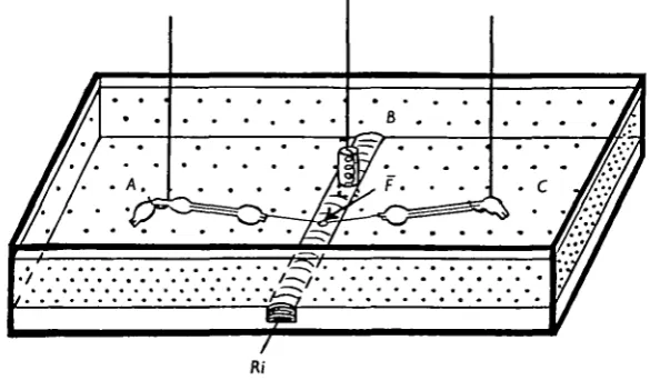
[image:3.451.78.371.306.477.2]The experimental chamber is shown in Fig. 1. It was filled with paraffin oil and crossed by a groove, 1 mm. deep and 07 mm. wide, in which the physiological saline flowed at a constant and regulated rate. A three-way tap (not shown) was placed
Fig. 1. Schematic representation of the recording cell and of the three electrodes used for experiments on an isolated cockroach giant axon. The Perspex chamber is filled with paraffin oil (stippled area). The physiological saline (.Rj) flows in a groove which also contains the central electrode B. The latter electrode is in contact with the physiological saline whereas the two lateral electrodes A and C are isolated from each other and from B by oil. The nerve cord, including the five last abdominal ganglia, is placed between the two lateral electrodes and the groove so that only a small area of the isolated axon remains in Ringer (F).
upstream. This allowed rapid replacement of the physiological saline either by isotonic KC1 (217 HIM) or by another test solution. An orifice in the lower part of the groove was connected to a glass tube by means of a rubber tubing. The raising or lowering of the tube thus effected a regulation of the liquid level in the chamber without perceptibly modifying the rate of flow of saline.
346 Y . PlCHON AND J . BOISTEL
(b) Electrodes
Three silver-silver chloride non-polarizing electrodes were used throughout the experiments. Their potential was lower than i mV. and they had an acceptable stability. The central electrode (B) consisted of a spiral coil of 1-3 mm. diameter chlorided silver wire. The two lateral electrodes (A and C) were made of hooked 0-4 mm. diameter chlorided silver wire placed each on a micromanipulator.
(c) Solutions
The 'modified Ringer solution' of Yamasaki & Narahashi (1959a) was used in these experiments. This solution has the following composition: NaCl, 210-2 HIM; KC1, 3'imM; CaClg, i-8 n m ; NaHjPO4, 0-2 mM; NaaHPO^ i-8mM;pH 7-2. This
saline proved to be satisfactory and allowed a prolonged survival of isolated cockroach giant axon.
(d) Electrical measurement and control apparatus
The electric devices were the same as those used by Schmidt & Stampfli (1966) and Adam, Schmidt, Stampfli & Weiss (1966) in their experiments on single nodes of Ranvier.
Changes in potential of the active membrane area in B were measured by means of
~s
Wn ImCommand signals
Outside
Inside
Fig. 2. Diagram of the electrically equivalent circuit of the experimental arrangement used for recording or stabilization of the membrane potential. R^j, is the axoplasmic resistance, R^ the resistance through oil. Em, R* and Cm are, respectively, the resting potential, the resistance
and the capacity of the active membrane area in B. R,,, Rt and R^ are the (very low) resistances of the A, B and C electrodes, i ^ and i ^ are the resistances of the axon in A and C. With the switch in the current—clamp position (i) the negative-feedback amplifier is connected between A and B and the stimulating current is injected via C. The membrane potential of the ' node' is recorded between the output (E) of the amplifier and ground. For voltage-clamp experiments the switch is set in position 2 so that the amplifier is connected between
A and C. Rectangular signals applied between B and D (a point inside the axoplasm maintained
a differential amplifier of high input impedance which was connected between electrodes A and B as shown in Fig. 2 (position 1 of the switch). Variations of the membrane potential (Vm) were brought about by the application of square pulses (delivered by Tektronix type 161 units) between the earth and the third electrode C. They were recorded at the amplifier output (E) on one beam of an oscilloscope (Tektronix type 502), the stimulating current (/m) being recorded on the other beam.
In the voltage-clamp experiments (position 2 of the switch), the amplifier was connected between the electrodes A and C and the potential of the active membrane area was abruptly changed to determined values, Vm, by the application of square pulses to the middle electrode B. Membrane currents Im were measured from the voltage drops, V^, across series resistances between D (a point inside the axoplasm in B, maintained near ground potential by feedback control) and the output of the amplifier (E). The current membrane current can be related to these resistances by the following equation:
7
C
()
where Re is the resistance of the C electrode, R^ the resistance of the membrane in C and R^ the longitudinal resistance of the axon between D and C. In most cases Rc and R^ were very small in relation to R^ and can be neglected. Equation (1) can therefore be written:
Im=^. (a)
Rax can be calculated knowing the axon diameter (d), the length of axon surrounded with oil (L) and the resistivity of the axoplasm (pax) '•
The values for R^ in our experiments were at least of 250 kD, taking L = o-i cm d = 50/4 and p^ = 46Q <5n.2 (Boistel, 1959). Membrane current density, Jm (mA./ cm2) was calculated from Jm after measurements of the diameter of the axon (d) and of the length of the active membrane (/):
The mean values for the resting membrane resistance (R^) and for the membrane capacitance (Cm) of the active membrane area ('the node') were respectively of
6 Mii and 1 nF., taking / = 100 /i, d = 50 ji, R (resistance x unit area of the surface membrane) = 942O cm.a (Pichon & Boistel, 1966) and C (capacity per unit area of the surface membrane) = 6-3 fiF. cm."2 (Yamasaki & Narahashi, 1959ft).
3 4 ^ Y . PlCHON AND J . BOISTEL
(e) Experimental procedure
Giant axons, with diameters ranging from 30 to 50 fi, were obtained from male cockroaches, Periplaneta americana, which had been reared at room temperature. The abdominal nerve cord was removed under dissecting microscope and immersed, ventral side uppermost, in a drop of physiological saline on a glass slide. The adhering tissue and tracheae associated with the connectives were carefully dissected away and the nerve sheath between the 4th and 5th ganglia was removed. Isolation of 2-3 mm. length of a single giant fibre was carried out using very fine needles, and the unused connective and fibres were cut near the ganglia.
The liquid level in the experimental chamber was then raised (by elevating the glass tube) and the whole preparation was carefully dipped through paraffin oil into the underlying physiological saline and placed on the two lateral electrodes A and C. Afterwards, the liquid level was lowered and the two electrodes were displaced until only a small area of isolated fibre remained in Ringer, the width of this 'node* being set under microscopic observation to approximately 100 ji.
Most experiments were carried out at room temperature, ranging from 20 to 23 ° C.
RESULTS
[a) Membrane potentials
The dissected fibre was mounted in the recording cell as described previously with the grid of the amplifier connected to ground and the gain control turned off. With
200 mV.
20 mV
[image:6.451.107.336.364.547.2]3 msec
Fig. 3. Recording of a membrane action potential (lower tracing) induced by a short-duration depolarizing pulse (upper tracing). Isolated cockroach giant axon. ao° C.
preparation. Great care was taken to avoid oscillations of the amplifier. Rectangular pulses were then applied and the membrane potentials were recorded.
A membrane action potential associated with a short depolarizing pulse is shown in Fig. 3. It is quite similar to those recorded by means of classical high-impedance amplifiers, either with microelectrodes or with external electrodes and the ' oil-gap' (Pichon & Boistel, 1966).
(b) Membrane currents
Measurements of membrane currents were carried out on giant axons giving normal action potentials for at least 10 min.
5 mA /cm :
40 mV.
[image:7.451.103.338.196.370.2]1 msec
Fig. 4. Voltage-clamp recording of the time course of the membrane currents (upper tracing), during a 26 mV. depolarizing pulse (lower tracing), with high sweep rate. Same axon as in fig. 3; 2 o ° C .
The amplifier-gain potentiometer was turned off and the BG product set to its maximum value (80 MHz). The output level was eventually readjusted while the switch was being connected in position 2 (voltage-clamp). The gain control at this time was then turned to its maximum value. Effective control of the membrane poten-tial was achieved when the active membrane area was sufficiently small. Widening of the ' node' was found to result in oscillations such as those reported by Tasaki & Bak (1957, 1958) and Tasaki & Spyropoulos (1958a, b). Similar observations have also been reported by Julian et al. (19626) for the lobster axon in 'sucrose gap'.
A typical current pattern associated with a step depolarization of 26 mV. is shown in Fig. 4. It will be seen that a transient membrane capacity current at the beginning of the voltage step is followed by an initial inward current (I{) which changes smoothly into an outward delayed current (7d). Another transient capacity current is visible
at the end of the voltage step.
This current pattern resembles those of membrane currents recorded in squid axon (Hodgkin et al. 1952), in node of Ranvier, (Dodge & Frankenhaeuser 1958) and in lobster axon (Julian et al. 1962 A).
35° Y. PlCHON AND J. BOISTEL
obtained. Higher depolarizations gave an initial inward current followed by a delayed outward current. The initial currents first increased with increasing pulses, reached a maximum and then decreased. Moreover, they reached their peak value sooner with larger impulses. These initial currents showed a reversal with higher levels of depolari-zation and became outwardly directed. The delayed outward currents increased gradually with the voltage of the depolarizations.
5 inA /cm ' [
40 mV. [
[image:8.451.104.342.160.374.2]1 msec.
Fig. 5. Series of recordings of the membrane currents (upper tracings) induced by depolarizing pulses (lower tracings). The clamping voltages are indicated in the upper left corner of each recording. The onset of the initial inward current occurs very early at room temperature (same axon as in Fig. 3 ; 200 C ) .
5 mA./cm
40 mV
[
f
1 msec.
[image:8.451.102.343.431.611.2]The increase of the initial currents was found to be graded with respect to impulse voltage as shown in Fig. 6.
It should be emphasized that, at room temperature, the maximum inward currents occurred very quickly (less than ioo /isec. for a 46 mV. depolarizing pulse at 200 C.) and that a correction for capacity was necessary. It was also necessary to subtract the non-specific leak current from the recorded values to obtain a correct picture of the ionic currents. This was done by subtracting from the records the appropriate fraction of the current produced by hyperpolarizing pulses. The current intensities at the peak of the inward current (open circles) and at the end of a 3-1 msec, duration pulse (filled circles) were plotted, after correction, against the membrane potential
+ 2
<
E
- 2
- 4
100 mV
Fig. 7. Plot of the ionic current characteristics against the membrane potential during clamp-ing pulses. I( = initial inward current, It = delayed outward current at the end of a 3-1 msec, duration pulse. The resting potential is taken as zero (same axon as in Fig. 3; 200 C).
during the pulse (Fig. 7). It will be seen that the initial inward currents first increased gradually, reaching a peak amplitude of about 4 mA./cm.2 for a 40 mV. depolarizing pulse (peak inward current), and then decreased with larger pulses and finally reversed to outward at about 108 mV. The curve relating the initial current and the pulse amplitude showed some divergence from the straight line in the region of largest depolarizations. The resistance of the membrane in the negative slope region of this curve was about —3-5^ cm.2, whereas in the positive slope region it was about 11 QcmA The delayed outward current increased continuously with the pulse amplitude.
352 Y . PlCHON AND J. BoiSTEL
5 mA /cm :
40 mV.
[image:10.451.104.340.49.250.2]1 msec.
Fig. 8. Superposed recordings of the membrane currents (upper tracings) induced by a 24 mV. clamping pulse when preceded by hyperpolanzing prepulses of long duration at different voltages (lower tracings). These recordings show a marked increase of the peak inward current. (Same axon as in Fig. 5; ao° C ) .
DISCUSSION
The action potentials recorded in current-clamp experiments with the 'oil-gap' technique were very similar to those previously recorded using a classical high-impedance amplifier and either microelectrodes (Boistel & Coraboeuf, 1954; Yama-saki & Narahashi, 1957) or external electrodes (Pichon & Boistel, 1966).
Satisfactory recording of the membrane currents under voltage-clamp conditions was achieved using the present technique. However, more accurate measurements of Rax, and thus, of the ionic current densities are needed for fully quantitative analysis. On the other hand ionic current flows were very rapid at room temperature (22-230 C ) , and on this account were partly disturbed on our records by capacity currents which were especially apparent at higher depolarizations. It is obviously desirable that graphical correction should be carried out when reliable values are required.
The initial ionic currents in the cockroach axon were smoothly graded with respect to the stepwise depolarizations. Their time course was S-shaped and they reached their peak values earlier with larger pulses than with smaller ones. The maximum inward current was reached with depolarizing voltage steps ranging from 40 to 50 mV. from the resting potential. The reversal of the early current from inward to outward was obtained for about 100 mV. depolarizations. This value agrees with the maximum amplitude of the membrane action potentials recorded in 'current-clamp'. The initial currents were larger when the main pulse was preceded by a long-duration hyper-polarizing prepulse, clearly showing that activation was not complete at the normal resting potential level.
were of the same order of magnitude as those predicted by Yamasaki & Narahashi (1959ft) (6*9 mA./cm.2). They are also in close agreement with a value of 910 V./sec. for the maximum rate of rise of membrane action potentials recorded by us with external electrodes (Pichon & Boistel, 1966), this value corresponding to a 57 mA./cm.2 maximum inward current.
The membrane currents recorded in the present investigations were, therefore, very similar to those previously reported for the squid axon, for the nodes of Ranvier and for the lobster giant axon. It seems likely that these currents in the cockroach axon are also related to the same ions as in previously studied structures (i.e. Na+ ions for the initial current and K+ ions for the delayed outward current). However, a possible contribution of other ions, for instance Ca2+ ions or Cl~ ions, to the ionic currents cannot be excluded at this time, and further investigations are needed. It is evident that this method will permit a better understanding of the mode of action of physical or chemical agents on the membrane of the insect axon.
Slight modifications of the experimental procedure, including cooling of the fibre, will permit an even better recording of the membrane currents and hence allow quantitative analysis of experimental data.
SUMMARY
1. An experimental method of recording and controlling the membrane potential of a small area of the membrane of the cockroach giant axon is described.
2. The recorded action potentials were essentially similar to those previously recorded by other methods.
3. The membrane currents resemble those reported for the squid axon, the node of Ranvier in frog nerve and the lobster giant axon.
4. Small cathodal polarizations gave only small outward currents; larger depolariza-tions (10-100 mV.) gave an initial inward current which changed into a delayed outward current.
5. The initial inward current attained a maximum with depolarizing pulses of 40-50 mV. and showed a reversed, outward, flow of about 100 mV.
6. Delayed outward currents increased continuously with increasing impulse voltage.
7. The initial inward current was larger when the pulse was preceded by an hyperpolarizing prepulse.
8. It is concluded that, although the early inward currents were in all probability related to Na+ ions and the delayed outward currents to K+ ions, the possible participa-tion of Ca*+ and Cl~ ions to the ionic currents cannot be excluded.
354 Y . PlCHON AND J. BOISTEL
REFERENCES
ADAM, K. R., SCHMIDT, H., STAMPFIJ, R. & WEISS, C. (1966) The effect of scorpion venom on single
myelinated nerve fibres of the frog. Br. J. Pharmac. Chemother. 36, 666-77.
BOISTEL, J. (1959). Quelques caracteristiques electriques de la membrane de la fibre nerveuse au repos d'un Insecte (Pertplaneta americana). C.r. Sianc. Soc. Biol. 153, 1009-13.
BOISTEL, J. (i960). Caracteristiques Fonctumnelles des Fibres Nerveuses et des Recepteurs Tactile* et
Oljactift da Insectes, 147 pp. Paris: Librairie Arnette.
BOISTEL, J. & CORABOEUF, E. (1954). Potentiels de membrane et potentiels d'action de nerf d'msecte recueillis a l'aide de microelectrodes intracellulairea. C.r. hebd. Sianc. Acad. Set., Paris 238, 3116-18. BOISTEL, J. & CORABOEUF E. (1958). R61e jou6 par les ions sodium dans la genese de l'activit£ electrique
du tissu nerveux d'insecte. C.T. hebd. Sianc. Acad. Set., Paris 347, 1781-3.
CHAPMAN, R A. (1966). The repetitive responses of isolated axons from the crab, Carcinus mamas. J.
Exp. Biol. 45, 475-88.
COLE, K. S. (1949). Dynamic electrical characteristics of the squid axon membrane. Archs Sci. phytiol. 3, 253-8.
DODGE F. & FRANKENHAEUSER, B. (1958). Membrane currents in isolated frog nerve fibre under voltage clamp conditions. J. Physiol., Lond. 143, 76—90.
DODGE, F. & FRANKENHAEUSER, B. (1959). Sodium currents in the myelinated nerve fibres of Xenopus
laevis investigated with the voltage clamp technique. J. Physiol., Lond. 148, 188-200.
FRANK, K., FUORTES, M, G. F. & NELSON, P. G. (1959). Voltage clamp of motoneuron soma. Science,
N. Y. 130, 38-9.
HAGIWARA, S. & SAITO, N. (1959a). Membrane potential change and membrane current in supra-medullary nerve cell of puffer. J. Neurophysiol. 33, 204-21.
HAGIWARA, S. & SAITO, N. (19596). Voltage-current relations in nerve cell membrane of Onchidtum
verruculatum. J. Physiol., Lond. 148, 161-79.
HODGKIN, A. L. & HUXLEY, A. F. (1952). A quantitative description of membrane current and its application to conduction and excitation in nerve. J. Physiol., Lond. 117, 500-44.
HODGKIN, A. L., HUXLEY, A. F. & KATZ, B. (1952). Measurement of current-voltage relations in the
membrane of the giant axon of Loligo. J. Phystol., Lond. 116, 424-48.
JULIAN, J. F., MOORE, J. W. & GOLDMAN, D. E. (1962a). Membrane potentials of the lobster giant axon
obtained by use of the sucrose-gap technique. J. gen. Physiol. 45, 1195—216.
JULIAN, F. J., MOORE, J. W. & GOLDMAN, D. E. (19626). Current-voltage relations in the lobster giant
axon membrane under voltage clamp conditions. J. gen. Physiol. 45, 1217-38.
KOPPENHOFER, E. & WEYMANN, D. (1965). Voltage-clamp am bespulten Ranvierschen Schniirring.
PflOgers Arch. ges. Phytiol. 383, 7.
MARMONT, G. (1949). Studies on the axon membrane. I. A new method. J. cell. comp. Phystol. 34, 351-82.
MOORE, J. W. NARAHASHI, T. & ULBRICHT, W. (1964), Sodium conductance shift in an axon internally
perfused with a sucrose and low potassium solution. J. Physiol., Lond. 173, 163—73.
NARAHASHI, T. (1963). The properties of insect axons. In Advances in Insect Physiology, vol. I, pp. 175-256. Eds. J. W. L. Beament, J. E. Treherne and V. B. Wigglesworth. London: Academic Press. NARAHASHI, T. (1965). The physiology of insect axons. In The Physiology of the Insect Central Nervous
System, pp. 1-20. Eds. J. E. Treherne and J. W. L. Beament. London: Academic Press.
NARAHASHI, T. (1966). Dependance of excitability of cockroach giant axons on external divalent cations.
Comp. Biochem. Physiol. 19, 759-^74.
NARAHASHI, T., DEGUCHI, T., URAKAWA, N. & OHKUBO, Y. (i960). Stabilization and rectification of
muscle fibre membrane by tetrodotoxin. Am. J. Physiol. 198 934-8.
NARAHASHI, T , MOORE, J W. & SCOTT, W. R. (1964). Tetrodotoxin blockage of sodium conductance
increase in lobster giant axons. J. gen. Physiol. 47, 965-74.
NARAHASHI, T. & YAMASAKI, T. (i960). Mechanism of the after-potential production in giant axons of the cockroach. J. Physiol., Lond. 151, 75-88.
NELSON, P. G. & FRANK, K. (1964). La production du potential d'action etudiee par la technique du voltage impost sur le motoneurone du chat. Actuaktis Neurophysiologiques, 5eme serie, pp. 15-35. Masson: Paris.
PICHON, Y. (1967). Application de la technique du voltage impost a l'£tude de la fibre nerveuse isolee d'insecte. J. Physiol., Paris 59, 282.
PICHON, Y. & BOISTEL, J. (1966). Etude de la fibre nerveuse isolee d'insecte. Enregistrement des poten-tiels de membrane et des potenpoten-tiels d'action de la fibre geante de Blatte. Pertplaneta americana L.
C.r. Sianc. Soc. Biol. 160, 1948-54.
SCHMIDT, H. & STAMPFLJ, R. (1966). Die Wirkung von TetraMthylammoniumchlorid auf den einzelnen
STAMPFLJ, R. (1954). A new method for measuring membrane potentials with external electrodes.
Experientia 10, 508-12.
TASAKI, I. & BAK, A. F. (1957). Oscillatory membrane currents of squid giant axon under voltage-clamp.
Science 126, 696-^.
TASAKI, I. & BAK, A. F. (1958). Current—voltage relations of single nodes of Ranvier as examined by voltage-clamp technique. J. Neuropkysiol. 21, 124-37.
TASAKI, I. & SPYROPOULOS, C. S. (1958a). Non-uniform response in the squid axon membrane under voltage-clamp. Am. J. Phynol. 193, 309-17.
TAaAKi, I. & SPYROPOULOS, C. S. (19586). Membrane conductance and current-voltage relation in the squid axon under voltage-clamp. Am. J. Pkysiol. 193, 318-27.
TAUC, L. & FRANK, K. (1962). Le neurone central des Mollusques £tudi£ avec la m&hode du 'voltage-clamp'. J. Phytiol., Parts 54, 415-16.
TOMITA, T. & WRIGHT, E. B. (1965). A study of the crustacean axon repetitive response: I. The effect of membrane potential and resistance. J. cell. comp. Pkystol. 65, 195-210.
TREHERNE, J. E. & MADDRELL, S. H. P. (1967). Axonal function and lomc regulation in the central nervous system of a phytophagous insect (Carauiua morosus). J. Exp. Biol. 47, 235-47.
YAMASAKI, T. & NAHAHASHI, T. (1957). Intracellular rrucroelectrode recordings of resting and action potentials from the insect axon and the effects of DDT on the action potential. Studies on the mechanism of action of insecticides. Botyu-Kagaku 22, 305—13.
YAMASAKI, T. & NARAHASHI, T. (1959a). The effects of potassium and sodium ions on the resting and action potentials of the cockroach giant axon. J. Insect Pkysiol. 3, 146-58.
YAMASAKI, T. & NARAHASHI, T. (19596). Electrical properties of the cockroach giant axon. J. Insect