J. Exp. Biol. (1967), 46, 297-305 2 9 7 With 7 text-figures
Printed in Great Britain
CHANGES IN BLOOD PRESSURE IN THE RAINBOW
TROUT DURING HYPOXIA
BY G. F. HOLETON AND D. J. RANDALL
Zoology Department, University of British Columbia, Vancouver 8, B.C. Canada
{Received 4 November 1966)
INTRODUCTION
The influence of hypoxia upon the heart rate and the rate and amplitude of breathing in the tench has been reported by Randall & Shelton (1963). Satchell (1961) de-monstrated that dogfish respond to anoxia by decreasing heart rate, temporarily increasing dorsal aortic blood pressure and increasing the mean and pulse pressure in the ventral aorta. These fish were restrained and anaesthetized. In this investigation the effects of hypoxia on certain parameters of the breathing and circulatory system were measured. Methods of sampling water and blood afferent and efferent to the gills are described. These methods permit an investigation of these factors in the intact, unanaesthetized, unrestrained but confined rainbow trout.
METHODS
The experiments were carried out on forty-nine rainbow trout (Salmo gairdneri). The fish were held in 500 gallon tanks. The experiments were carried out at the same temperature as that of the water in the holding tanks, which varied between 9 and 190 C. during the period of the experiments, but never by more than o-6° C. during any single experiment.
A fish was anaesthetized in 1:10,000 tricaine methanesulphonate (MS-222), and placed ventral side up in a cloth hammock (Smith & Bell, 1964). Water containing 1:15,000 MS-222 was perfused over the gills by means of a recirculating pump, in either the normal or reverse direction of water flow. Cooling the recirculating water greatly extended the period for which the fish could be maintained on the operating table.
While under anaesthetic the fish were cannulated at the following sites:
1. Buccal cavity cannulation
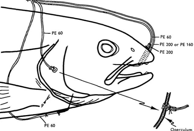
A hole was punched in the cartilaginous portion of the snout in the midline using a no. 16 needle (Saunders, 1961). 80 cm. of PE 200 or 160 polyethylene tubing was passed through this hole. The end of the tubing was heat-flared to anchor the tubing on the inside of the snout (Fig. 1) and cotton thread was wrapped around the tubing where it emerged from the snout to prevent inward movement of the cannula.
2. Opercular cavity cannulation
against the operculum. When the PE 90 was secured with cotton thread, the cannula was anchored firmly in position (Fig. 1). PE 60 rather than PE 200 tubing was used for the opercular cannula so as not to impair movement of the operculum.
3. Dorsal aortic cannulation
The dorsal aorta was cannulated as described by Smith & Bell (1964). 80 cm. of PE 60 tubing, tipped with the end of a no. 21 huber point hypodermic needle was used (Fig. 1).
PE60
PE 200 or PE 160 PE 200
PE 60
[image:2.451.70.388.175.389.2]Operculum Fig. i. Details of the head of a trout showing relative positions of the cannulae
4. Ventral aortic cannulation
The ventral aorta of the trout is barely visible from the lateral view as it passes through the isthmus. It was possible to pierce the ventral aorta with a no. 21 short-bevel hypodermic needle bent at an angle of 6o° about 1-2 cm. back from the point, by passing the needle through the median ventral surface of the animal and observing its entry from the lateral position. The needle was attached to 80 cm. of PE 60 poly-ethylene tubing, which when in position (Fig. 1) was anchored with two stitches placed 1 and 2 cm. posterior to the point of emergence of the cannula from the isthmus.
The coronary artery, which lies on the ventral surface of the aorta, was so small and slippery that it was seldom pierced by the cannula. The cannula passed between the paired stenohyoideus muscles, but the breathing movements did not noticeably affect the position of the cannula.
The aortic cannulae were filled with heparinized Courtland saline (Wolf, 1963) and the ends were plugged with tapered stainless-steel pins.
Changes in blood pressure in the rainbow trout during hypoxia 299
13 l./min. capacity. The volume of the recirculation system was 12-23 1- Cannulae passed out of the tube via one of a pair of exit tubes, and the fish usually assumed a position near the inlet of the tube, facing upstream.
All experiments consisted of sealing the respirometer and allowing the fish to consume the available oxygen. The rate of oxygen uptake by the fish was monitored either by the unmodified Winkler method or by using a Beckman 325814 Oxygen Macro Electrode mounted in a Modular Cuvette and connected to a Beckman Model 160 Physiological Gas Analyser. Pressure in the buccal and opercular cavities was
120
a so
40
40 80 120 Environmental Po (mm. Hg)
160
Fig. 2. T h e effect of hypoxia upon breathing and heart rate. Each circle represents an averaged value for all determinations within a 10 mm. Hg interval of environmental oxygen tension. T h e vertical bars represent plus or minus 2 standard errors.
recorded by connecting the respective cannulae to Statham P23AA pressure ducers. Dorsal and ventral aortic cannulae were connected to Statham P23BB trans-ducers to record blood pressures. Alternatively the aortic cannulae were connected to a Sanborn model 268 B differential pressure transducer and the blood pressure drop across the gills measured. The output from the transducers was recorded continually on four channels of a Beckman Offner Type R Dynograph.
Heart and breathing rates were obtained from the pressure recordings.
Thus changes in a variety of parameters of the circulatory and respiratory systems were recorded during hypoxia, which developed as the fish consumed the available oxygen in the recirculated water. This method is a convenient way of decreasing environmental oxygen, for the rate oxygen decreases is proportional to the rate of oxygen uptake by the fish. The method has the disadvantage of a buildup of CO2 in
the water concommitant with the oxygen decrease. However, because of the high solubility of CO2 in water the changes in partial pressure of CO2 during the experiment
were of the order of only 1-2 mm. Hg.
Dorsal
aorta
-, 70
Changes in blood pressure in the rainbow trout during hypoxia 301
recordings during the presence or absence of an implanted ventral aortic cannula. The ventral aortic pressures were similarly not affected by the dorsal aortic cannulation. It can therefore be presumed that the cannulations themselves have little effect on blood pressure. The respirometer tube, rather than the cannulae, restricted the move-ments of the fish, and in general, the fish rested quietly on the bottom of the tube.
120 r
8 0
40
Systolic
I I
40 80 120 Environmental Po , (mm. Hg)
160
Fig. 4. T h e effect of hypoxia upon systolic and pulse pressures in the ventral aorta. T h e vertical bars represent plus or minus 2 standard errors.
60 r
60 40
20
Pulse
0 40 80 120 160 Environmental PO a (mm. Hg)
RESULTS
The changes in heart and breathing rates and breathing amplitude in the trout during hypoxia (Fig. 2) were similar to those reported for the tench (Randall & Shelton, 1963). In the trout, however, the onset of bradycardia developed at a much higher oxygen level than in the tench.
Differential blood pressure records Higher resistance. Lower resistance.
10 mm. Hg
0 mm. Hg
1-1 4 2 0 % 0-80
1 0
—*
•60 0 4 0 0 •20
-1
q A
• •
1 1
^ o
•
1
_ ^ "
• \ B
1 *
40 60 80 100 120 140
Environmental Po (mm. Hg).
Fig. 6. An analysis of differential blood pressure records (ventral aorta-dorsal aorta) to determine changes in vascular resistance to blood flow through the gills during hypoxia. The length of time for the pressure to drop 10 mm. Hg (taken at the 10 mm. Hg level) is pro-portional to the resistance to flow through the gills. Only records where the flatter phase of the differential pulse record passed the 10 mm. Hg level were used. Tangent lines were drawn to touch the curves at the 10 mm. Hg mark. While absolute values are illustrated in the graph, it is a relative increase in the time interval that reflects an increase in vascular resistance. In six out of the eight records examined there was an increase in vascular resistance with hypoxia (A), while in the remaining two records the results indicated no change in vascular resistance during hypoxia (B).
Both ventral aortic pulse and systolic pressure increased during hypoxia (Figs. 3 and 4); the increase being more marked at lower environmental POi. Ventral aortic blood pressure often decreased when the oxygen level was reduced below 40 mm. Hg, and this decrease was symptomatic of the onset of respiratory and cardiac collapse.
Changes in blood pressure in the rainbow trout during hypooda 303
and 5), as did the differential pressures between the dorsal andventral aortae. An increased differential pulse pressure was also observed.
The ratio of dorsal aortic to ventral aortic blood pressure remained uniform through-out the experiment.
In six out of eight records examined the rate of drop in the differential pressure (after each heart beat) indicated that there is an increase in gill resistance with de-creasing PQ2 in the medium (Fig. 6).
-50 mm. Hg
0 mm. Hg
F.W. 8 sec. Ventral aorta, 23 Aug. 1965
/ V
70 mm. Hg
30 mm. Hg
F.W. 8 sec. Dorsal aorta, 26 Aug. 1965
/"V- Mil
1 sec
-40 mm. Hg
• 0 mm. Hg
4
F.W. 10 sec. Differential, 2 Sept. 1965
[image:7.451.91.364.161.437.2]1 sec.
Fig. 7. The effect upon the heart rate and blood pressures of the rainbow trout of flushing fresh oxygenated water into the respirometer at the end of an experiment when the environ-mental water in the respirometer was of low oxygen content.
Flushing the respirometer with fresh water at the end of each experiment resulted in a rapid increase in heart rate (Fig. 7); the rate tripled within two or three beats when the oxygenated water reached the fish. This cardio-acceleration was associated with an increase in ventral aortic and dorsal aortic systolic blood pressure and a decrease in pulse pressure. Changes in ventral aortic pressure were more marked than those in dorsal aortic pressure and, in one instance, a 308 g. female developed a ventral aortic blood pressure of 128 mm. Hg. Aortic pressures and rate and amplitude of breathing declined slowly and returned to their initial values within 90 min. following the intro-duction of fresh water to the recirculation system.
DISCUSSION
are generally higher than blood pressures recorded by other authors in other species oi fish (Mott, 1951; Burger & Bradley, 1951; Robertson et al. 1966). As suggested by Stevens & Randall (1967) these discrepancies can be related to the method of recording and the physiological state of the animal during each experiment.
An increase in blood pressure can result from either an increase in cardiac output or an increase in peripheral resistance to blood flow. If the increase in blood pressure recorded during hypoxia is related to an increase in cardiac output, then there must be at least a fourfold to sixfold increase in stroke volume in the face of the decreased heart rate. Although there may be an increase in stroke volume during hypoxia, a change of such magnitude seems unlikely. Thus a change in the vascular resistance to blood flow must be at least in part responsible for the increase in blood pressure in the ventral and dorsal aortae.
An analysis of the data (Fig. 6) indicated that there was an increase in resistance to blood flow through the gills. The ratio of the dorsal aortic and ventral aortic pressures was uniform even in the face of increasing blood pressure. This indicates that there are concomitant changes in the vascular resistance of the gill and general body circulation maintaining the constant ratio between pressures in the ventral and dorsal aortae. Thus it would seem that there are increases in resistance to blood flow in both the respiratory and systemic circulations in fish during hypoxia. The increased vascular resistance in the gills may be associated with a shunting of blood through an alternate pathway (Steen & Kruysse, 1964) which decreases the diffusion distance between the blood and water, or increases the area of the gills involved in gas exchange.
The introduction of oxygenated water to the hypoxic fish produces much larger changes in ventral aortic than dorsal aortic blood pressure. This indicates that post hypoxia is associated with either increased resistance to blood flow through the gills or decreased systemic peripheral resistance to blood flow. Analysis of the differential pulse pressure was not possible because of the large discrepancy between mean pres-sures before and after the introdutiocn of oxygenated water at the termination of the
experiment.
SUMMARY
1. Methods for cannulating the ventral aorta of the trout, which permit the measure-ment of blood pressure in the unanaesthetized, unrestrained, intact fish are described. 2. Rate and amplitude of breathing and blood pressure in the dorsal and ventral aortae increase during hypoxia. These changes are associated with a marked brady-cardia.
3. There are increases in vascular resistance to blood flow in both respiratory and systemic circulations during hypoxia.
4. Post hypoxia is associated with large increases in ventral aortic and dorsal aortic pressures.
Changes in blood pressure in the rainbow trout during hypoxia 305
REFERENCES
BRETT, J. R. (1964). The respiratory metabolism and swimming performance of young sockeye salmon.
J. Fish. Res. Bd Can. 21 (5), 1183-226.
BURGER, J. W. & BRADLEY, S. E. (195I). The general form of the circulation in the dogfish (Squalus
acanthias) J. cell. comp. Physiol. 37, 389-402.
GREENE, C. W. (1904). Physiological studies of the chinook salmon. Bull. U.S. Bur. Fisheries 24,
431-47-MOTT, J. C. (1951). Some factors affecting the blood circulation in the common eel. (Anguilla anguilla).
J. physiol. 114, 387-98.
RANDALL, D. J. & SHELTON, G. (1963). The effects of changes in environmental gas concentrations on the breathing and heart rate of a teleost fish. Comp. Biochem. Physiol. 9, 229—39.
RANDALL, D. J., SMITH, L. S. & BRETT, J. R. (1965). Dorsal aortic blood pressures recorded from rain-bow trout (Salmo gairdneri). Can. J. Zool. 43, 863-72.
ROBERTSON, O. H., KRUPP, M. A., THOMPSON, N., THOMAS, S. F., & HANE, S. (1966). Blood pressure
and heart weight in immature and spawning Pacific salmon. Am. J. Physiol. 256, 957-64. SATCHELL, G. H. (1961). The response of the dogfish to anoxia. J. exp. Biol. 38, 531-43. SAUNDERS, R. L. (1961). The irrigation of gills in fishes I. Can. J. Zool. 39, 637-53.
SMITH, L. S. & BELL, G. R. (1964). A technique for prolonged blood sampling in free swimming salmon.
J. Fish. Res. Bd Can. 21 (4), 711-17.
STEEN, J. B. & KRUYSSE, A. (1964). The respiratory function of the teleosteen gills. Comp. Biochem.
Physiol. 12, 127-42.
STEVENS, E. D. & RANDALL, D. J. (1967). Changes in blood pressure, heart rate and breathing rate during moderate swimming activity in rainbow trout. J. Exp. Biol. 46, 307—15.