With 6 text-figures Printed in Great Britain
EVIDENCE FOR AN AUDITORY FUNCTION
OF THE SWIMBLADDER IN THE COD
BY OLAV SAND AND PER S. ENGER
Institute of Zoophysiology, University of Oslo, Oslo, Norway
{Received 26 February 1973)
INTRODUCTION
Fish flesh has nearly the same acoustic properties as sea water. In a sound field a fish will thus be effectively acoustically transparent, and vibrate with the same phase and amplitude as the surrounding water particles. The denser otoliths, which are in close contact with the sensory epithelium in the ear, will lag behind, thereby creating a shear movement of the sensory hairs (Pumphrey, 1950; Griffin, 1955; de Vries, 1956). This is the adequate stimulus for the hair cells in the acoustico-lateralis system in vertebrates. The otolith/hair-cell system in fish thus responds to the kinetic part of a sound wave (particle velocity or particle displacement) rather than the sound pressure.
Because of the high compressibility of gas compared to water, the surface of a gas-filled swimbladder will show much larger displacement amplitudes when ex-posed to sound than the water particles in the absence of a bladder. If these swim-bladder displacements are converted into an amplified movement of the otolith, the swimbladder may improve hearing by functioning as a pressure/displacement trans-former in a sound field. The system will then respond to sound pressure, although the end organ itself is still sensitive to particle motion.
The swimbladder no doubt plays a role as an accessory hearing organ in the Ostari-physi. In these fish an anterior chamber of the swimbladder is linked to the sacculi through a chain of small bones, the Weberian ossicles. Poggendorf (1952) showed that the hearing sensitivity decreased drastically when this bony connexion was destroyed, and he furthermore demonstrated that Ictalurus ( = Atniurus) nebulosus is sensitive to sound pressure, and not to the kinetic part of the sound. The function of the Weberian apparatus has been reviewed by Alexander (1966).
Other forms of close association between the swimbladder and the ear have been reported for the families Anabantidae, Balistidae, Clupeidae, Engraulidae, Holo-centridae, Moridae, Mormyridae, Notopteridae, Ophiocephalidae, Sciaenidae and Sparidae (Jones & Marshall, 1953; Alexander, 1966; van Bergeijk, 1967; Tavolga, 1971). From anatomical considerations it is likely that the swimbladder has an auditory function in these groups.
were only obtained in the near-field, where the displacement amplitude is relatively large. In the cod, on the other hand, microphonic potentials were recorded in the far-field, where the displacements are relatively small. These differences were attributed to an accessory role of the swimbladder in the hearing of cod. Their data from cod, however, did not show that this fish was sensitive to sound pressure rather than to the kinetic part of the sound (Chapman & Hawkins, 1973), and the difference in sensitivity in the two species could thus not be attributed conclusively to the functioning of a pressure/displacement transformer in cod.
Chapman & Hawkins (1973), on the other hand, found sound pressure, and not particle motion, to be the relevant auditory stimulus in cod for frequencies above about 50 Hz. These authors measured acoustic thresholds in cod, and varied the ratio be-tween displacement and sound pressure by changing the distance to the sound source. Their data indicate, though indirectly, that the swimbladder in cod is important for hearing over the frequency range from 50 Hz to the upper frequency limit. Measuring threshold values, Poggendorf (1952) had previously shown Ictalurus to be pressure-sensitive even with the Weberian ossicles destroyed, and this also indicates that the swimbladder might be utilized in hearing in fish without a specialized apparatus like the Weberian ossicles.
Chapman & Sand (1973) studied the hearing in flatfish, which lack a swimbladder. By supplying these fish with an artificial swimbladder consisting of a small gas-filled toy balloon placed just beneath the fish head, the hearing sensitivity was markedly increased and the audible frequency range was extended. However, a natural swim-bladder may only improve the hearing if the sensory cells in the ear have a suitable orientation. These cells must be sensitive to vibrations radiating from the swimbladder, and this turned out to be the case in haddock (Melanogrammus aeglefinus) (Enger
et al. 1973).
Although there is indirect evidence to show that the swimbladder might be utilized in the hearing of an unspecialized fish, direct confirmation is so far lacking. The present investigation was undertaken to obtain direct evidence of an auditory function of the swimbladder in cod. An electro-physiological technique, as described by Enger & Andersen (1967), was chosen, and saccular microphonic potentials were measured as a function of the swimbladder volume. The experiments were performed at 6 m depth in the sea. This was done to obtain reasonably good free-field conditions. The acoustics in small tanks are complicated (Parvulescu, 1964) and the displacement for a given sound pressure may be far above the values for free-field conditions. This will tend to decrease the importance of a swimbladder in hearing.
„ . , MATERIALS AND METHODS
FtStl
Auditory function of the swimbladder
407Voltmeter Oscillator
Amplifier
Oscilloscope
Voltmeter
Hydrophone •
[image:3.451.67.391.41.405.2]Preamplifier
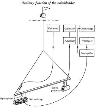
Fig. 1. Sketch of the experimental arrangement. See text for details.
Operation
The fish were anaesthetized with an intraperitoneal injection of approximately 40 mg sodium-iso-amyl-ethyl-barbiturate per kg body weight (Keys & Wells, 1930), which gives an anaesthesia lasting about 10 h. The skull was exposed and the elec-trodes—a pair of stainless-steel wires (0-3 mm diameter) insulated with a diamel coating except for a tiny area at the tips—were implanted through small holes in the skull. One electrode tip was placed between the brain and the sacculus, as close as possible to the saccular sensory epithelium, whereas the other wire was shorter and acted as the indifferent electrode. The distal part of the electrode pair was bent at right angles and fixed to the skull by dental cement (Fleck, Mizzy, Inc.). The con-necting wires were attached to the skin by sutures just behind the incision. Gas was removed from, or injected into, the swimbladder by use of a hypodermic syringe.
Experimental
10 msec
Fig. 2. Oscillographic recordings of the saccular microphonic potentials (upper trace) evoked by background noise (A) compared to the microphonic potentials generated by a 300 Hz tone of 22 dB with (B) and without (C) gas in the swimbladder. Sound recordings on lower beam. Note the pronounced decrease in the microphonic potentials caused by emptying the bladder.
was suspended from a surface float. The experiments were conducted at 6 m depth about 20 m from the shore. The fish was then 4 m above the bottom and the distance between the fish and loudspeaker kept at about 2 m. Signals from the fish were ampli-fied by a pre-ampHfier (Tektronix, 122) displayed on a storage oscilloscope (Tek-tronix, 564) and measured with a vacuum-tube voltmeter (Bruel & Kiaer, 2409). The microphonic potentials in this paper are given in dB referred to 1 jiY rms.
Sinusoidal sound stimuli were generated by an oscillator (Phillips, GM 2308), amplified through a power amplifier (Quad II) and fed into the sound projector.
Sound pressures were measured by a calibrated hydrophone (Atlantic Research Corp., LC 54) fitted with pre-amplifier and connected to a vacuum-tube voltmeter (Bruel & Kiaer, 2409). Sound pressures are given in dB referred to 1 /iBar rms.
RESULTS
Saccular microphonic potentials for fish with normal swimbladder were obtained for frequencies from 50 to 600 Hz. The microphonic potentials showed a steep cut-off towards higher frequencies and we were unable to generate sound sufficiently intense to evoke microphonics at frequencies below 50 Hz. The frequency of the micro-phonic potentials were usually twice that of the sound. However, the two peaks corresponding to one cycle of the sound were frequently of different amplitude. In order to compare different microphonic potential amplitudes, these were therefore measured by an integrating a.c. voltmeter.
Auditory function of the swimbladder
409
3.32
28 24 20 o 16 D,
1 . 2
£
400 Hz
o Full a Half full
' Empty
300 Hz 100 Hz
[image:5.451.50.405.44.213.2]16 24 32 - 8 0 8 16 24 32 0 8 16 24 Sound pressure (dB re 1 /ibar)
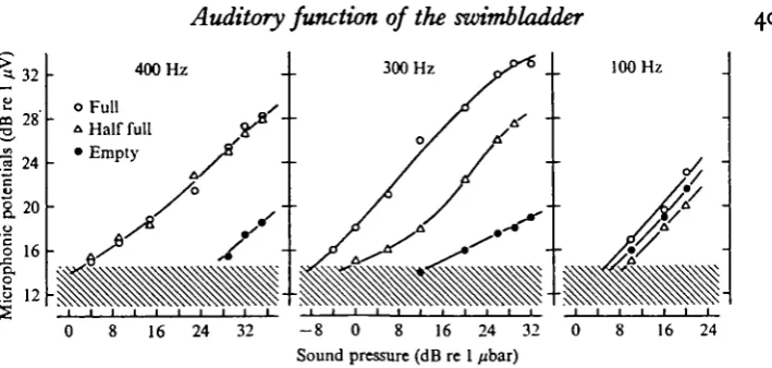
Fig. 3. Amplitude of saccular microphonic potentials as a function of sound pressure for three different frequencies and swimbladder volumes. Frequency and swimbladder state are given in the graph. Hatched area indicates the level of electric background noise. Note that the existence of gas in the swimbladder has no effect on the microphonic potentials at 100 Hz whereas this effect is marked at 300 and 400 Hz.
Fig. 3 gives data from another fish and shows the microphonic potentials as a function of sound pressure for three frequencies. In this fish the measurements at 6 m depth were first taken with an empty bladder, after 18 ml of gas had been removed at the surface. A diver then re-injected 18 ml gas, at 6 m depth, into the bladder, which thus gained its former volume. To ensure that the gas filled the bladder and not the body cavity, the needle was stitched in position after the bladder was initially emptied and the needle-tip remained in position inside the collapsed bladder. The needle was removed after re-filling the bladder, and microphonic potentials were then measured. Further recordings were taken with a swimbladder volume of 10 ml, after the diver had removed 8 ml of gas.
Fig. 3 clearly shows that the amplitude of the microphonic potentials depends on the volume of the swimbladder gas at the two highest frequencies. At 300 Hz the microphonic potentials evoked by a given sound pressure increased in proportion to the gas content of the swimbladder. However, at 400 Hz no difference in microphonic potential amplitudes was detected when the swimbladder volumes were 18 and 10 ml respectively, but with an empty bladder the microphonic potentials were far below these values. At 100 Hz, on the other hand, no significant difference in the micro-phonic potentials for the different swimbladder volumes was obtained.
Assuming that a definite potential amplitude corresponds to the auditory thresholds, independent of frequency, relative values of auditory thresholds can be obtained. To compare the effect of the gas volume on the microphonic potentials at different frequencies the sound pressure necessary to evoke a certain amplitude of the micro-phonic potentials was measured. These sound-pressure values will then indicate the relative auditory threshold at each frequency, and relative audiograms may be con-structed.
•Z 20
2
a 16
•a
I 12
•a
c
3
o
1/3
4
-50 100 200
Frequency (Hz)
[image:6.451.86.352.60.370.2]400
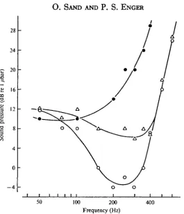
Fig. 4. Relative audiograms showing the sound pressure necessary to evoke microphonic potentials just above the noise level a« a function of frequency. Values for three different swim-bladder volumes are included. Same fish and symbols as in Fig. 3. Note that for all fre-quencies above about 100 H i the existence of gas in the swimbladder has a positive effect on the microphonic potentials.
these potentials are independent of the gas content at lower frequencies. The differ-ence in microphonic potentials generated with 18 and 10 ml swimbladder volumes was negligible above 400 Hz, while for frequencies between 100 and 400 Hz the relative auditory thresholds were lower for the greatest swimbladder volume. At 300 Hz the sound pressure necessary to evoke the threshold microphonic potential was about 34 dB higher for the empty bladder compared to the full bladder of 18 ml. The cor-responding value for the bladder volume of 10 ml was about 15 dB. With gas in the bladder, the relative auditory thresholds were lowest at 200-300 Hz. No such mini-mum values were detected with an empty bladder.
Auditory function of the swimbladder
4 1 142
38
34
30
26
I-
2
3 O DO
18 - A
14 "
™ - I J
100 200 400 800 100 Frequency (Hz)
[image:7.451.51.383.57.321.2]200 400 800
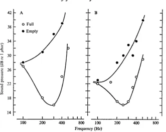
Fig. 5. Relative audiograms for two cods, including values for two different swimbladder volumes.
DISCUSSION
The summed extracellular current from several hair cells, due to the receptor potentials, contributes to the microphonic potentials recorded with our extracellular macroelectrodes (Flock, 1971). The microphonic potentials are thus ultimately connected with the excitation of auditory nerve fibres. Comparison of microphonic potentials at different swimbladder states therefore gives good indication of a possible connexion between hearing ability and swimbladder state.
-a
24
20
16
12
•a
3
c
I I I
50 100 200 Frequency (Hz)
400
12
0 H
- 3
- 4 a
- 8 c.
•12 I
- 1 6
- 2 0
- 2 4
Fig. 6. Average relative audiogram for full swimbladdere compared to two behavioural audiograms for cod (dotted line, Buerkle, 1967; broken line, Chapman & Hawkins, 1973). Thresholds are presented as absolute sound pressure in the behavioural audiograms (right ordinate), whereas in the relative audiogram absolute values are converted to dB re value at the most sensitive frequency in each fish (left ordinate). The symbols (#), (O) and (A) indicate the cods presented in Fig. 4, 5 A and B, respectively.
and increase with swimbladder volume, and thus with fish size (see Chapman & Sand,
1973)-The effect swimbladder gas in the cod has on the relative auditory thresholds is in good agreement both with the decrease in auditory thresholds and the extension of the audible frequency range which Chapman & Sand (1973) observed in flat-fish after introducing an artificial swimbladder. We conclude that the swimbladder in cod, and probably in all fish possessing a swimbladder, acts as a pressure/displacement transformer, and thus increases the hearing sensitivity for all frequencies from a lower limit to the upper frequency cut-off for hearing.
Auditory function of the swimbladder 413
f upper frequency cut-off corresponds fairly well in the two types of audiograms, thus
indicating that the upper limit of the audible frequencies may be determined by in-adequacy of the peripheral auditory apparatus. The differences between the two be-havioural audiograms, both in regard to shape and absolute threshold values, are mainly due to the high level of background noise in Buerkle's experiments. The thresholds reported by him were thus heavily masked, and the audiogram below 140 Hz was related to the background aquarium noise. Reports on other fish (see for instance Offutt (1968) for data on goldfish) have also shown that different techniques and experimental conditions may result in different audiograms for the same species. The general shape of the microphonic and behavioural audiograms for cod given in Fig. 6 are therefore in reasonably good agreement. Enger & Andersen (1967), on the other hand, recorded saccular microphonic potentials in cod at frequencies up to 1000 Hz, which is about one octave above the upper frequency cut-off in this species. However, these authors worked at shallow depths, where the relation between particle displacement and sound pressure is higher than for free-field conditions (Banner,
1968). This may explain the difference between their results and ours.
SUMMARY
1. Saccular microphonic potentials in cod (Gadus morhua) were recorded by means of implanted electrodes during sound stimulation with different swimbladder volumes. To obtain acceptable acoustic conditions, the experiments were conducted at 6 m depth in the sea.
2. Swimbladder volume had no effect on the microphonic potentials at 100 Hz, whereas its effect was marked at higher frequencies. The sound pressure necessary to evoke microphonic potentials just above the electric background noise was about 20 dB higher for the empty bladder, compared to the full bladder at 300 Hz.
3. The microphonic potentials are ultimately linked to excitation of eighth-nerve fibres, and it is concluded that the existence of gas in the cod swimbladder has a positive effect on hearing sensitivity for all frequencies from a lower transient frequency to the upper frequency limit of hearing. Gas in the swimbladder will furthermore extend the audible frequency range.
4. The upper frequency limit for the saccular microphonic potentials is in good agreement with the upper audible frequency limit determined from behavioural audiograms for cod, thus indicating that this frequency cut-off may be due to inade-quacy of the peripheral auditory apparatus.
VAN BERGEIJK, W. A. (1964). Directional and nondirectiona] hearing in fish. In Marine Bio-Acoustics (ed. W. N. Tavolga), pp. 281-99. Oxford: Pergamon Press.
VAN BERGEIJK, W. A. (1967). The evolution of vertebrate hearing. Contrib. Sensory Pkysiol. 3, 1-49. BUERKLE, U. (1967). An audiogram of the Atlantic cod, Gadus morhua L. J. Fish. Res. Bd Can. 34,
2309-19.
CHAPMAN, C. J. & HAWKINS, A. D. (1973). A field study of hearing in the cod, Gadus morhua L. J. comp. Physiol. (In the press.)
CHAPMAN, C. J. & SAND, O. (1973). Field studies of hearing in two species of flatfish Pleuronectes platesta (L.) and Limanda limanda (L.) (family Pleuronectidae). Comp. Biochem. Physiol. (In the press.) DE VRIES, H. (1956). Physical aspects of the sense organs. Prog. Biophys. biophys. Chem. 6, 207-64. ENGER, P. S. & ANDERSEN, R. (1967). An electrophysiological field study of hearing in fish. Comp.
Biochem. Physiol. 23, 517-25.
ENGER, P. S., HAWKINS, A. D., SAND, O. & CHAPMAN, C. J. (1973). Directional sensitivity of the saccular microphonic potentials in the haddock. J. Exp. Biol. 59, 425-33.
FLOCK, A. (1971). The lateral line organ mechanoreceptors. In Fish Physiology (ed. W. S. Hoar and D. J. Randall), vol. v, pp. 241-63. New York: Academic Press.
GRIFFIN, D. R. (1955). Hearing and acoustical orientation in marine animals. Deep-Sea Res. 3, Suppl., 406-17.
JONES, F. R. H. & MARSHALL, N. B. (1953). The structure and function of the teleostean swimbladder. Biol. Rev. 38, 16-83.
KEYB, A. B. & WELLS, N. A. (1930). Amytal anesthesia in fishes. J. Pharmac. exp. Ther. 39, 115-28. OFFUTT, G. C. (1968). Auditory response in the goldfish. J. Aud. Res. 8, 391—400.
PARVULESCU, A. (1064). Problems of propagation and processing. In Marine Bio-Acoustics (ed. W. N. Tavolga), pp. 87-100. Oxford: Pergamon Press.
POGGENDORF, D. (1952). Die absoluten H8rschwellen des Zwergwelses (Amiurus nebulosus) und Beitrflge zur Physik des Weberschen Apparates der Ostariophysen. Z. vergl. Physiol. 34, 222-57. PUMPHREY, R. J. (1950). Hearing. Symp. Soc. exp. Biol. 4, 3-18.
SAND, O. & HAWKINS, A. D. (1973). Acoustic properties of the cod swimbladder. J. exp. Biol. 58, 797-820..