Roger A. Hyman 1 Jon H. Edwards S. J. Vacirca Harry L. Stein
Received July 18, 1984; accepted after revision October 30, 1984.
Presented at the annual meeting of the American Society of Neuroradiology, Boston, June 1984.
, All authors: Department of Radiology, Cornell University Medical College, New York, NY 10021, and North Shore University Hospital, 300 Commu-nity Dr., Manhasset, NY 11030. Address reprint requests to R. A. Hyman.
AJNR 6:229-236, March/April 1985 0195-6108/85/0602-0229 © American Roentgen Ray Society
0.6 T MR Imaging of the
Cervical Spine:
Multislice and
Multiecho Techniques
During a 6 month period, 50 patients with signs and symptoms referable to the cervical spine were studied with a 0.6 T superconducting magnetic resonance (MR) imaging unit. The last 23 of these 50 patients were studied with combined multislice and multiecho techniques. In 38 of the 50 patients, abnormalities were demonstrated on MR images. Intramedullary lesions included syringomyelia (three cases), primary tumors (two), metastatic neoplasm (one), cord atrophy secondary to trauma (one), and multiple sclerosis (one). Intradural, extramedullary lesions included two neurofibromas and two Chiari malformations. The rest of the lesions were extradural: degenerative changes (10), spinal stenosis with cord compression (five), disk degeneration and/or herniation (five), postoperative changes (four), metastases to bone/epidural disease (three), and neurofibromatosis (one). Two patients had more than one abnormality. The MR findings were compared with available routine radiographs, computed tomographic (CT) scans with and without metrizamide, and myelograms. MR imaging was consistently better than routine CT scanning in the detection of lesions of the spinal cord and in directly imaging the effects on the spinal cord of extrinsic abnormalities such as spinal stenosis. Metrizamide-enhanced CT scanning detected all cases of syringomyelia, but it involved an invasive procedure. Myelography alone was slightly less sensitive and considerably less specific than MR in detecting intramedullary lesions and in distinguishing cord neoplasms from syringomyelia. Multislice, multiecho techniques with up to 240 msec echo times (TEs) were particularly helpful in the detection and characterization of extradural processes. The addition of this technique to standard spin-echo 500 msec repetition-time (TR), 30 msec TE sagittal and transverse multi slice studies and trans-verse 2000 msec TR, 60 msec TE multi slice studies provides an examination of the cervical spine that optimizes the detection rate when the disease process is unknown. After plain radiography, MR is the imaging procedure of choice for screening suspected disease of the cervical spinal canal.
During the past 5 years, rapidly progressing technologic advances with magnetic resonance (MR) imaging have given rise to moderate- and high-field-strength superconducting units capable of producing high-quality clinical images. Early clinical experience with resistive and low-field-strength superconducting units in the cervical spine has been reported [1-4). We used a 0.6 T superconducting system to study the cervical spine and compared the results of these examinations with computed tomography (CT) with and without metrizamide, myelography, and routine radiography. Multislice and multiecho techniques introduced during the
latter part of the study resulted in considerable reduction in examination time.
Subjects and Methods
230 HYMAN ET AL. AJNR:6, March/April 1985
1
2
were examined in a 28.5-cm-diameter head coil in addition to the body coil.
One major and several minor software changes occurred during the study period. Because of these changes, the first 27 patients
were studied using single-slice techniques, while the final 23 patients had both multislice and multiecho studies, also using two-dimensional techniques. The latter group of patients provided us with the oppor-tunity to compare the efficacy of multislice and multiecho techniques with the single-slice techniques used in the initial group.
Several different pulse sequences were used during the study period. All patients underwent an initial sagittal spin-echo (SE) 500 msec repetition-time (TR), 30 msec echo-time (TE) either single- or multislice. Five mm offsets were used to optimize cord localization and imaging. In addition to this relatively T1-weighted sagittal SE series, all patients underwent a relatively T2-weighted sagittal SE
series as well. Initially, these consisted of 1000 msec TRs and 60 or 120 msec TEs. When multislice/multiecho software was added, 2000 msec TRs and 60, 90, or 120 msec TEs were used, or 2240 msec TRs and 30-240 msec TEs at 30 msec intervals.
Some patients with demonstrated anatomic lesions also underwent inversion recovery (IR) studies with 1500 msec TRs, 450 msec inversion times (Tis), and 30 msec TEs. Transverse sections were
frequently obtained as well. A transverse T1-weighted sequence was used when a syrinx was suspected, and a transverse T2-weighted
series was obtained when extradural disease was the clinical
consid-eration. The imaging time varied with a number of factors, including TR and the number of sequences per buffer; time is related to both of these in a linear fashion. Slice thicknesses of 10 mm were used
for both single- and multislice examinations. There were no gaps between slices in multislice studies.
The 50 patients were 10-81 years old. There were 32 males and 18 females. The MR studies were compared with available routine radiographs of the cervical spine, CT scans with and without
metri-zamide, and myelograms. All CT studies at our institution were carried
out on a Picker 1200 unit. Transverse sections of 2, 3, 5, or 10 mm thicknesses were used depending on the clinical circumstances.
Results
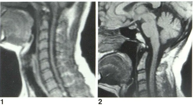
Twelve of the 50 patients studied with MR demonstrated no abnormalities. The images obtained with the 55 cm body coil (fig. 1) had an inherently lower signal-to-noise (S/N) ratio
Fig. 1.-Sagittal SE 500/30 image. Normal study using body coil. Inherently lower SIN ratio compared with head-coil examination in fig. 2.
Fig. 2.-SE 500/30. Normal study in head coil. Low signal from CSF on this pulse sequence provides excellent contrast with spinal cord. As cortical bone provides little if any signal, apparent width of CSF compartment as well as anulus fibrosus is exagger-ated by adjacent cortical bone.
than those obtained with the 28.5 cm head coil (fig. 2). However, because of the constraints imposed by the shoul-ders, only a limited amount of the cervical spine could be imaged using the head coil. For this reason, routine exami-nation of the cervical spine currently must use the body coil for complete imaging. The head coil can be used successfully to evaluate the craniocervical junction and to evaluate lesions of the upper cervical spine when patient positioning permits.
Thirty-eight of the 50 patients studied demonstrated 40 abnormalities (table 1). Two patients exhibited more than one abnormality. Both patients with Chiari malformations had other associated abnormalities: syringomyelia in one case and degenerative changes without cord compression in the other.
Intramedullary Lesions
[image:2.612.55.386.88.268.2]Fig. 3.-Syringomyelia. A-C, SE 500/30 images. A, Fluid-filled cavity (arrow) in spinal cord is seen to extend into upper thoracic region. Chiari malformation is present also. e, Coronal study in same patient shows syrinx and no communication with fourth ven-tricle. C, In head coil. Syrinx and associated Chiari malformation are shown to better advantage. D, De-layed metrizamide CT scan. Central part of cord shows metrizamide within it at 6 hr, consistent with diagnosis of syringomyelia.
A
c
TABLE 1: Cervical Spine Abnormalities Demonstrated by MR
Imaging in Symptomatic Patients
Location of Lesion: Type No. (n = 38)
Intramedullary:
Syringomyelia . . . 3 Cord neoplasms . . . . . . 3
Cord atrophy ... 1
Multiple sclerosis. . . . . 1
Subtotal . . . 8 Intradural, extramedullary:
Neurofibroma .. ... . . . 2
Chiari malformation . ... . _--=2 _ _
Subtotal 4
Extradural:
Degenerative changes . . . . 1 0
Spinal stenosis with cord compression 5
Disk degeneration and/or herniation . . . 5
Postoperative Changes 4
Metastases to bone/epidural disease .. . . 3 Neurofibromatosis . . . . . . . 1
Subtotal 28
Total 40
8
o
high signal from the spinal cord and the relatively low signal emanating from the adjacent cerebrospinal fluid (CSF) space.
Three intramedullary neoplasms were studied: two gliomas and one metastasis from carciroma of the lung. All three
lesions demonstrated increased signal on T2-weighted image
series, indicative of a prolonged T2 relaxation time with
re-spect to normal spinal cord. All three lesions were identifiable
as mass lesions on T1-weighted images as well, but abnormal signal intensity was not apparent.
A 45-year-old woman with an undiagnosed cervical mye-lopathy was examined after a workup at another institution.
T1- and T2-weighted sagittal series were performed initially.
A mass lesion in the cervical cord was readily apparent (fig. 4A). With an SE 1000/60 series (fig. 48), increased signal
was noted within the lesion. This was even more apparent on an SE 1000/120 study (not shown) at the cost, however, of decreased SIN ratio.
A linear area of decreased signal intensity was seen in the
center of the lesion on all pulse sequences. If this represented
[image:3.612.228.555.86.442.2] [image:3.612.52.295.508.726.2]232 HYMAN ET AL. AJNR:6, March/April 1985
A
8
Fig. 4.-lntramedullary metastasis from lung carcinoma. A, SE 500/30. Lower cervical cord is enlarged;
central part appears to contain cavity (arrow). Loss of normal marrow signal from T1 and T5 vertebral bodies represents bone metastases. B, SE 1000/60. Increase in signal from lesion. Other findings essentially unchanged.
Fig. 5.-Spinal cord atrophy. Sagittal single-slice study. Spinal cord is narrowed opposite previously traumatized C5 vertebral body. Decreased signal in-tensity from body of C5.
made. Cavitation within tumors on MR studies has been reported [2], and a tumor giving rise to a syrinx is a well
recognized phenomenon. Thus the ability of MR imaging to
detect abnormal signal from a tumor when a cavity is seen is of significance in distinguishing between syrinx alone and syrinx in conjunction with cord neoplasm.
The 21-year-old man whose MR study is shown in figure S sustained trauma to his cervical region 3 years before this examination. Posttraumatic atrophy of the spinal cord was apparent in the lower cervical region. There was decreased signal intensity from the body of CS. However, traumatic bone changes were shown more clearly on CT scans and plain films.
One unproven intramedullary lesion in our series was be-lieved to represent multiple sclerosis. An MR study in a
42-year-old man demonstrated a region of increased signal
inten-sity in the midcervical region associated with equivocal cord
enlargement on a T2-weighted image. This lesion was
indis-tinguishable from a cord neoplasm on both MR imaging and myelography. 8ecause the patient was clinically a multiple sclerosis suspect, an MR study of the brain was performed.
This demonstrated several periventricular areas of increased
signal intensity on a T2-weighted series; multiple sclerosis of
the cord was diagnosed, and restudy after a suitable interval was planned.
The relative sensitivity and specificity of MR with respect to other imaging techniques in evaluating intramedullary le-sions is summarized in table 2. Myelography alone was
slightly less sensitive and considerably less specific than MR in detecting intramedullary lesions and in distinguishing cord neoplasms from syringomyelia in the five cases studied by
both methods.
TABLE 2: Relative Sensitivities and Specificities of MR Imaging, Initial and Delayed Metrizamide CT, and Myelography
in the Evaluation of Intramedullary Lesions
Study
MR imaging: No. detected Specificity. . . Metrizamide CT: No. detected Specificity. Myelography: No. detected Specificity.
Syringomyelia
3 3
3 ?3
3
o
Cord
Neoplasms
3 3
2
o
Intradural, Extramedullary Lesions
Atrophy!
Trauma
Multiple Sclerosis
1
o
1
o
Four lesions were detected in this group: two neurofibro-mas and two Chiari malformations. A 41-year-old man dem-onstrated widening of the left neural foramen between C2 and C3 on routine oblique radiographs of the cervical spine. The T2-weighted SE 2000/60 multislice sequence was carried out in sagittal and coronal planes. In the coronal plane at the level of the neural foramen (fig. 6A) a region of sharply demarcated increased signal intensity was demonstrated. Somewhat more posteriad on the same multislice sequence,
the lesion was seen displacing the spinal cord to the right and
compressing it (fig. 68). The lesion was clearly distinguished from the spinal cord and was well seen extending extradurally as well. A myelogram demonstrated the intradural extramed-ullary part of the mass, but did not adequately demonstrate
[image:4.612.59.559.85.315.2] [image:4.612.315.559.412.521.2]Fig. 6.-Neurofibroma. A, Coronal SE 2000/60 image from multislice series. Well circumscribed le-sion within left neural foramina at C2-C3 level. e,
More posterior coronal section. High-signal lesion displaces spinal cord to right. Extradural part of lesion is imaged as well as intradural component.
A
For evaluation of the Chiari malformation (fig. 3C) MR is the imaging method of choice [5]. The T1-weighted SE 500/30 sequence gives rise to optimal contrast between the tonsillar ectopia and the adjacent CSF without the necessity for an invasive procedure. While this diagnosis can be made readily
by metrizamide CT, myelography, and even on occasion with
high-resolution plain CT, MR is simpler, noninvasive, has multiplanar capabilities, and involves no radiation.
Extradural Lesions
In our series, 28 of 40 lesions were extradural in location.
It was in this group of lesions that T2-weighted multislice and multiecho techniques were most advantageous. Sagittal
stud-ies using a 2240 msec TR were most common. Three
contig-uous midline sagittal sections of about 10 mm thicknesses
were obtained, and data from eight echoes for each section
were collected and displayed for a total of 24 images. Scan-ning time for one entire series was 10.1 min. Degenerative
changes without cord compression were demonstrated in 10
patients. Five patients had degenerative changes associated with spinal stenosis and spinal cord compression. Using an SE 500/30 technique, the effect of a stenotic lesion on the cord itself could be identified in a 69-year-old man (fig. 7 A). While the spinal cord was seen well with this technique, and the compression clearly delineated at the C5-C6 level and suggested at C3-C4 as well, the signal had to be increased from the CSF to fully evaluate the effect of the spinal stenosis on the thecal sac. The multiecho series in this patient
accom-plished this objective using a 2240 msec TR. The third echo
(90 msec TE) illustrated in figure 7B showed the extent of extradural involvement to considerably better advantage. With the CSF white, the effect of the adjacent cortical bone on the spinal canal was readily apparent. With increasing TEs,
the contrast between CSF and cord increased, although the
B
SIN ratio decreased. The fifth echo (150 msec TE) illustrated this contrast well (fig. 7C), although some detail was lost. From our experience in examining the cervical spines of 23 patients, echoes beyond the sixth, with a 180 msec TE, rarely
provided significant information because of the extremely
poor SIN ratio at 210 and 240 msec. Echoes at 150 and 180
msec did occasionally produce diagnostically useful images.
We found that multislice, multiecho techniques provided information of greater significance in patients with stenotic lesions than that obtained with plain films or CT. While the
lateral cervical spine view demonstrated degenerative
changes and retrolisthesis (fig. 70), the direct effect of the stenosis on the cord could not be ascertained, nor could this information be obtained directly with CT unless metrizamide was used. Myelography was judged to yield the most infor-mation in our stenosis group, but at the cost of an invasive
procedure and hospitalization not required with MR.
The inability to detect signal from cortical bone is a draw
-back of MR that we believe has been overstated. It is often possible to obtain significant information about bony struc-tures, both cortical and medullary, with MR imaging. A sagittal T2-weighted SE sequence in one of our patients with spinal stenosis (fig. 8) clearly demonstrated the loss of height and
abnormal shape of the C3-C6 vertebral bodies, irregular
contour of their anterior and posterior margins consistent with
degenerative changes, and irregular signal from the medullary
portion as compared with nearby vertebral bodies. In addition,
abnormal signal from the C3-C6 disk spaces and
compres-sion of the subarachnoid space are apparent. A myelogram confirmed the cord compression before surgical intervention.
Disk degeneration and/or herniation was detected in five
patients. While MR imaging was more sensitive to disk
de-generation, demonstrating decreased signal from the involved
[image:5.614.228.555.86.313.2]234
HYMAN ET AL. AJNR:6, March/April 1985c
o
Postoperative changes were detected in four patients. One patient with a prior extensive laminectomy (fig. 9) demonstra-ted the soft-tissue changes and absence of spinous proc-esses associated with prior surgery.
Three patients with metastases to bone and/or epidural disease were included in the study group. Metastases to bone were detected as regions of decreased signal intensity within the otherwise high-signal bone narrow (figs. 4A and 10). Epidural disease was detectable as well.
A final patient in the extradural group demonstrated the cutaneous lesions of neurofibromatosis on the anterior and posterior chest wall. He was studied to exclude the presence of spinal stenosis or intraspinal neurofibroma, neither of which was present. The ability to image cutaneous lesions is
iIIus-Fig. 7.-Spinal stenosis. A, SE 500/30. Compres-sion of spinal cord confirmed at C5-C6 and sug
-gested at C3-C4. B, Multiecho SE 2240/90 image.
Full extent of extradural compression is more appar-ent on T2-weighted image with increased CSF signal. C, Fifth echo (SE 2240/150). Increased contrast be-tween spinal cord and CSF; decreased SIN ratio
compared with earlier. echoes. 0, Lateral cervical spine film. Bony changes and retrolisthesis at C5-C6; direct information about spinal cord is not pro-vided.
trative of the capabilities of MR to image soft-tissue pathol-ogy, even when the cervical cord and adjacent structures constitute the region targeted by the examination.
Discussion
8
9
Fig. 8.-Extensive bony changes in C3-C6 vertebral bodies; cord compre s-sion.
Fig. 9.-Sagittal SE 500/30 image. Extensive soft-tissue and bony changes
posterior to spinal cord in patient who underwent prior extensive cervical laminectomy.
Fig. 1 ~.-Sagittal SE 500/30 image. Vertebral bodies of C7 and T1 dem
-demonstrates this degree of specificity, and even delayed metrizamide CT can result in difficulty in diagnosing a syrinx with associated tumor.
The recent availability of multislice, multiecho techniques,
with up to eight echoes used in many of our examinations, has made a significant contribution to MR imaging of the cervical spine, particularly with respect to imaging extradural disease processes. Cord compression associated with spinal stenosis, as well as extradural defects associated with cervi-cal spondylosis, herniated nucleus pulposus, metastases, and postoperative changes, is more efficiently demonstrable with multislice, multiecho MR, which can be carried out as an outpatient screening procedure without the necessity for hos-pital admission or invasive procedures. Extradural processes that are more subtle, such as entities that lead to minor nerve-root compression, but not myelopathy, are better evaluated at this time with thin-section high-resolution CT, metrizamide CT, or myelography. At the present time, this is true of small extradural defects associated with herniated nucleus pulpo-sus or lateral recess stenosis with root-sleeve abnormalities. The ability to perform thin-section high-resolution transverse MR imaging using surface coils applied directly to the neck may, however, change this situation in the near future. Re-cently, the feasibility of thin-section MR techniques using both two- [6] and three- [7] dimensional multislice techniques has been described.
Several other advantages of MR with respect to CT and myelography are evident. The cervical spinal cord and adja-cent brainstem and cerebellar tonsils can be imaged directly
10
on strate decreased signal compared with vertebral bodies above and below,
consistent with metastases. Soft tissue with high signal between vertebral bodies and cord represents epidural disease. Signal from nucleus pulposus is intact between C7 and T1, consistent with metastasis and unlike what would be expected in infection.
without injecting contrast agents or using ionizing radiation. Noncontrast CT will rarely demonstrate the cervical spinal cord as distinct from the thecal sac below the C2 level. The ability to perform a multi planar examination without moving the patient is of considerable benefit, particularly in elderly,
severely ill, or traumatized patients. MR is already more sensitive than other imaging methods in identifying degener
-ative disks, although myelography and CT remain more sen
-sitive in detecting herniated nucleus pulposus. Finally, numer-ous pulse sequence strategies are available to optimize the examination in varying clinical circumstances. Current disad-vantages include the inability to completely assess bony integrity after trauma compared with CT. Nonetheless, the effect of the trauma on the neuraxis is better evaluated with MR imaging. Subtle effects on exiting nerve roots are difficult
to detect with current techniques. Although cortical bone
gives rise to little if any signal, long TEfTR SE techniques will show strong signal from both CSF and medullary bone. The
low-signal region between the two represents cortical bone in part, and its position and thickness is identifiable. Major destruction associated with metastases will usually involve
either the epidural space or marrow cavity of the vertebral body and can be detected with MR.
Examination Design
While it is not difficult to suggest an appropriate single
[image:7.612.53.557.81.316.2]236 HYMAN ET AL. AJNR:6, March/April 1985
would optimize the detection rate of all entities when the disease process is unknown. At 0.6 T, an SE 500/30 se-quence shows the cervical spinal cord in contrast to the low signal from the CSF. We routinely begin a cervical spine MR study with a sagittal multislice SE 500/30 series. This is followed by a 0.5 cm offset of the above. If a syrinx is suspected, a transverse multislice examination with this pulse sequence is also obtained. These sequences require only 2.3 min of scanning time with two sequences per buffer.
Techniques using long TR intervals of at least 2000 msec as well as multiecho techniques using up to eight echoes with TEs between 30 and 240 msec are particularly helpful in the identification and characterization of extradural processes.
After the T1-weighted series described, we perform a sagittal 2240 msec TR, 30-240 TE study. This sagittal sequence includes three continuous 10 mm sections with eight echoes from each, and requires 10.1 min of scanning time with two sequences per buffer. If an extradural process is suspected,
a transverse multislice 2000 msec TR, 60 or 90 msec TE series lasting 9.0 min is added. If all five sequences described above are performed, scanning time is under 30 min, and a relatively complete examination has been obtained.
We believe that a 2000 msec TR is a minimum at 0.6 T in order to increase the signal in the CSF to the point where extradural disease is optimally detected. It is possible that at higher field strengths, where one sequence per buffer may be used, a somewhat longer TR may be warranted for optimum detection of extradural processes, without excessive scan-ning time.
MR of the cervical spine at moderate and high field strengths is an easy-to-perform, well tolerated, noninvasive
examination with no known side effects [8]. When a complete examination using various pulse sequences and projections as described above is carried out, we believe it to be the screening procedure of choice, after plain radiography, when disease of the cervical spinal canal is suspected. If the ex-amination is unrevealing and clinical circumstances so dictate,
CT and/or myelography should then be performed.
REFERENCES
1. Modic MT, Weinstein MA, Pavlicek W. Nuclear magnetic reso-nance imaging of the spine. Radiology 1983;148:757-762
2. Han JS, Kaufman B, EI Yousef SJ, et al. NMR imaging of the spine. AJNR 1983;4:1151-1159, AJR 1983;141 :1137-1146 3. Norman D, Mills CM, Brant-Zawadzki M, Yeates A, Crooks lE,
Kaufman L. Magnetic resonance imaging of the spinal cord and canal: potentials and limitations. AJNR 1983;5:9-14, AJR 1983;141 :1147-1152
4. Modic MT, Weinstein MA, Pavlicek W, Boumphrey F, Starnes D,
Duchesneau PM. Magnetic resonance imaging of the cervical spine: technical and clinical observations. AJNR 1984;5: 15-22,
AJR 1983;141:1129-1136
5. DelaPaz Rl, Brady T J, Buonanno FS, et al. Nuclear magnetic resonance (NMR) imaging of Arnold-Chiari type-1 malformation with hydromyelia. J Comput Assist Tomogr 1983;7:123-129
6. Hyman RA. Thin section imaging techniques. Presented at the Vanderbilt University MRI users meeting, Nashville, June 1984
7. Crooks lE, Mills CM, Hoenninger JC, et al. Thin section MRI at 0.35 T. Presented at the annual meeting of the American Society of Neuroradiology, Boston, June 1984
8. Budinger TF. Nuclear magnetic resonance (NMR) in vivo studies:
known thresholds for health effects. J Comput Assist Tomogr