Shoki Takahashi1
Fumihiko Hoshino2
Kazuo Uemura3 Akira Takahashi4 Kiyohiko Sakamoto 1
Received June 13, 1988; accepted after revision
October 25, 1988.
' Department of Radiology, Tohoku University
School of Medicine, 1-1 Seiryocho, Sendai-shi, 980, Japan. Address reprint requests to S. Takahashi.
2
Division of Radiology, Kohnan Hospital, Sendai, Japan
3 Division of Radiological Sciences, Research In -stitute of Brain and Blood Vessels, Akita, Japan.
4
Division of Neurosurgery, Kohnan Hospital,
Sendai, Japan.
AJNR 10:563-568, May/June 1989 0195-6108/89/1003-0563
© American Society of Neuroradiology
563
Accessory Middle Cerebral Artery:
Is It a Variant Form of the Recurrent Artery
of Heubner?
. . ... •. .- . . . -..___ .. ; •. "·.P,n~}~
:
~-~
' .'
.
-
·:'
.~:;;,'
·_~--
'.
' . ·_·--:
...~
·...
~ ~:~:;::~~~~
Fourteen accessory middle cerebral arteries demonstrated on angiography were reviewed relative to the pertinent literature. The anomalous vessel follows a fairly constant pattern in terms of origin, course, and distribution, and it frequently gives rise to basal perforating arteries. It is suggested that the vessel represents a persistent anastomosis between the anterior and middle cerebral arteries over the tuberculum olfactorium, the anastomosis being a predecessor of the recurrent artery of Heubner in phylogenetic development. Among various descendants of the anastomotic channels could be included both the normal recurrent artery of Heubner and the accessory middle cerebral artery, with or without basal perforating arteries.
We believe the simultaneous presence of the recurrent artery of Heubner and the
accessory middle cerebral artery on one side may represent the persistence of two anastomoses, one as the recurrent artery of Heubner and the other as the accessory middle cerebral artery; this is similar to the situation of duplication of the recurrent artery of Heubner.
Ever since it was first proposed by Teal et al. [1] in 1973, the term accessory middle cerebral artery (accessory MCA) has generally been restricted to the anomalous artery that arises from the anterior cerebral artery (ACA) to supply the cortex in the distribution of the middle cerebral artery (MCA); that branch arising from the internal carotid artery has been called duplication of the MCA.
Accessory MCA was first used by Crompton [2] in 1962 to describe a vessel that passes into the sylvian fissure with the MCA, supplying the cortex in the distribution of this artery; the term included both the duplication and the accessory MCA as currently defined. Of 347 dissected brains, Crompton found 10 duplications of the MCA (2.9%) and one accessory MCA (0.3%). In 1964, Jain [3] dissected 300 brains and found eight accessory MCAs (2.7%) and two duplications of the MCA (0.7%).
564 TAKAHASHI ET AL. AJNR:10, MayfJune 1989
We believe the question remains open concerning the relationship between the accessory MCA and the RAH, and
we suspect they share some common, if not identical,
devel-opmental mechanism. This idea prompted us to review our cases of accessory MCA demonstrated on angiography and the relevant literature.
Materials and Methods
We reviewed 400 consecutive carotid angiograms in 200 patients,
all of which were obtained stereoscopically. The anomalous artery
was identified in eight patients (4%), a single accessory MCA in
seven, and paired accessory MCAs in one (2.25% of hemispheres). One patient had duplication of the MCA on the opposite side and
another had anomalous temporal arteries bilaterally. Five more
ac-cessory MCAs were collected (some of the angiograms were not
stereoscopic). In all, we reviewed angiographic findings in a total of 14 accessory MCAs; in all but one study, subtraction was used to
identify their origin, course, and distribution, focusing especially on whether the vessel had perforators arising from it and whether the
RAH was present in addition to the accessory MCA.
A
B
Results
Every accessory MCA in our series was seen on angiog-raphy to originate from the ACA near the level of the anterior communicating artery, to pass laterally through the cistern in the region of the anterior perforated substance, and to go to the insular region, finally supplying the cortex in the distribu-tion of the MCA. Although the extent of the distribution was not identified accurately in all cases on angiography, it seemed to vary to some degree according to the vessel's diameter, but all of them appeared to supply the insula, the lateral orbital surface, and the lateral aspect of the frontal lobe.
Perforating arteries were found to arise from six of 14 accessory MCAs (Figs. 1-3), while no perforators were seen to arise from three accessory MCAs (Fig. 4). When this vessel was opacified on the opposite carotid angiogram, it was not difficult to decide whether or not it had a perforating artery (Figs. 1, 3, and 4). It was difficult, however, when the anom-alous vessel was opacified only on carotid angiograms on the same side, especially without stereoscopic observation (Fig. 2). In the other five cases, it was not determined whether
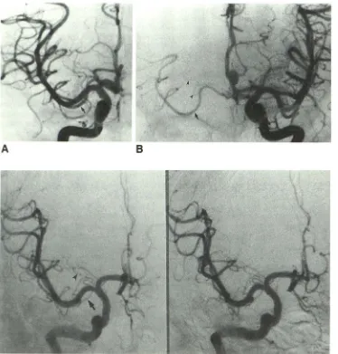
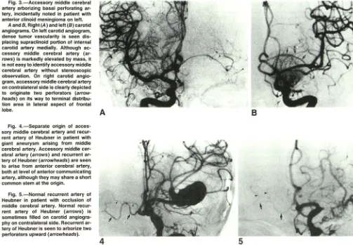
Fig. 1.-Accessory middle cerebral artery arborizing basal perforating ar-tery.
A, Right carotid angiogram shows accessory middle cerebral artery
(ar-row), which is superimposed on hori-zontal portions of anterior and middle cerebral arteries. Whether or not ac-cessory middle cerebral artery arbor-izes basal perforators could not be de-termined from this view.
B, Left carotid angiogram shows
ac-cessory middle cerebral artery (arrow) on opposite side. Anomalous vessel is clearly shown to give rise to basal
per-forator (arrowheads) at anterior perfo-rated substance and to extend to insula and then to lateral surface of frontal lobe on right side.
[image:2.612.53.427.341.732.2]AJNR:10, MayfJune 1989 ACCESSORY MIDDLE CEREBRAL ARTERY 565
Fig. 3.-Accessory middle cerebral artery arborizing basal perforating ar-tery, incidentally noted in patient with anterior clinoid meningioma on left.
A and 8, Right (A) and left (8) carotid angiograms. On left carotid angiogram, dense tumor vascularity is seen dis-placing supraclinoid portion of internal carotid artery medially. Although ac-cessory middle cerebral artery (ar-rows) is markedly elevated by mass, it is not easy to identify accessory middle cerebral artery without stereoscopic observation. On right carotid angio-gram, accessory middle cerebral artery on contralateral side is clearly depicted to originate two perforators (arrow-heads) on its way to terminal distribu-tion area in lateral aspect of frontal lobe.
A
B
Fig. 4.-Separate origin of acces-sory middle cerebral artery and recur-rent artery of Heubner in patient with giant aneurysm arising from middle cerebral artery. Accessory middle cer-ebral artery (arrows) and recurrent ar-tery of Heubner (arrowheads) are seen to arise from anterior cerebral artery, both at level of anterior communicating artery, although they may share a short common stem at the origin.
Fig. 5.-Normal recurrent artery of Heubner in patient with occlusion of middle cerebral artery. Normal recur-rent artery of Heubner (arrows) is sometimes filled on carotid angiogra-phy on contralateral side. Recurrent ar-tery of Heubner is seen to arborize two perforators upward (arrowheads).
4
perforating arteries arose only from the MCA proper or also from the accessory MCA. One accessory MCA was seen in a patient with an anterior clinoid meningioma that displaced the anomalous vessel upward (Fig. 3). The RAH was seen to arise independently from the ACA on the same side as the accessory MCA in two cases (Fig. 4), although the possibility was not ruled out that they might share a short common stem in the region of origin from the ACA trunk.
Our results on perforating arteries are given in Table 1 along with the cases found in the literature. In all, approxi-mately 44% of accessory MCAs arborize perforating arteries, while 27% do not have perforating arteries. Independent RAHs are simultaneously present with the accessory MCAs in approximately 13% of cases.
Discussion
Although it is quite unnatural in a hemodynamic sense to detour to the terminal distribution area, nearly all accessory MCAs follow very constant courses, originating from the ACA, usually at the level of the anterior communicating artery (like the RAH); passing laterally through the cistern in the region of the anterior perforated substance; and finally supplying the insula, lateral orbital surface, and lateral aspect of the frontal lobe. This constancy suggests a certain common
develop-.,..
mental basis for the various forms of this anomalous artery. In fact, a striking similarity is seen between the RAH and the proximal part of the accessory MCA with the perforators that branch from it; this resemblance prompted us to undertake this study (compare Figs. 1 and 5). Tran-Dinh [16] also noted the morphologic similarity between them and is the only author to support the idea of Handa et al. [ 4] that the accessory MCA represents a variant of the RAH.
Phylogenetically, the RAH represents the one remnant of the anastomotic channels over the tuberculum olfactorium, between the ACA and the original stem of the MCA, as seen in marsupials and ungulates [17]. Because an enlarging neo-cortex in primates causes pyriform branches of the MCA to grow outward, the medial half of this anastomotic channel enlarges and the ACA supplies the anteromedial striate branches, while the communication with the MCA eventually disappears. Thus, the ACA eventually supplies regions once fed by the MCA via the enlarged anastomotic channel of the RAH (Fig. 6) [18].
[image:3.612.50.553.78.427.2]plana-566 TAKAHASHI ET AL. AJNR:10, MayfJune 1989
TABLE 1: Accessory Middle Cerebral Artery (MCA) and Its Perforator
Reference Accessory
MCA
Crompton [2] (1962) 1
Jain [3] (1964) 8
Handa et al. [ 4] (1968) 2
Krayenbuhl and Yasargil [12] (1968) 1
Handa et al. [5] (1970) 2
Teal et al. [1] (1973) 3
Watanabe and Togo [9] (1974) 4
Ito et al. [7] (1975) 6
Yasargil and Smith [8] (1976) 1
Waga et al. [13] (1977) 1
Ito et al. [14] (1981) 1
Umansky et al. [11] (1984) 2
Marinkovic et al. [15] (1986) 3
Tran-Dinh [16] (1986) 3
This study 14
Total 52
Note.-RAH = recurrent artery of Heubner.
Anastomotic channel between ACA and MCA
Striate branches
Pyriform branches of MCA
A
8
Present
0 1 2 0 2 0 2 1 0 0 1 2 3 3 6
23 (44)
No.(%)
Perforator Independent
RAH Absent
0 7 0 0 0 0 2 2 0 0 0 0 0 0 3
14 (27)
Unclear
1 0 0 1 0 3 0 3 1 1 0 0 0 0 5
15 (29)
Dwindled portion of the anastomotic channel
MSA
RAH
0 1 0 0 0 0 0 2 1 0 0 1 0 0 2
7 (13)
Fig. 6.-Phylogenetic development of the recurrent artery of Heubner (RAH). (Reprinted from [18], with permission.)
A, Basal surface of marsupial forebrain. Note anastomotic channels between anterior cerebral artery (ACA) and original stem of middle cerebral artery (MCA), one of which is predecessor of recurrent artery of Heubner (RAH).
B, Basal surface of primate forebrain. Continuation of anterior cerebral artery channel enlarges while that of middle cerebral artery eventually disappears,
thus completing formation of recurrent artery of Heubner.
OB
=
olfactory bulb; OC=
optic chiasm; OT=
olfactory tubercle; ICA=
internal carotid artery; MSA=
medial striate arteries; LSA=
lateral striate arteries; PCA=
posterior cerebral artery.tion that the RAH represents one remnant of some anasto-motic channels (Fig. 7).
[image:4.615.57.563.101.304.2]Duplication and absence of the RAH have been described
[20-22]. Gomes et al. [23] reported the presence of a double
RAH in 12% and its absence in only 3% of cases. In the study
[image:4.615.51.562.329.618.2]AJNR:10, MayfJune 1989 ACCESSORY MIDDLE CEREBRAL ARTERY
567
or triplication of the RAH may represent persistence of the anastomotic channels over the tuberculum olfactorium, not inconsistent with the phylogenetic explanation. From this point of view, existence of an independent RAH on the same side as the accessory MCA (13% in our series) would not
Olf.Tr
.
Temp
.
L.
Fig. 7.-Perforation sites of basal perforating arteries in anterior perfo·
rated substance. (Adapted from [19], with permission.) Medial striate
arteries, from horizontal (A 1) portion of anterior cerebral artery, enter brain
through posteromedial part and lateral striate arteries, from sphenoidal
(M1) portion of middle cerebral artery, enter brain through posterolateral
part of anterior perforated substance; branches of recurrent artery of
Heubner enter full mediolateral extent but are confined predominantly to
anterior half of anterior perforated substance. A 1 = perforation site of
perforators from A 1 portion of anterior cerebral artery; GR = gyrus rectus;
IHF = interhemispheric fissure; Ll = limen insulae; LOS = lateral olfactory
stria; M 1 = perforation site of perforators from M 1 portion of middle
cerebral artery; MOS = medial olfactory stria; Olf. Tr. =olfactory tract; ON
= optic nerve; O.T. = optic tract; POL = posterior orbital lobule; RAH =
perforation site of perforators from recurrent artery of Heubner; Temp. L.
= temporal lobe.
A
ACAE xtracerebral anastomosis
between RAH and LSA
_._ ...
R..,A._Hi..._
..
~
.
...
·
·
·
.. ·
·
·
.
l.s,q
ICA
refute the idea that they share a developmental origin. This
idea is also supported by the fact that most patients with an
accessory MCA do not have an RAH, since cadaveric hemi
-spheres with no RAH are quite rare [15, 23]. The simultaneous
presence of the RAH and the accessory MCA on one side
may in fact represent the persistence of two anastomoses,
one as the RAH and the other as the accessory MCA; this
situation is similar to that of duplication of the RAH.
The RAH has frequently been described to arborize
fron-toorbital and olfactory branches almost consistently [15, 20,
22-25]; for example, in the microsurgical study of Gomes et
al. [24], an olfactory branch of the RAH was found in 91% of
60 hemispheres. The latter vessel may represent the remnant of the original stem of the MCA in the phylogenesis.
Further-more, Kaplan and Ford [24] suggested that extracerebral
anastomoses exist between the RAH and lateral striate
arter-ies. They were observed by Gomes et al. [23] in 9% of
postmortem brains, which also supports the phylogenetic
explanation.
The lateral striate arteries that arise from the MCA originate
either from the proximal stem or from one of the secondary
vessels into which the proximal stem divides (Fig. 8A). Whether or not some lateral striate arteries arise from the
secondary branches depends on how soon the major stem becomes subdivided [24]. In one-half of the hemispheres in
the series of Rosner et al. [19], some lateral striate arteries
arose from the secondary branches. When this occurs it is usually from the most rostrally placed branch [24].
If an extracerebral anastomosis remains between the RAH and lateral striate artery, which arises from the secondary
(rostral) branch of the MCA, and the communication between the secondary branch and the major stem of the MCA dis-appears, the accessory MCA will be formed and give rise to
ACA
Ace. MCA with basal perforators
ICA
B
Fig. B.-Formation of accessory middle cerebral artery (Ace. MCA) by persistence of one of the anastomotic channels between anterior (ACA) and middle (MCA) cerebral arteries over tuberculum olfactorium.
A, Normal recurrent artery of Heubner (RAH). Some lateral striate arteries (LSA) frequently arise from one of the secondary branches of the middle
cerebral artery. When this occurs, it is usually from the most rostrally placed branch. An extracerebral anastomosis occasionally may be present between
recurrent artery of Heubner and lateral striate artery.
B, Accessory middle cerebral artery with basal perforators. If extracerebral anastomosis remains between recurrent artery of Heubner and lateral striate artery, which arises from secondary (rostral) branch of middle cerebral artery, and communication between rostral branch and major stem of middle
cerebral artery subsequently disappears, accessory middle cerebral artery will be formed that gives rise to perforators and terminates in frontal part of
[image:5.612.52.296.148.312.2] [image:5.612.56.560.458.637.2]568 TAKAHASHI ET AL. AJNR:10, MayfJune 1989
Anastomotic channels over tuberculum olfactorium
//\~
RAH with Ace. MCA r--A-e-c-. M-C-A--,
RAH extracerebral with without
anastomoses perforators perforators
'---r--' ~
Normal Variation
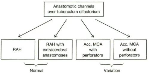
Fig. 9.-Various descendants of anastomotic channels over tuberculum olfactorium between anterior and middle cerebral arteries. RAH
=
recurrentartery of Heubner; Ace. MCA
=
accessory middle cerebral artery.perforators and then terminate in the frontal part of the MCA distribution (Fig. 88). This idea is consistent with the fact that approximately 44% of accessory MCAs have been found to have perforators; but it is inconsistent with the significant number of anomalous vessels not arborizing any perforators
(27%). In this regard, the anomalous vessel cannot be consid-ered just a variant of the RAH itself, despite the striking similarity, because the RAH by definition should have basal
perforators. This question could be resolved only if there were
some channels without perforators among those anasto-moses. Thus, the accessory MCA with or without perforating
arteries could be considered a persistent form of various descendants of anastomotic channels between the ACA and MCA over the tuberculum olfactorium, one of which is also a normal predecessor of the RAH (Fig. 9).
REFERENCES
1. Teal JS, Rumbaugh CL, Bergeron RT, Segall HD. Anomalies of the middle cerebral artery: accessory artery, duplication, and early bifurcation. AJR
1973;118:567-575
2. Crompton MR. The pathology of ruptured middle-cerebral aneurysms with
special reference to the differences between the sexes. Lancet 1962;2:
421-425
3. Jain KK. Some observations on the anatomy of the middle cerebral artery. Can J Surg 1964;7:134-139
4. Handa J, Seta K, Honda H. Die akzessorische A. cerebri media. ROFO
1968;1 08:539-541
5. Handa J, Shimizu Y, Matsuda M, Honda H. Accessory middle cerebral
artery. Report of further two cases. Clin Radio/1970;21 :415-416
6. Stabler J. Two cases of accessory middle cerebral artery, including one with an aneurysm at its origin. Br J Radio/1970;43:314-318
7. Ito J, Sa to T, Arai H, Honda H. Accessory middle cerebral artery and duplication of the middle cerebral artery. Rinsho Hoshasen 1975;20: 449-457
8. Yasargil MG, Smith RD. Association of middle cerebral artery anomalies
with saccular aneurysms and moyamoya disease. Surg Neurol 1976;6:
39-43
9. Watanabe T, Togo M. Accessory middle cerebral artery. Report of four cases. J Neurosurg 1974;41 :248-251
10. Miyazaki Y, Tsuruta J. Clinical studies on the congenital anomalies of the intracranial arteries associated with cerebral aneurysm. Hokkaido lgaku Zasshi 1977;52: 111-123 (Jpn).
11. Umansky F, Juarez SM, Dujovny M, et al. Microsurgical anatomy of the proximal segments of the middle cerebral artery. J Neurosurg 1984;
61:458-467
12. Krayenbi.ihl HA, Yasargil MG. Cerebral angiography, 2nd ed. London:
Butterworth, 1968: 58-60
13. Waga S, Kojima T, Morooka Y, Sakakura M. Aneurysm of the accessory
middle cerebral artery. Surg Neuro/1977;8:359-360
14. Ito J, Washiyama K, Hong Kim C, lbuchi Y. Fenestration of the anterior cerebral artery.' Neuroradiology 1981;21 :277-280
15. Marinkovic S, Milisavljevic M, Kovacevic M. Anatomical bases for surgical approach to the initial segment of the anterior cerebral artery. Microana-tomy of Heubner's artery and perforating branches of the anterior cerebral artery. Surg Radio/ Anat 1986;8:7-18
16. Tran-Dinh H. The accessory middle cerebral artery-a variant of the recurrent artery of Heubner (A. centralis longa)? Acta Anat (Basel)
1986;126: 167-171
17. Abbie AA. The morphology of the fore-brain arteries, with especial refer-ence to the evolution of the basal ganglia. J Anat 1934;68:433-470 18. Takahashi S, Goto K, Fukasawa H, et al. Computed tomography of cerebral
infarction along the distribution of the basal perforating arteries. Part 1:
Striate arterial group. Radiology 1985;155: 107-118
19. Rosner SS, Rhoton AL, Ono M, Barry M. Microsurgical anatomy of the anterior perforating arteries. J Neurosurg 1984;61 :468-485
20. Ahmed DS, Ahmed RH. The recurrent branch of the anterior cerebral artery. Anat Rec 1967;157:699-700
21. Leeds NE. The striate (lenticulostriate) arteries and the artery of Heubner. In: Newton TH, Potts DG, eds. Radiology of the skull and brain, vol. 2. St. Louis: Mosby, 1974:1527-1539
22. Perlmutter D, Rhoton AL. Microsurgical anatomy of the anterior
cerebral-anterior communicating-recurrent artery complex. J Neurosurg 1976;
45:259-272
23. Gomes F, Dujovny M, Umansky F, et al. Microsurgical anatomy of the recurrent artery of Heubner. J Neurosurg 1984;60: 130-139
24. Kaplan HA, Ford DH. The brain vascular system. Amsterdam: Elsevier,
1966:41-63
25. Kribs M, Kleihues P. The recurrent artery of Heubner. A morphological
study of the blood supply of the rostral basal ganglia in normal and pathological conditions. In: Zulch· KJ, ed. Cerebral circulation and stroke.
[image:6.612.51.300.79.201.2]