Characterization of Intracranial Berry Aneurysms
J. K. A. Hope, J. L. Wilson, and F. J. Thomson
PURPOSE: To assess the accuracy of three-dimensional CT angiography (CTA) in the detection and characterization of intracranial aneurysms and to help determine its role as a screening test for aneurysms in the asymptomatic population and as an adjunct to angiography in subarachnoid hemorrhages and in the follow-up of untreated aneurysms. METHODS: In a blinded, prospective study, the 3-D CTA studies in 80 patients with symptomatic aneurysms were analyzed for the presence and morphology of aneurysms. Angiography or surgery acted as the control. RESULTS: Ninety-four aneurysms were found in 63 patients. Negative findings at angiography were noted in 17. Sensitivity and specificity of 3-D CTA for all aneurysms, all patients, and aneurysms 5 mm or smaller were 90.4% and 50%, 98.4% and 82.4%, and 78.8% and 51.9%, respectively. CONCLU-SION: Three-dimensional CTA may have a role in noninvasive screening for asymptomatic aneurysms in the general population, but caution is advocated when data obtained from symp-tomatic patients are extrapolated to the asympsymp-tomatic population who harbor smaller aneurysms. Also, 3-D CTA may be useful as an adjunct to angiography in the characterization of berry aneurysms and in the follow-up of untreated aneurysms.
Index terms: Aneurysm, computed tomography; Aneurysm, intracranial; Computed tomography, three-dimensional
AJNR Am J Neuroradiol17:439 –445, March 1996
The high morbidity and mortality of sub-arachnoid hemorrhages (SAH) caused by rup-ture of berry aneurysms (1, 2) and the low sur-gical risk associated with elective clipping of these aneurysms (1, 3–5) make it desirable to have a screening test to identify berry aneu-rysms before they rupture in “at-risk” groups. At-risk groups include, among others, patients with adult polycystic kidney disease and family groups (6 – 8), as well as patients with a clinical suspicion for aneurysmal rupture but with neg-ative computed tomography (CT) findings and negative results from cerebrospinal fluid sampling.
Catheter angiography is the most accurate means of identifying and excluding intracranial
berry aneurysms in these groups; however, it carries a small but defined morbidity that in-cludes stroke (6, 9 –11). This morbidity and the logistics associated with the performance of catheter angiography have led to the search for alternative techniques that include high-res-olution CT with thin axial sections (12–14), magnetic resonance (MR) angiography (15), and three-dimensional CT angiography (CTA) (16).
The purpose of this study was to determine the accuracy of 3-D CTA as a screening method for the groups at risk. This accuracy was tested by using 3-D CTA studies performed as part of the routine workup of patients presenting to our institution with subarachnoid hemorrhages or other features suggesting a berry aneurysm. This is a valuable group to assess because the occurrence rate of aneurysms is high in this group and because all of these patients have undergone angiography or surgery, which can be used for correlation.
A second purpose of the study was to assess the role of 3-D CTA as an adjunct to
angiogra-Received April 28, 1995; accepted after revision September 21. From the Department of Neuroradiology, Auckland (New Zealand) Hospital.
Address reprint requests to Dr J. K. A. Hope, Department of Neurora-diology, Auckland Hospital, Park Road, Auckland, New Zealand.
AJNR 17:439–445, Mar 1996 0195-6108/96/1703–0439
qAmerican Society of Neuroradiology
phy in the investigation of berry aneurysms and to examine its role in the follow-up of incidental aneurysms.
Subjects and Methods
Image Generation
We used the Somatom DRH (Siemens AG, Erlangen) CT scanner and a fast-scan protocol (310 mAs, 125 kV, 7 section acquisitions per minute) to obtain sections of 253 2 mm with a 1-mm overlap beginning 40 seconds after the start of a continuous intravenous infusion of 120 mL of iohexol (1 mL/s). The scan plane was parallel to the roof of the sella turcica, with the lowest section 1 mm below the floor of the sella. Image reconstruction was delayed until acquisition was completed; then the first and last images were reconstructed and additional scans were obtained as necessary. The field of view was centered on the sella and was coned to exclude the skull vault and to include the circle of Willis and the principle branches out to the prox-imal A2, M2, and P2 segments. This imaging volume nor-mally excludes the proximal vertebrobasilar arteries and the distal portions of the anterior and middle cerebral ar-teries. Reconstructed images were transferred via Ethernet to a workstation, where a trilinear interpolation algorithm produced an isotropic data set of 60 sections (voxel size, 0.4 mm linear). This procedure took 2.5 minutes.
By using this data set, we produced multiplanar recon-structions in the coronal, axial, and sagittal planes (30 seconds per plane). These reconstructions were viewed as a movie sequence (window, 200 Hounsfield units [HU]; center, 100 HU).
Three-dimensional CTA images were reconstructed at 68 increments by using the techniques of maximum-intensity projection (11 minutes) and surface-image re-construction (8 minutes). The result was a movie se-quence of 60 images per rotation about both horizontal and vertical axes. (A simple bone removal program was written to edit out unwanted bone detail from the CT image files before the reconstruction of the maximum-intensity projection images; this required another 4 minutes of re-construction time.) The images obtained were then trans-ferred to video tape and analyzed. The acronym3-D CTA refers here to both maximum-intensity projection and sur-face-rendered 3-D reconstructions, as well as to the mul-tiplanar reconstruction images.
Patient Selection
Subjects who came to our unit for investigation of a suspected berry aneurysm were eligible for inclusion in the study if they had undergone both catheter angiography and 3-D CTA during the same hospital stay and before surgery on any aneurysm that might have been discov-ered.
Catheter angiograms and 3-D CTA were obtained and interpreted prospectively and independently by two inves-tigators; each investigator was blinded to the results of the
other arm of the study. Patients were excluded if blinding was compromised.
The following information was collected on each pa-tient: age, sex, diagnosis, and time from presentation to 3-D CTA and catheter angiography. The 3-D CTA and catheter angiogram were examined separately in a blinded fashion for the presence and location of any aneurysm; if an aneurysm was found, its maximum lumen size, neck size, orientation, and vascular relationships were noted. Investigators also looked for mural calcification and intra-aneurysmal thrombus.
Results
From October 1992 through April 1994, 145 patients underwent catheter angiography as part of the investigation of a subarachnoid hem-orrhage and/or a suspected intracranial berry aneurysm. Eighty-eight of these patients also underwent 3-D CTA (57 did not undergo this procedure because of restlessness or schedul-ing difficulties). Of the 88, 4 were subsequently excluded because of loss of blinding and an-other 4 were excluded because of movement degradation of the images. Thus, 80 patients were eligible for analysis (3-D CTA was per-formed within 48 hours of catheter angiography in 65 of the 80).
Of the 80 patients studied, 54 were women and 26 were men. Age was accurately docu-mented in 74 of these 80 patients (mean age, 49.2 years; range, 23 to 75 years).
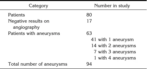
[image:2.612.316.555.100.212.2]Ninety-four aneurysms were identified in 63 patients either at angiography or at surgery (Table 1). Of the 62 symptomatic aneurysms, 3-D CTA correctly identified 60 (96.8%). Of the 32 incidental aneurysms, 3-D CTA correctly identified 25 (78.1%). Three-dimensional CTA correctly predicted a negative result in 14 of the 17 patients with normal results on angiography. There were 9 false-negative 3-D CTA results and 14 false-positive 3-D CTA results (Table 2).
TABLE 1: Angiographic and surgical findings
Category Number in study
Patients 80
Negative results on angiography
17
Patients with aneurysms 63
41 with 1 aneurysm 14 with 2 aneurysms
Using catheter angiography (or surgical mea-surement in one patient) as the reference
stan-dard, 3-D CTA (a) correctly identified 16
(100%) of 16 aneurysms larger than 11 mm, 43 (95.6%) of 45 aneurysms measuring between 6 and 10 mm, 19 (86.4%) of 22 aneurysms mea-suring between 4 and 5 mm, and 7 (63.6%) of 11 aneurysms smaller than 3 mm; (b) correctly showed maximum aneurysm size to within 1 mm in 81% and to within 2 mm in 90.5% (infor-mation available in 71 patients); and (c) re-vealed aneurysm neck size to within 2 mm in 95% (information available in 42 patients).
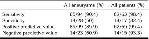
Sensitivity, specificity, positive predictive value, and negative predictive value for all an-eurysms are listed in Table 3. For 3-D CTA to be an effective screening test for aneurysms in the
general population, it must correctly give a neg-ative result for people without aneurysms and correctly identify at least one aneurysm in those with solitary or multiple aneurysms. In our 80 patients, 3-D CTA had a sensitivity of 98.4% (62/63), a specificity of 82.4% (14/17), a pos-itive predictive value of 95.4% (62/65), and a negative predictive value of 93.3% (14/15) (Table 3).
Discussion
Accuracy of 3-D CTA in Aneurysm Detection
The sensitivity of 3-D CTA for all aneurysms was 90.4% (85/94). This compares favorably with the 82% recorded for CT infusion scanning (14), but it appears no better than the 90.8% recorded when reformations are also performed (13). This suggests that 3-D CTA offers no ad-ditional advantage over the latter technique; certainly, for symptomatic aneurysms in these series, this is true (96.8% vs 98.5%). For inci-dental aneurysms, however, 3-D CTA is more accurate (78.1% vs 67%); despite the longer reconstruction time, this may justify its use when one is looking for smaller aneurysms.
Some limitations of 3-D CTA in the detection of aneurysms are disclosed by an analysis of the 14 false-positive results and 9 false-negative results (Table 2). These limitations include its susceptibility to motion artifacts (2 false-positive results) and the possibility of confusing high-attenuation blood clot with intravascular contrast material in an aneurysm (2 false-pos-itive results), especially when surface rendered techniques are used.
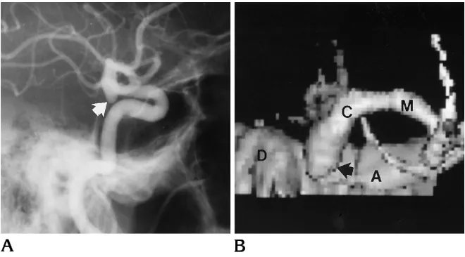
In addition, its inherent low spatial resolution (caused in part by our decision to use a 2-mm section thickness, which gives improved low contrast resolution at the expense of reduced spatial resolution) makes it difficult to (a) dif-ferentiate tight vascular loops from small aneu-rysms (three false-positive results) (Fig 1) and (b) differentiate an infundibulum of the posterior communicating artery from an aneurysm (four false-positive results) (Fig 2). Similar difficulties have been recorded by others (12). The other three false-positive results occurred in the group for whom angiographic results were negative.
[image:3.612.58.295.101.270.2]Thirteen of 14 false-positive findings mea-sured 3 mm or less. The probability of rupture is low for aneurysms of this size (17–20), so this
TABLE 3: Value of 3-D CTA in identifying aneurysms and patients with aneurysms
All aneurysms (%) All patients (%)
Sensitivity 85/94 (90.4) 62/63 (98.4)
Specificity 14/28 (50) 14/17 (82.4)
Positive predictive value 85/99 (85.9) 62/65 (95.4) Negative predictive value 14/23 (60.9) 14/15 (93.3)
[image:3.612.58.298.355.418.2]Note.—CTA indicates CT angiography; sensitivity 5number of true-positive results/(number of true-positive results 1 number of false-negative results); specificity5number of true-negative results/ (number of true-negative results1number of false-positive results); positive predictive value5number of true-positive results/(number of true-positive results1number of false-positive results); and negative predictive value5number of negative results/(number of true-negative results1number of false-negative results).
TABLE 2: Aneurysm location and detection by 3-D CTA
Site and type No. of aneurysms
3-D CTA
T1 F2 F1
Carotid, cavernous 1 . . . 1 . . .
Carotid, ophthalmic 4 4 . . . 1
Carotid, PCo A/A Chor 25 22 3 4
Carotid, Bifurcation 6 6 . . . .
A1 1 . . . 1 1
ACo complex 19 18 1 . . .
A2 and Distal 2 . . . 2 1
M1 2 2 . . . 3
M1/M2 28 28 . . . 4
Basilar tip 4 4 . . . .
Basilar/superior cerebellar 1 1 . . . .
Vertebral/PICA 1 . . . 1 . . .
Total 94 85 9 14
result suggests that the index of suspicion used in analyzing the images may have been too high. However, use of a lower index of suspicion would have led to a higher false-negative rate for the 1 symptomatic and 10 asymptomatic aneurysms smaller than 3 mm.
In the false-negative group (n 5 9), three aneurysms were missed because they were out-side the imaging volume (one aneurysm of the posterior inferior communicating artery and two distal anterior cerebral aneurysms). At present, our technique includes more than 96% of aneu-rysm sites (21–23) but it should be possible with helical scanning to extend the imaging volume and thus reduce this source of error.
Four aneurysms were missed because they were close to the skull base and were difficult to identify against the bony background (Fig 3). The two remaining false-negative results in-cluded one aneurysm in the cavernous portion
of the internal carotid artery and a 2-mm aneu-rysm that projected from the inferior aspect of the A1 segment of the anterior cerebral artery. Finally, size was an important determinant of the six false-negative results within the imaging volume. Four of these measured 4 mm or less. The sensitivity of MR angiography for detec-tion of aneurysms (85.7%) has been tested in a small series (15); that technique holds promise as a screening test for aneurysms. However, a larger number of patients must be studied be-fore firm conclusions can be drawn.
Use of 3D-CTA for Noninvasive Screening of Groups at Risk
The role of 3-D CTA, when it is used as a screening test, is not to identify every aneurysm but to identify every patient with one or more aneurysms (since identification of at least one Fig 2. A, Lateral projection carotid
an-giogram shows infundibulum (arrow) at origin of posterior communicating artery.
B, Corresponding surface-rendered 3-D CTA that identified the infundibulum but misinterpreted it as an aneurysm (ar-row).Aindicates anterior clinoid process; C, distal internal carotid artery;D, dorsum sella; andL, lesser wing of sphenoid.
Fig 1. A, Surface-rendered 3-D CTA of circle of Willis, viewed from above, shows false-positive aneurysms arising from the superior aspect of both internal carotid arteries (arrows). Note the irregu-lar appearance of the left middle cerebral artery and branches, produced by sub-arachnoid blood clot misregistering as ves-sel lumen.
[image:4.612.223.558.85.470.2]indicates the need for four-vessel angiography in that patient). Three-dimensional CTA must also have a low false-positive rate in those who have no aneurysms. The sensitivity, specificity, positive predictive value, and negative predic-tive value of 3-D CTA are better when applied to all patients than when applied to all aneurysms (Table 3). These figures suggest that 3-D CTA has a role as a noninvasive screening test for asymptomatic aneurysms (6); however, there are limitations to extrapolating data derived from our symptomatic patients to patients in at-risk populations who have asymptomatic aneurysms, and these limitations need to be emphasized. For example, asymptomatic rysms are smaller than symptomatic aneu-rysms (17, 18, 24). Any proposed screening test for aneurysms must be able to identify these smaller aneurysms. In our series, 3-D CTA had a sensitivity of 78.8% and a specificity of 51.9% for aneurysms measuring 5 mm or less. These results would yield a net benefit of 1.5 years difference in survival if they were ap-plied to a screened population of patients younger than 25 years, with an occurrence rate of aneurysms exceeding 30% and a surgical complication rate of 1% or less (6). This sensi-tivity and this specificity appear to be less sat-isfactory than those for MR angiography (15), but the mean size of aneurysms in that MR an-giography study (15) was 11.6 mm, and only 6 of the 21 aneurysms studied were 5 mm or smaller.
Other limitations include the possibility of bias introduced by the localizing nature of sub-arachnoid hemorrhage on CT (25) and the po-tential bias introduced by the 57 patients who
did not undergo 3-D CTA. We analyzed both these issues and determined that they had no significant effect on our results.
A final potential limitation of this study is the small number of patients who had negative
find-ings at angiography (n 5 17). We have used
positive predictive value and negative predic-tive value (two important indexes in evaluating screening tests) to describe the accuracy of 3-D CTA in predicting the presence and absence of an aneurysm. Although the number of patients is large enough to give confidence to the posi-tive predicposi-tive values found, a larger number of true negatives (n 5 14) would be required to increase confidence in the negative predictive values obtained.
3-D CTA as a Supplement to Angiography
Despite the sensitivity of 3-D CTA and CT infusion techniques in the detection of acutely ruptured aneurysms (13), these methods are unlikely at present to replace catheter angiog-raphy in the primary workup of subaranchoid hemorrhages, primarily because of their poor spatial resolution, which limits the definition of surgically important vessels such as the anterior choroidal artery. However, 3-D CTA may re-place angiography in patients with intracerebral hematomas and rapidly declining neurologic function (14), and it may be able to screen for aneurysms in patients with oculomotor palsy (12).
An important role of 3-D CTA is as an adjunct to angiography, because it can provide surgi-cally important information about the shape and direction of an aneurysm and its relation to Fig 3. A, Lateral projection carotid an-giogram shows a small posterior commu-nicating artery aneurysm (arrow) directed inferiorly from the internal carotid artery.
[image:5.612.57.387.86.269.2]adjacent vessels (16) (Fig 4). In this setting, maximum-intensity projection is usually prefer-able because surface-rendered 3-D CTA uses a threshold of 70 to 100 HU that may not allow
the differentiation of clotted blood from
contrast-enhanced aneurysm lumen (two false-positive results in our study). Maximum-inten-sity projection images are not threshold-depen-dent, and this characteristic may be useful in larger aneurysms to display the relationship be-tween intraluminal thrombus and the aneurysm
neck or lumen and to identify mural calcifica-tion and its relacalcifica-tionship to the neck (Fig 5).
Follow-up of Incidental Aneurysms
Incidental aneurysms are now commonly found because of the widespread availability of CT and MR (8). In the absence of surgery, follow-up is desirable because such aneurysms tend to grow (26). This ordinarily requires an-giography; however, the abilities of 3-D CTA to Fig 5. A, Anterioposterior projection, left carotid angiogram shows posterior communicating artery aneurysm (arrow) directed laterally from origin. Note absence of calcification.
B, Maximum-intensity projection 3-D CTA, coned to left side of circle of Willis and viewed from above, shows relationship of aneurysm lumen and calcification in aneurysm wall (black and white solid arrows) to neck of aneurysm (open arrow). The cap of thrombus within the partially thrombosed aneurysm is also visible (arrowheads).Aindicates anterior clinoid process;c, internal carotid artery;D, left side of dorsum sella;m, middle cerebral artery; anda, anterior cerebral artery.
C, Surface-rendered 3-D CTA (shallow Townes projection) of the same aneurysm (arrow) shows the limitations imposed by the inability to differentiate calcification from opacified blood in aneurysm lumen. The thrombus in the aneurysm is not seen because its density falls below the prescribed threshold.
Fig 4. A, Steep Townes projection from vertebral angiogram shows basilar tip aneurysm with loculus directed to the left.
[image:6.612.58.559.86.267.2] [image:6.612.59.556.314.495.2]portray aneurysm morphology accurately (16) and to display aneurysm size and neck size to within 2 mm (90.5% and 95%, respectively), coupled with a low risk of contrast-related com-plications (27–29), suggest it can replace arte-riographic follow-up in these patients. It has one additional advantage over MR imaging: 3-D CTA allows follow-up of incidental aneurysms in patients who have previously undergone clip-ping of a symptomatic aneurysm.
References
1. Phillips LH II, Whishnani JP, O’Fallan WM, et al. The unchanging pattern of subarachnoid haemorrhage in a community.Neurology 1980;30:1034 –1040
2. Drake CG. Management of cerebral aneurysm.Stroke1981;12: 273–283
3. Jain KK. Surgery of intact intracranial aneurysms.J Neurosurg 1974;40:494 – 498
4. Salazar JL. Surgical treatment of asymptomatic and incidental intracranial aneurysms.J Neurosurg1980;53:20 –21
5. Samson DS, Hodosh RM, Kemp Clark W. Surgical management of unruptured asymptomatic aneurysms.J Neurosurg1977;46:731– 734
6. Levey AS, Pauker SG, Kassirer JP. Occult intracranial aneurysms in polycystic kidney disease: when is cerebral arteriography indi-cated?N Engl J Med1983;308:986 –994
7. Chyatte D. The epidemiology, genetics and clinical behavior of intracranial aneurysms. In: Awad IA, ed.Current Management of Cerebral Aneurysms. New York, NY: McGraw Hill, 1993:1–20 8. Leblanc R. Unruptured aneurysms, multiple aneurysms, familial
aneurysms, and aneurysms in pregnancy. In: Awad IA, ed. Cur-rent Management of Cerebral Aneurysms. New York, NY: McGraw Hill, 1993;277–295
9. Mani RL, Eisenberg RL, McDonald EJ Jr, et al. Complications of catheter cerebral angiography: analysis of 5000 procedures, parts I–III.AJR Am J Roentgenol1978;131:861– 874
10. Peret G, Nishioka H. Report on the Cooperative Study of Intracra-nial Aneurysms and Subarachnoid Haemorrhage, section IV: ce-rebral angiography and the analysis of the diagnostic value and complications of carotid and vertebral angiography in 5484 pa-tients.J Neurosurg1966;25:98 –114
11. Grzyska U, Freitag J, Zeumer H. Selective cerebral intraarterial DSA: complication rate and control of risk factors. Neuroradiol-ogy1990;32:296 –299
12. Teasdale E, Statham P, Straiton J, et al. Non-invasive radiological investigation for oculomotor palsy.J Neurol Neurosurg Psychiatry 1990;53:549 –553
13. Schmid UD, Steiger HJ, Huber P. Accuracy of high resolution computed tomography in direct diagnosis of cerebral aneurysms. Neuroradiology1987;29:152–159
14. Newell DW, LeRoux PD, Dacey RG, et al. CT infusion scanning for the detection of cerebral aneurysms.J Neurosurg1989;71:175– 179
15. Ross JS, Masaryk TJ, Modic MT, et al. Intracranial aneurysms: evaluation by MR angiography.AJNR Am J Neuroradiol1990;11: 449 – 450
16. Aoki S, Sasaki Y, Machida T, et al. Cerebral aneurysms: detection and delineation using 3-D CT angiography.AJNR AM J Neurora-diol1992;12:1115–1120
17. de la Monte SM, Moore GW, Monk MA, et al. Risk factors for the development and rupture of intracranial berry aneurysms.Am J Med1985;78:957–964
18. McCormick WF, Acosta-Rua GJ. The size of intracranial saccular aneurysms: an autopsy study.J Neurosurg1970;33:422– 427 19. Crompton MR. Mechanism of growth and rupture in cerebral berry
aneurysms.Br Med J1966;1:1138 –1142
20. Freytag E. Fatal rupture of intracranial aneurysms: survey of 250 medico legal cases.Arch Pathol1966;81:418 – 424
21. Newton TH, Potts DG.Radiology of the Skull and Brain, vol 2, book 4:Angiography: Specific Disease Processes. St Louis, Mo: CV Mosby, 1974:2449
22. Yasargil MG.Microneurosurgery. New York, NY: Georg Thieme Verlag, 1984; 1:299
23. Marks PV, Hope JKA, Cluroe AD, et al. Racial differences between Maori and European New Zealanders in aneurysmal subarachnoid haemorrhage.Br J Neurosurg1993;7:175–182
24. Chason JL, Hindman WM. Berry aneurysms of the circle of Willis: results of a planned autopsy study.Neurology1958;9:41– 44 25. Drake CG, Girin JP. The surgical treatment of subarachnoid
hem-orrhage with multiple aneurysms. In: Morlet TP, ed.Current Con-troversies in Neurosurgery. Philadelphia, Pa: WB Saunders, 1976: 274 –278
26. Allcock JM, Canham PB. Angiographic study of the growth of intracranial aneurysms.J Neurosurg1976;45:617– 621 27. Palmer FJ. The RACR Survey of Intravenous Contrast Media
Re-actions Final Report.Australas Radiol1988;32:426 – 428 28. Katayama H, Yamaguchi K, Kozuka T, et al. Adverse reactions to
ionic and nonionic contrast media: a report from the Japanese Committee on the Safety of Contrast Media.Radiology1990;175: 621– 628