http://www.scirp.org/journal/ojog ISSN Online: 2160-8806
ISSN Print: 2160-8792
Comparative Study between Endometrial
Resection and Electrocoagulation in Patients
with Abnormal Uterine Bleeding
Leonardo Vieira Elias
1*, Daniel Spadoto-Dias
1, Nilton José Leite
1, Flávia Neves Bueloni-Dias
1,
Gustavo Filipov Peres
1, Carlos Roberto Padovani
2, Rogério Dias
1,3#1Department of Gynecology and Obstetrics, Botucatu Medical School, São Paulo State University—UNESP, São Paulo, Brazil 2Department of Biostatistics, Botucatu Institute of Biosciences, São Paulo State University—UNESP, São Paulo, Brazil 3The Gynecological Endoscopy and Family Planning Sector, Botucatu Medical School Hospital, São Paulo State
University—UNESP and Principal Investigator, São Paulo, Brazil
Abstract
Objective: To compare clinical outcomes between two first-generation en-dometrial ablation techniques. Design: Prospective comparative coorte. Setting: Tertiary public hospital, university teaching center. Seventy-three patients with abnormal uterine bleeding unresponsive to clinical treatment submitted to endometrial ablation from October 2011 to September 2013. Methods and Main Outcome Measures: Patients were assigned to either monopolar U-shaped electrode resection with rollerball electrocoagulation (group A, n = 36) or rollerball electrocoagulation alone (group B, n = 37). Mean follow-up length was 359 (280 - 751) and 370 days (305 - 766) in groups A and B, respectively. Bleeding pattern, associated symptoms, fail-ure/success rates were assessed 30, 90, 180 and 360 days post-procedure. Findings: Patient characteristics were similar in both groups (P ≥ 0.05). Surgery duration (mean of 48.5 [±12.0] vs. 31.9 [±5.6] min, P < 0.001) and medium distention use (5.700 mL vs. 3.500 mL, P < 0.01) were decreased in group B. Post-ablation clinical improvement was considerable in both groups. Vaginal discharge incidence after the procedure was lower in group B (30.5% vs. 8.1%, P < 0.05). Hysterectomy rate was 9.6%. Overall success rate was 86.1% and 88.1% in groups A and B, respectively. Conclusions: Endometrial ablation using rollerball electrocoagulation alone may be con-sidered safer than resection with rollerball electrocoagulation, which re-quires shorter surgical time and less distention medium, and is associated with lower postoperative vaginal discharge incidence. Success rate did not
#Adjunct Professor III and Principal Investigator. How to cite this paper: Elias, L.V.,
Spado-to-Dias, D., Leite, N.J., Bueloni-Dias, F.N., Peres, G.F., Padovani, C.R. and Dias, R. (2017) Comparative Study between Endo-metrial Resection and Electrocoagulation in Patients with Abnormal Uterine Bleeding. Open Journal of Obstetrics and Gynecology, 7, 312-325.
https://doi.org/10.4236/ojog.2017.73033
Received: February 4, 2017 Accepted: March 19, 2017 Published: March 22, 2017
Copyright © 2017 by authors and Scientific Research Publishing Inc. This work is licensed under the Creative Commons Attribution International License (CC BY 4.0).
http://creativecommons.org/licenses/by/4.0/
Open Access
statistically differ between groups, but study parameters in absolute values and percents were superior in group B.
Keywords
Uterine Bleeding, Hysteroscopy, Surgical Procedures, Endometrial Ablation Techniques, Patient Satisfaction
1. Introduction
Abnormal uterine bleeding (AUB) is defined as any deviation from the normal menstrual cycle pattern including changes in the frequency, duration or volume of blood flow [1] [2]. Excessive menstrual blood loss, which can be accompanied by other symptoms, very often interferes with a woman’s physical and mental well-being bringing limitations to daily life activities and changes in social beha-vior that might reduce health-related quality of life—HRQOL [3] [4] [5].
AUB affects one woman in five, and most commonly occurs around menarche and menopause [3] [5] [6]. In the general population, AUB prevalence is esti-mated at 11% - 13% and increases with age, reaching 24% in women aged 36 - 40 years [6] [7]. The management for AUB has been widely investigated over the past years with the goals of stopping bleeding or promoting regular menstrual cycles with adequate flow volume, taking into account the patient’s desire for future fertility [1]. Initial medical therapy includes the use of non-steroidal anti- inflammatory drugs (NSAIDs), antifibrinolytic drugs, combination oral contra-ceptives (COC) or oral progestins, levonorgestrel intrauterine system (LNG- IUS), and hormone replacement therapy during climacterium [6] [8]. However, when medications are not effective, surgical treatment might be indicated.
In the past, the first option was curettage, which has a reasonable diagnostic performance, but is of little therapeutic value. The final and definitive treatment is hysterectomy, especially in women without reproductive desire. Over the last two decades, diagnostic tools have significantly advanced and new therapy mod-alities have been developed. These new procedures are less invasive and offer the possibility of preserving the uterus and avoiding the morbidity related to hyste-rectomy, which varies between 5.2% to 9.4%, reaching up to 40% in some series
[9] [10] [11]. One of the alternatives to hysterectomy is endometrial ablation,
which destroys the endometrium using different sources of energy [12].
demon-strated to improve quality of life even among obese women (BMI > 30 kg/m2)
and those with coagulopathies [17]-[22]. Nonetheless, between 9.3% and 13.6% of the patients undergoing endometrial ablation may need retreatment consist-ing from medical therapy or repeat endometrial ablation to hysterectomy, at in-tervals ranging from 1 to 8 years [23] [24] [25] [26].
Although a large number of studies have compared first-generation with second-generation ablation techniques, comparisons between the first-generation procedures endometrial resection and electrocoagulation using rollerball alone were performed by a limited number of studies [13] [27] [28] [29] [30]. The type of electrical current used in the procedure may interfere with surgical outcome as the currents used in endometrial resection and rollerball coagulation differ in maximum peak tension and modulation [31]. Thus, this study consisted of a prospective comparative analysis between clinical outcomes of two different first-generation techniques in AUB patients unresponsive to medical therapy.
2. Material and Methods
This prospective analytic study included AUB patients who underwent endome-trial ablation at the Gynecological Endoscopy and Family Planning Sector of Botucatu Medical School, São Paulo State University—UNESP, from October 2011 to September 2013. The study was approved by the institutional Committee of Research Ethics in 2011 September 5, process number 3966-2011, and written informed consent was obtained from all participants.
Eligibility criteria for endometrial ablation were as follows: abnormal uterine bleeding unresponsive to medical treatment for at least 12 months, absence of pregnancy desire; normal oncologic colpocytology; no genital infection on phys-ical examination, having undergone transvaginal ultrasonographic examination and office hysteroscopy with endometrial biopsy to rule out malignant processes and hysterometric measurement ≤ 12 cm. The presence of benign conditions such as endometrial polyps and submucosal fibroids < 2 cm did not contraindi-cated the inclusion in the study [7]. Patients with cardiovascular problems as well as those who refused to participate were excluded. During the study period 80 women adequately fulfilled the eligibility criteria and were selected for screening. After proper counseling about the purposes of research and interven-tions to be performed, only 73 women agreed to participate and were included in the evaluation.
The procedure was performed under spinal anesthesia and no endometrial preparation prior to surgery was carried out. Antisepsis was performed using 10% povidone-iodine in aqueous solution. Cervical dilatation to Hegar number 9 was performed for the easy passage of the resectoscope with no leaking of the distention medium through the cervix. Depending on availability, either 3% sor-bitol or 5% mannitol was used as distention medium. Prophylactic antibiotic therapy with cefazolin 2 grams I.V. was administered 30 minutes before surgery according to our operative room routine.
one of the two techniques surveyed and previously prepared without the prior knowledge of the medical team was opened by the surgeon in order to select the type of surgery to be performed. Ablations were performed using a monopolar Karl Storz 26040 SL gynecologic continuous flow resectoscope with 30˚ Storz Hopkins II optic, U-shaped cutting loop and rollerball 5 mm. A Hamou Endo-mat 263310 20 (Karl Storz, Tuttlingen, Germany) was used to keep pressure sim-ilar to average arterial pressure and constant flow at 400 mL/min. A WEM SS-501S electrosurgical generator (Covidien, Dublin, Ireland) with the power of electrodes adjusted to 110 volts, 100 W of intensity for cutting and 60 W for coagulation, blend 1 monopolar current, was used in group A. In group B, a 60 W coagulation fulguration current was applied.
The rollerball electrode was used to coagulate around the tubal ostia and mar-gin of the inner cervical os, determined as the inferior resection limit. The elec-trode was then switched to the U-shaped loop, set at the same current as in group A. The laterals were removed from the tubal ostia to the isthmus, and the posterior and anterior walls were resected. Hemostasis was attained moving a rollerball electrode set at coagulation current over the entire uterine cavity and bleeding sites. In group B, a rollerball electrode was used to cauterize the laterals, from the tubal ostia to the isthmus, and the posterior and anterior walls. Intra-uterine lesions such as polyps and submucosal fibroids found in group B pa-tients were previously removed using the U-shaped loop electrode with a cutting current of 100 W, blend 1.Patients stayed in hospital for 12 - 24 hours after sur-gery for observation of bleeding and recovery from anesthesia, and were in-structed to resume normal daily activities after 3 days. Based on each patient’s menstrual calendar, menstrual pattern was assessed over a mean period of 12 months (30, 90, 180, and 360 days) after the surgical procedure.
Urinary tract infection was considered to be present if both urinalysis and urine culture were positive in patients with clinical signs suggestive of urinary infection within 30 days after surgery. Surgical site infection was diagnosed in the presence of lower abdominal pain and/or uterine or cervical tenderness on bimanual examination performed 30 days after the procedure. Any vaginal dis-charge with positive Whiff test and vaginal pH change was also highlighted in the first postoperative revaluation. Satisfaction/success was defined as reduced or infrequent menstrual flow, absence of menstruation and/or normal menstrual pattern, as reported by the patient. Dissatisfaction/failure was characterized as abnormal uterine bleeding recurrence or need for further surgical treatment (endometrial re-ablation or hysterectomy) during the follow-up period.
the test of Dunn or the test of Bonferroni for multiple comparisons in indepen-dent groups [32] [33] [34] [35]. Data analysis was performed using commercially available software (SPSS for Windows, version 21.0; SPSS, Inc., Chicago, IL), with significance level set at 5%.
3. Results
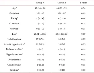
During the study period, 73 endometrial ablations were performed. Participants were submitted to endometrial resection + rollerball coagulation (group A, n = 36) or rollerball electrocoagulation alone (group B, n = 37). Mean follow-up length was 359 days (280 - 751) in group A and 370 days (305 - 766) in group B. Clinical/epidemiological characteristics were similar between groups, except for a higher mean number of children in group B (Table 1).
[image:5.595.210.538.446.701.2]No significant differences between groups were observed regarding ultraso-nographic variables and hysterometric measures routinely taken after diagnostic hysteroscopy (Table 2). The incidence of hysteroscopic findings was similar in both groups (P ≥ 0.05). To estimate the likelihood of successful hysteroscopic fi-broid removal, the classification system described by Lasmar et al. was used, taking into account fibroid size, topography, extension of the base in relation to the uterine wall, penetration into the myometrium, and location on lateral walls (STEPW) [36]. Lasmar score ranged from 2 to 6 in group A, and from zero to 6 in group B (Table 2).
Table 1. Clinical and epidemiological data on the 73 patients who underwent either en-dometrectomy with rollerball coagulation (Group A, 36 patients) or rollerball coagulation alone(Group B, 37 patients).
Group A Group B P value
Agea 45 (34 - 54) 44 (33 - 55) 0.54
Gestationa 3 (0 - 6) 3 (1 - 11) 0.89
Paritya 2 (0 - 6) 3 (1 - 8) 0.04
C-sectiona 1 (0 - 4) 1 (0 - 4) 0.71
Abortiona 0 (0 - 3) 0 (0 - 3) 0.91
BMIb 28.84 (±5.72) 28.64 (±5.73) 0.89
Tubal ligationc 17 (47.2) 20 (54) 0.05
Arterial hypertensionc 12 (33.3) 20 (54) 0.05
Diabetes mellitusc 3 (8.3) 4 (10.8) 0.05
Hypothyroidismc 4 (11.1) 2 (5.4) 0.05
Dyslipidemiac 5 (13.9) 2 (5.4) 0.05
Coagulopathyc 4 (11.1) 3 (8.1) 0.05
Smokingc 5 (13.9) 10 (27) 0.05
aMann-Whitney non-parametric test. Mean value; minimum and maximum within parentheses; b
Stu-dent’s t test for independent groups. Mean values; standard deviation within parentheses; cGoodman
Table 2. Analysis of transvaginal ultrasonography and diagnostic hysteroscopy parame-ters between endometrectomy with rollerball coagulation (Group A, 36 patients) and rol-lerball coagulation alone (Group B, 37 patients).
Group A Group B P value Longitudinal uterinal measure in cm (TVUS)a 9.06 (±1.09) 9.11 (±0.93) 0.86
Uterine volume in cm3 (TVUS)a 152.71 (±61.3) 148.44 (±56.04) 0.76
Fibroid (TVUS)c 17 (47.2) 18 (48.6) 0.05
Submucous fibroid (TVUS)3 9 (25) 5 (13.5) 0.05 Hysterometry (cm)a 8.33 (±1.12) 8.5 (±1.26) 0.55 Endometrial polyp (OH)b 14 (38.9) 18 (48.6) 0.05 Polyp size in mm (OH)a 19.25 (±12.26) 14.2 (±8.6) 0.26 Number of polyps (OH)b 1 (0 - 7) 1 (0 - 6) 0.71 Endocervical polyp (OH)c 1 (2.8) 4 (10.8) 0.05 Submucous fibroid (OH)c 5 (13.9) 6 (16.2) 0.05 Fibroid size in cm (OH)c 2 (1.5 - 7) 1.5 (0.8 - 4) 0.05 Lasmar et al. scorec 3 (2 - 6) 4 (0 - 6) 0.05
aStudent’s t test for independent groups. Mean; standard deviation within parentheses; bMann-Whitney
non-parametric test. Absolute values; percent within parentheses; cGoodman test. Absolute number;
per-cent within parentheses; TVUS: Transvaginal ultrasonography; OH: Office hysteroscopy.
Surgery duration (group A = 48.5 min. [±12.0] and group B = 31.9 min [±5.6], P < 0.001) was shorter and the volume of distention medium used during sur-gery (group A = 5.700 mL [2.000 - 9.000] and group B = 3.500 mL [1.250 - 12.000], P < 0.01) was lower in group B. No difference in fluid deficit was noted between groups (Table 3). At the time of surgery, endometrial pattern was simi-lar in both groups, with 27.8% of the patients in group A, and 35.1% of those in group B having undergone preoperative uterine curettage due to exuberant se-cretory endometrium.
Histopathological examination revealed statistical similarities between groups
(Table 3). Whereas 5 patients from group A (13.9%) had endometrial
hyperpla-sia without atypia, 3 patients in group B (8.1%), also diagnosed with endometrial polyp, showed endometrial hyperplasia with atypia present in one case (2.7%) (P ≥ 0.05). This patient later underwent hysterectomy, which confirmed the pres-ence of a lesion restricted to the endometrium. Adenomyosis, associated with endometrial polyp, was found in only 3 patients (8.1%) from group B. There were no cases of surgical site infection and the incidence of vaginal discharge af-ter procedure was lower in group B (30.5% vs. 8.1%, P < 0.05) (Table 3).
Table 3. Surgical procedure data in patients undergoing endometrectomy with rollerball coagulation (Group A, 36 patients) and rollerball coagulation alone (Group B, 37 pa-tients).
Group A Group B P value
Time (minutes)a1 48.5 (±12.0) 31.9 (±5.6) <0.001 HB/In (mL)a2 5700 (2000 - 9000) 3.500 (1.250 - 12.000) <0.01 HB/Retained (mL)b 300 (0 - 3000) 200 (0 - 600) 0.23 Proliferative endometrium (SH)c 12 (33.3) 15 (40.5) 0.05 Secretory endometrium (SH)c 22 (61.1) 17 (45.9) 0.05 Uterine curettage (before SH)c 10 (27.8) 13 (35.1) 0.05 Proliferative endometrium (AP)c 10 (27.8) 4 (10.8) 0.05 Secretory endometrium (AP)c 16 (44.4) 10 (27) 0.05 Atrophic endometrium (AP)c 2 (5.6) 3 (8.1) 0.05
Polyp (AP)c 14 (38.9) 20 (54) 0.05
Poly size in mm (AP)a1 18.75 (±11.57) 18.07 (±9.86) 0.88
Fibroid (AP)c 4 (11.1) 9 (24.3) 0.05
Fibroid size in cm (AP)a1 3.33 (±2) 3.42 (±1.39) 0.93
Hyperplasia (AP)c 5 (13.9) 3 (8.1) 0.05
Atypic hyperplasia (AP)c 0 1 (2.7) 0.05
Polyp hyperplasia (AP)c 0 2 (5.4) 0.05
Adenomyosis (AP)c 0 3 (8.1) 0.05
Urinary tract infection (UTI)c 1 (2.8) 2 (5.4) 0.05 Vaginal dischargec 11 (30.5) 3 (8.1) <0.05
a1Student’s t test for independent groups. Mean; standard deviation within parentheses; a2Student’s t test
for independent groups. Median; minimum and maximum values within parentheses; bMann-Whitney
non-parametric test. Median; minimum and maximum values within parentheses; cGoodman test.
Ab-solute number; percent within parentheses; HB: Hydric balance; SH: surgical hysteroscopy; AP: Anato-mopathology.
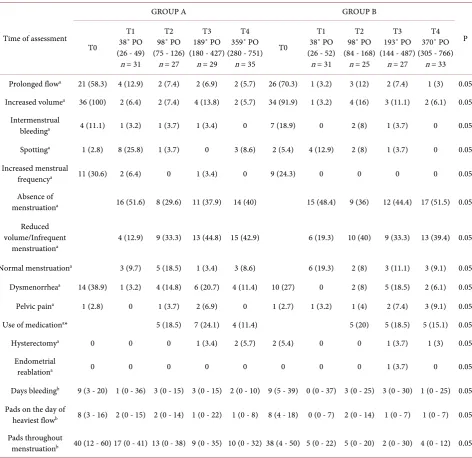
Table 4. Pre- and post-procedure clinical parameters in group A (36 patients) and group B (37 patients).
Time of assessment
GROUP A GROUP B
P T0
T1 38˚ PO (26 - 49)
n = 31 T2 98˚ PO (75 - 126)
n = 27 T3 189˚ PO (180 - 427)
n = 29 T4 359˚ PO (280 - 751)
n = 35 T0
T1 38˚ PO (26 - 52)
n = 31 T2 98˚ PO (84 - 168)
n = 25 T3 193˚ PO (144 - 487)
n = 27 T4 370˚ PO (305 - 766)
n = 33
Prolonged flowa 21 (58.3) 4 (12.9) 2 (7.4) 2 (6.9) 2 (5.7) 26 (70.3) 1 (3.2) 3 (12) 2 (7.4) 1 (3) 0.05 Increased volumea 36 (100) 2 (6.4) 2 (7.4) 4 (13.8) 2 (5.7) 34 (91.9) 1 (3.2) 4 (16) 3 (11.1) 2 (6.1) 0.05
Intermenstrual
bleedinga 4 (11.1) 1 (3.2) 1 (3.7) 1 (3.4) 0 7 (18.9) 0 2 (8) 1 (3.7) 0 0.05 Spottinga 1 (2.8) 8 (25.8) 1 (3.7) 0 3 (8.6) 2 (5.4) 4 (12.9) 2 (8) 1 (3.7) 0 0.05 Increased menstrual
frequencya 11 (30.6) 2 (6.4) 0 1 (3.4) 0 9 (24.3) 0 0 0 0 0.05
Absence of
menstruationa 16 (51.6) 8 (29.6) 11 (37.9) 14 (40) 15 (48.4) 9 (36) 12 (44.4) 17 (51.5) 0.05 Reduced
volume/Infrequent
menstruationa 4 (12.9) 9 (33.3) 13 (44.8) 15 (42.9) 6 (19.3) 10 (40) 9 (33.3) 13 (39.4) 0.05
Normal menstruationa 3 (9.7) 5 (18.5) 1 (3.4) 3 (8.6) 6 (19.3) 2 (8) 3 (11.1) 3 (9.1) 0.05
Dysmenorrheaa 14 (38.9) 1 (3.2) 4 (14.8) 6 (20.7) 4 (11.4) 10 (27) 0 2 (8) 5 (18.5) 2 (6.1) 0.05
Pelvic paina 1 (2.8) 0 1 (3.7) 2 (6.9) 0 1 (2.7) 1 (3.2) 1 (4) 2 (7.4) 3 (9.1) 0.05 Use of medicationa* 5 (18.5) 7 (24.1) 4 (11.4) 5 (20) 5 (18.5) 5 (15.1) 0.05 Hysterectomya 0 0 0 1 (3.4) 2 (5.7) 2 (5.4) 0 0 1 (3.7) 1 (3) 0.05
Endometrial
reablationa 0 0 0 0 0 0 0 0 1 (3.7) 0 0.05
Days bleedingb 9 (3 - 20) 1 (0 - 36) 3 (0 - 15) 3 (0 - 15) 2 (0 - 10) 9 (5 - 39) 0 (0 - 37) 3 (0 - 25) 3 (0 - 30) 1 (0 - 25) 0.05 Pads on the day of
heaviest flowb 8 (3 - 16) 2 (0 - 15) 2 (0 - 14) 1 (0 - 22) 1 (0 - 8) 8 (4 - 18) 0 (0 - 7) 2 (0 - 14) 1 (0 - 7) 1 (0 - 7) 0.05 Pads throughout
menstruationb 40 (12 - 60) 17 (0 - 41) 13 (0 - 38) 9 (0 - 35) 10 (0 - 32) 38 (4 - 50) 5 (0 - 22) 5 (0 - 20) 2 (0 - 30) 4 (0 - 12) 0.05
aGoodman test. Absolute number; percent within parentheses; bNon-parametric repeated measures anova in independent groups followed by the
Dunn test. Median, minimum and maximum within parentheses; *Non-hormonal anti-inflammatory agent (NHAA), tranexamic acid, combination oral contraceptives or oral progestins; Flow duration (d)—prolonged > 8 days; Flow volume (mL)—increased > 80 mL; Increased menstrual frequency: < 24 days; Reduced volume/Infrequent menstruation: < 4.5 days/> 38 days; Normal menstruation: 24 - 38 days duration; variation of 2 - 20 days per men-strual cycle in 12 months; flow duration of 4.5 - 8 days; volume of 5 - 80 mL (Munro et al., 2012); T: Time elapsed; PO: Postoperative.
Hemoglobin levels significantly improved in both groups, increasing from 12.72 g/dL (±1.96) to 14.09 g/dL (±1.15) (P < 0.01) in group A, and from 12.59 g/dL (±1.6) to 13.62 g/dL (±0.82) (P < 0.01) in group B, on days 189 (180 - 427) and 193 (144 - 487), respectively. However, there were no differences between pre- and post-procedure hematocrit levels between groups (P ≥ 0.05). One group B patient, who had a history of pulmonary thrombosis, had to stay longer in hospital due to postoperative acute deep vein thrombosis of a lower limb. Addi-tionally, an obese patient (BMI = 35 kg/m2) from group A, who underwent
abla-tion combined with myomectomy, had water intoxicaabla-tion. The rate of satisfac-tion/success was 86.1% in group A and 88.6% in group B, with no significant difference between groups (P ≥ 0.05).
4. Discussion
Endometrial ablation was first performed by Goldrath and colleagues in 1981. However, the efficacy of the procedure was established in 1983 by De Cherney and Polan, who reported that of 11 patients treated with ablation, 6 remained amenorrheic for a sustained period [37] [38]. The technique of endometrial re-section includes removal of the functional and basal layers of the endometrium, as well as underlying 2 - 3 mm myometrium. Predictive factors of successful en-dometrial ablation, defined as reduction or absence of menstrual flow, include age older than 40 - 45 years and uterus with a volume under 200 cm3 without
intramural fibroid. In contrast, history of menstrual pain, adenomyosis, tubal li-gation (association with post ablation tubal lili-gation syndrome), and parity greater than five are associated with failure [23] [39].
Endometrial ablation success rate has been reported to be around 80% - 90%
[7] [39]. Shavell et al. found that, through five years of follow-up, hysterectomy
was performed subsequently to endometrial ablation in 13.4% of the cases, pri-marily due to persistent bleeding and pelvic pain [24]. Longinotti et al., in 8-year follow-up of 754 women, reported a 26% probability of hysterectomy subsequent to endometrial ablation, with pelvic pain being the main reason for the proce-dure in 22% of the cases [25]. According to the literature, reduced bleeding or infrequent menstruation is achieved in 48% - 60%, absence of menstruation in 20% - 48%, and normal menstrual pattern in 2% - 20% of the cases, while failure occurs in 2% - 11% [13] [16] [37] [39] [40].
In our study, there was no significant difference in menstrual pattern between groups 30, 90 and 180 days after endometrial ablation. By the end of the study period, no differences were observed between groups regarding menstrual ab-sence (40% vs. 51.5%), reduced bleeding or infrequent menstruation (42.9% vs. 39.4%), and normal menstrual pattern (8.6% vs. 9.1%), in agreement with pre-vious reports.
Intraopera-tively, 2 patients from group B (5.4%), who were undergoing endometrial abla-tion combined with myomectomy, required switch to hysterectomy because of technical difficulties and heavy intraoperative bleeding.
During postoperative follow-up, 5 patients (3 from group A and 2 from group B) underwent hysterectomy, and 1 patient from group B had an endometrial reablation. The reasons for hysterectomy were persistent AUB and incapacitat-ing dysmenorrhea in the 3 patients from group A, and persistent severe pelvic pain in both patients from group B. Endometrial reablation was performed in one group B patient due to persistent heavy menstrual bleeding [7]. The proce-dure was followed by improved clinical condition and patient satisfaction.
Five patients from group A and 2 patients from group B underwent endome-trial ablation combined with polypectomy [7]. Histopathological examination showed simple hyperplasia with no atypia in the polyp. Therefore, they were clinically treated with medroxyprogesterone for an initial period of 6 months, and are still being followed up [42]. In the only case where histopathological examination revealed atypical complex hyperplasia, hysterectomy was per-formed and the patient’s condition has evolved satisfactorily since then.
The patient with a history of pulmonary thromboembolism (group B) had to stay in hospital for longer. Despite adequate preoperative preparation, she had acute deep vein thrombosis of a lower limb. However, her condition evolved ex-tremely well and she was discharged 13 days later with absent menstruation and still on warfarin. Consistently with the literature, the only case of water intoxica-tion was seen in an obese patient undergoing endometrial ablaintoxica-tion and myo-mectomy using 3% sorbitol as distention medium [7] [43]. This group A patient showed a BMI of 35 kg/m2 and a immediate postoperative sodium plasma level
of 116 mEq/L. Clinical support measures were taken, and the patient was dis-charged in good condition 4 days later.
Vaginal discharge is described as a normal clinical complaint within the first 30 days after endometrial ablation, provided that fever and fetid odor are absent. In this study, the incidence of vaginal discharge during the early postoperative period was higher in group A than in group B. However, good resolution was observed in all cases after clinical treatment with oral imidazole derivatives.
highlight is the fact that our institution is a university center where novice doc-tors are instructed in minimally invasive techniques which can explain some of the observed complications and also the amount of distension medium used to complete the procedures.
5. Conclusion
Endometrial ablation using rollerball electrocoagulation alone, which is easier to perform, may be considered safer. Besides taking shorter time in surgery and requiring a smaller amount of distention medium, it is associated with a lower postoperative vaginal discharge incidence. The rate of clinical success did not statistically differ between groups. However, study parameters, both in absolute values and percents, were superior in group B.
Funding
This study was supported by São Paulo Research Foundation (FAPESP)—Grant 2011/13034-5.
Conflict of Interest Statement
The authors have no commercial, proprietary, or financial interest in the prod-ucts or companies describes in this article. All authors have no other conflict of interest.
References
[1] Fraser, I.S., Critchley, H.O. and Munro, M.G. (2007) Abnormal Uterine Bleeding: Getting Our Terminology Straight. Current Opinion in Obstetrics and Gynecology, 19, 591-595. https://doi.org/10.1097/01.aids.0000299801.42415.8a
[2] Munro, M.G., Critchley, H.O. and Fraser, I.S. (2012) The FIGO Systems for No-menclature and Classification of Causes of Abnormal Uterine Bleeding in the Re-productive Years: Who Needs Them? American Journal of Obstetrics and Gyne-cology, 207, 259-265. https://doi.org/10.1016/j.ajog.2012.01.046
[3] NICE Guideline 44. (2011) Heavy Menstrual Bleeding. ISBN 978-1-904752-35-6.
http://www.nice.org.uk.
[4] Cote, I., Jacobs, P. and Cumming, D.C. (2003) Use of Health Services Associated with Increased Menstrual Loss in the United States. American Journal of Obstetrics and Gynecology, 188, 343-348. https://doi.org/10.1067/mob.2003.92
[5] Shapley, M., Jordan, K. and Croft, P.R. (2004) An Epidemiological Survey of Symp-toms of Menstrual Loss in the Community. British Journal of General Practice, 54, 359-363.
[6] Marret, H., Fauconnier, A., Chabbert-Buffet, N., Cravello, L., Golfier, F., Gondry, J., et al. (2010) Clinical Practice Guidelines on Menorrhagia: Management of Abnor-mal Uterine Bleeding before Menopause. European Journal of Obstetrics and Gy-naecology Reproductive Biology, 152, 133-137.
https://doi.org/10.1016/j.ejogrb.2010.07.016
https://doi.org/10.1016/S1701-2163(15)30288-7
[8] Cooper, K., Lee, A., Chien, P., Raja, E., Timmaraju, V. and Bhattacharya, S. (2011) Outcomes Following Hysterectomy Or Endometrial Ablation for Heavy Menstrual Bleeding: Retrospective Analysis of Hospital Episode Statistics in Scotland. An In-ternational Journal of Obstetrics and Gynaecology, 118, 1171-1179.
https://doi.org/10.1111/j.1471-0528.2011.03011.x
[9] Varol, N., Healey, M., Tang, P., Sheehan, P., Maher, P. and Hill, D. (2001) Ten-Year Review of Hysterectomy Morbidity and Mortality: Can We Change Direction? Aus-tralian and New Zealand Journal of Obstetrics and Gynaecology, 41, 295-302.
https://doi.org/10.1111/j.1479-828X.2001.tb01231.x
[10] Kafy, S., Huang, J.Y., Al-Sunaidi, M., Wiener, D. and Tulandi, T. (2006) Audit of Morbidity and Mortality Rates of 1792 Hysterectomies. Journal of Minimally Inva-sive Gynecology, 13, 55-59. https://doi.org/10.1016/j.jmig.2005.10.003
[11] Aniuliene, R., Varzgaliene, L. and Varzgalis, M. (2007) A Comparative Analysis of Hysterectomies. Medicina (Kaunas), 43, 118-124.
[12] Wortman, M. (2016) Hysteroscopic Endometrial Resection versus Laparoscopic Supracervical Hysterectomy Hysterectomy—A Flawed Conclusion. Journal of Mi-nimally Invasive Gynecology, 23, 288-289.
https://doi.org/10.1016/j.jmig.2015.09.027
[13] Papadopoulos, N.P. and Magos, A. (2007) First-Generation Endometrial Ablation: Roller-Ball vs Loop vs Laser. Best Practice & Research Clinical Obstetrics & Gynae-cology, 21, 915-929. https://doi.org/10.1016/j.bpobgyn.2007.03.014
[14] Kumar, V., Chodankar, R. and Gupta, J.K. (2016) Endometrial Ablation for Heavy Menstrual Bleeding. Women’s Health (London, England), 12, 45-52.
https://doi.org/10.2217/whe.15.86
[15] Ghazizadeh, S., Bakhtiari, F., Rahmanpour, H., Davari-Tanha, F. and Ramezanza-deh, F.A. (2011) Randomized Clinical Trial to Compare Levonorgestrel-Releasing Intrauterine System (Mirena) vs Trans-Cervical Endometrial Resection for Treat-ment of Menorrhagia. International Journal of Women’s Health, 3, 207-211. [16] Penninx, J.P., Herman, M.C., Mol, B.W. and Bongers, M.Y. (2011) Five-Year
Fol-low-Up after Comparing Bipolar Endometrial Ablation with Hydrothermablation for Menorrhagia. Obstetrics & Gynaecology, 118, 1287-1292.
https://doi.org/10.1097/AOG.0b013e318236f7ed
[17] Hoaglin, D.C., Filonenko, A., Glickman, M.E., Wasiak, R. and Gidwani, R. (2013) Use of Mixed-Treatment-Comparison Methods in Estimating Efficacy of Treat-ments for Heavy Menstrual Bleeding. European Journal of Medical Research, 18, 17.
https://doi.org/10.1186/2047-783X-18-17
[18] Donohoe, C.L., Feeney, C., Carey, M.F. and Reynolds, J.V. (2011) Perioperative Evaluation of the Obese Patient. Journal of Clinical Anesthesia, 23, 575-586.
https://doi.org/10.1016/j.jclinane.2011.06.005
[19] El-Nashar, S.A., Hopkins, M.R., Barnes, S.A., Pruthi, R.K., Gebhart, J.B., Cliby, W.A., et al. (2010) Health-Related Quality of Life and Patient Satisfaction after Global Endometrial Ablation for Menorrhagia in Women with Bleeding Disorders: A Follow-Up Survey and Systematic Review. American Journal of Obstetrics and Gynecology, 202, 348 e1-348 e7.
[20] Madsen, A.M., El-Nashar, S.A., Hopkins, M.R., Khan, Z. and Famuyide, A.O. (2013) Endometrial Ablation for the Treatment of Heavy Menstrual Bleeding in Obese Women. International Journal of Gynecology & Obstetrics, 121, 20-23.
https://doi.org/10.1016/j.ijgo.2012.10.024
Ab-lation in Women with Inherited Bleeding Disorders. Haemophilia, 18, 413-420.
https://doi.org/10.1111/j.1365-2516.2011.02712.x
[22] Kouides, P.A. and Kadir, R.A. (2007) Menorrhagia Associated with Laboratory Ab-normalities of Hemostasis: Epidemiological, Diagnostic and Therapeutic Aspects. Journal of Thrombosis and Haemostasis, 5, 175-182.
https://doi.org/10.1111/j.1538-7836.2007.02494.x
[23] Thomassee, M.S., Curlin, H., Yunker, A. and Anderson, T.L. (2013) Predicting Pel-vic Pain after Endometrial Ablation: Which Preoperative Patient Characteristics Are Associated? Journal of Minimally Invasive Gynecology, 20, 642-647.
https://doi.org/10.1016/j.jmig.2013.04.006
[24] Shavell, V.I., Diamond, M.P., Senter, J.P., Kruger, M.L. and Johns, D.A. (2012) Hysterectomy Subsequent to Endometrial Ablation. Journal of Minimally Invasive Gynecology, 19, 459-464. https://doi.org/10.1016/j.jmig.2012.03.013
[25] Longinotti, M.K., Jacobson, G.F., Hung, Y.Y. and Learman, L.A. (2008) Probability of Hysterectomy after Endometrial Ablation. Obstetrics & Gynaecology, 112, 1214- 1220. https://doi.org/10.1097/AOG.0b013e31818c1766
[26] Matteson, K.A., Abed, H., Wheeler 2nd, T.L., Sung, V.W., Rahn, D.D., Schaffer, J.I., et al. (2012) A Systematic Review Comparing Hysterectomy with Less-Invasive Treatments for Abnormal Uterine Bleeding. Journal of Minimally Invasive Gyne-cology, 19, 13-28. https://doi.org/10.1016/j.jmig.2011.08.005
[27] Boujida, V.H., Philipsen, T., Pelle, J. and Joergensen, J.C. (2002) Five-Year Fol-low-Up of Endometrial Ablation: Endometrial Coagulation versus Endometrial Re-section. Obstetrics & Gynaecology, 99, 988-992.
https://doi.org/10.1097/00006250-200206000-00007
[28] Furst, S.N., Philipsen, T. and Joergensen, J.C. (2007) Ten-Year Follow-Up of En-dometrial Ablation. Acta Obstetricia et Gynecologica Scandinavica, 86, 334-338.
https://doi.org/10.1080/00016340601089701
[29] Onoglu, A., Taskin, O., Inal, M., Sadik, S., Simsek, M., Akar, M., et al. (2007) Com-parison of the Long-Term Histopathologic and Morphologic Changes after Endo-metrial Rollerball Ablation and Resection: A Prospective Randomized Trial. Journal of Minimally Invasive Gynecology, 14, 39-42.
https://doi.org/10.1016/j.jmig.2006.06.027
[30] Bongers, M.Y. (2015) Hysteroscopy and Heavy Menstrual Bleeding (To Cover TCRE and Second-Generation Endometrial Ablation). Best Practice & Research Clinical Obstetrics & Gynaecology, 29, 930-939.
https://doi.org/10.1016/j.bpobgyn.2015.03.011
[31] Singh, S., Best, C., Dunn, S., Leyland, N., Wolfman, W.L., Clinical Practice—Gy- naecology Committee, et al. (2013) Abnormal Uterine Bleeding in Pre-Menopausal Women. Journal of Obstetrics and Gynaecology Canada, 35, 473-479.
https://doi.org/10.1016/S1701-2163(15)30939-7
[32] Zar, J.H. (2010) Biostatistical Analysis. 5th Edition, Prentice-Hall/Pearson, Upper Saddle River, xiii, 944 p.
[33] Goodman, L.A. (1964) Simultaneous Confidence Intervals for Contrasts among Multinomial Populations. Annals of Mathematical Statistics, 35, 716-725.
https://doi.org/10.1214/aoms/1177703569
[34] Goodman, L.A. (1965) On Simultaneous Confidence Intervals for Multinomial Proportions. Technometrics, 7, 247-254.
https://doi.org/10.1080/00401706.1965.10490252
[36] Lasmar, R.B., Barrozo, P.R., Dias, R. and Oliveira, M.A. (2005) Submucous Myo-mas: A New Presurgical Classification to Evaluate the Viability of Hysteroscopic Surgical Treatment—Preliminary Report. Journal of Minimally Invasive Gynecolo-gy, 12, 308-311. https://doi.org/10.1016/j.jmig.2005.05.014
[37] Goldrath, M.H., Fuller, T.A. and Segal, S. (1981) Laser Photovaporization of Endo-metrium for the Treatment of Menorrhagia. American Journal of Obstetrics and Gynecology, 140, 14-19. https://doi.org/10.1016/0002-9378(81)90251-9
[38] DeCherney, A. and Polan, M.L. (1983) Hysteroscopic Management of Intrauterine Lesions and Intractable Uterine Bleeding. Obstetrics and Gynecology, 61, 392-397. [39] Takahashi, W.H., Lopes, R.G., Depes, D. de B. and Martins e Castello Branco, H.K.
(2012) Results of Hysteroscopic Endometrial Ablation after Five-Year Follow-Up. Revista Brasileira de Ginecologia e Obstetrícia, 34, 80-85.
https://doi.org/10.1590/S0100-72032012000200007
[40] Loffer, F.D. (1988) Laser Ablation of the Endometrium. Obstetrics and Gynecology Clinics of North America, 15, 77-89.
[41] Smithling, K.R., Savella, G., Raker, C.A. and Matteson, K.A. (2014) Preoperative Uterine Bleeding Pattern and Risk of Endometrial Ablation Failure. American Journal of Obstetrics and Gynecology, 211, 556.e1-556.e6.
[42] Pontes, A., Traiman, P., Franco, M., Nahás, E.A.P., Dias, R. and De Luca, L.A. (2000) Clinical Treatment and Follow-Up of Endometrial Hyperplasia. Revista Bra-sileira de Ginecologia e Obstetrícia, 22, 325-331.
https://doi.org/10.1590/S0100-72032000000600002
[43] Fonseca, M. de F., Andrade Junior, C.M., Nogueira, E. de A., Sessa, F.V. and Crispi, C.P. (2015) Predictors of Fluid Intravasation during Operative Hysteroscopy: A Preplanned Prospective Observational Study with 200 Cases. Revista Brasileira de Ginecologia e Obstetrícia, 37, 24-29.
https://doi.org/10.1590/S0100-720320140005139
List of Abbreviations
AUB: Abnormal Uterine Bleeding;
FIGO: International Federation of Gynecology and Obstetrics; HRQOL: Health-Related Quality of Life;
NDAIDs: Nonsteroidal Anti-Inflammatory Drugs; COC: Combination Oral Contraceptives;
LNG-IUS: Levonorgestrel Intrauterine System; BMI: Body Mass Index;
PBLAC: Pictorial Blood Loss Assessment Chart; TVUS: Transvaginal Ultrasonography;
Submit or recommend next manuscript to SCIRP and we will provide best service for you:
Accepting pre-submission inquiries through Email, Facebook, LinkedIn, Twitter, etc. A wide selection of journals (inclusive of 9 subjects, more than 200 journals)
Providing 24-hour high-quality service User-friendly online submission system Fair and swift peer-review system
Efficient typesetting and proofreading procedure
Display of the result of downloads and visits, as well as the number of cited articles Maximum dissemination of your research work
Submit your manuscript at: http://papersubmission.scirp.org/