Myelodysplastic syndrome (MDS) associated with EBV
infection in a pediatric patient
Esther Manor1*, Yonat Shemer-Avni2, Sophia Boriakovski1, Michael Kafka3, Lipa Bodner4, Joseph Kapelushnik5
1Soroka Medical Center, Faculty of Health Sciences, Institute of Human Genetics, Ben-Gurion University of the Negev, Beer-Sheva,
Israel
2Soroka Medical Center, Laboratory for Viral Diagnosis, Faculty of Health Sciences, Ben-Gurion University of the Negev, Beer-Sheva,
Israel
3Soroka Medical Center, Department of Hematology, Faculty of Health Sciences, Ben-Gurion University of the Negev, Beer-Sheva,
Israel
4Soroka Medical Center, Department of Oral and Maxillofacial Surgery, Faculty of Health Sciences, Ben-Gurion University of the
Negev, Beer-Sheva, Israel
5Soroka Medical Center, Department of Pediatric Hemato-Oncology, Faculty of Health Sciences, Ben-Gurion University of the Negev,
Beer-Sheva, Israel Email: *manore@bgu.ac.il
Received 30 December 2012; revised 31 January 2013; accepted 10 February 2013
ABSTRACT
Background: Epstein Barr Virus (EBV) is associated with different kinds of tumors. In the present study we tried to understand its role in pediatric MDS of a 4-year-old girl with EBV infection and MDS with refractory anemia and monosomy 7. Procedures: The work-up included: hematological tests; serology for IgM, IgG antibodies to EBV; PCR for EBV; cytoge-netics of bone marrow (BM), and FISH analysis of BM and blood; immunohistochemistry-LMP1 ex-pression on BM smears. Results: Hematological fol- low up showed constant mild dysplastic changes mostly in the erythroid lineage. PCR for EBV showed positive results in the nasopharygeal smears as well as in blood 15 weeks after disease onset. Cytogenetic analysis showed monosomy 7 in all the mitoses of BM sample. Fluorescence in situ hybridization (FISH) showed monosomy 7% in 57% of the cells, followed by a decreased tendency in the percentage of mono- somy 7 cells in both BM and blood. Immunohisto- chemistry for EBV-latent membrane protein 1 (LMP-1) on the patient’s BM smears, 21 weeks post disease onset, showed 9% positive cells, 80% of them carried monosomy 7. Conclusion: The parallel oc-currence of the EBV infection and MDS, as well as the continuous EBV PCR positive and monosomy 7, support the possibility that they are related.
Keywords:Pediatric; MDS; EBV Infection
1. INTRODUCTION
Myelodysplastic syndrome (MDS) represents a hetero- geneous clonal hematopoetic stem cell disorder charac- terized by ineffective hematopoiesis and increased risk of transformation to MDS-related leukemia. MDS is un- common in children and accounts for less than 5% of all hematopoetic neoplasms [1].
Monosomy 7 is the most common chromosomal aber-ration in MDS [2-6]. It implies a rather poor prognosis that is associated with high risk of transformation to acute leukemia [7]. The age distribution is 2 - 5 years with male predominance of 70% - 80% [8]. There are also reports on familial MDS with monosomy 7. Familial monosomy 7 is defined when at least two siblings are found to carry monosomy 7 associated with pre-leukemic or leukemic manifestations [9].
Epstein Barr Virus (EBV) is one of the causative agents of infectious mononucleosis and the first de- scribed human tumor virus. EBV was discovered in Burkitt’s lymphoma tumor cells, characterized by t(8,14), t(2,8), and t(8,22) [10]. It has also been found to be asso-ciated with post-transplant lymphoproliferative disorders (PTLD) and with malignancies such as, Hodgkin’s lym-phoma (HD) and nasopharyngeal carcinoma (NPC) [11,12].
EBV’s latent membrane protein (LMP1) gene is con-sidered the main viral oncogene driving cell growth, promoting metastases, apoptotic resistance, and immune modulation, including blockade of interferon alpha-in- duced antiviral signaling [13].
Infection with EBV can produce symptoms closely resembling those seen in patients with MDS. This raises
a question regarding the diagnosis of malignant disease in cases of MDS in children with EBV infection [14-16] and the role of EBV in MDS pathogenesis: Does EBV infection mimic childhood MDS? Does it trigger or en-hance childhood MDS onset? Is EBV the etiology of MDS or is MDS onset with EBV infection just coinci-dental?
Here we present results that might support the possi-bility that EBV plays a role in pediatric MDS of a 4- year-old girl with monosomy 7.
2. CASE REPORT AND RESULTS
A 4-year-old girl was admitted to the pediatric depart- ment with suspected anemia, after experiencing two epi-sodes of fever about 3 weeks earlier. Upon admittance she was weak and pale with a diagnosis of infectious mononucleosis by the family doctor, although no tests were undertaken. According to her mother’s anamnesis, she had been suffering on and off from viral and bacte-rial infections.
Hematology: Peripheral blood count showed a white blood count (WBC) of 4400/μl with no blasts, Hb of 4.9 g/dl, and platelet count of 175,000/μl. The clinical im-pression was of erythroblastopenia of childhood, proba-bly related to viral infection. Serial transfusions of packed cells elevated the Hb to 6.4 g/dl. Peripheral blood count was followed and transfusions of packed cells were given due to the anemia.
Serology: The work-up revealed positive antibodies to cytomegalovirus (CMV) IgM and IgG, with high avidity. Antibodies to EBV (viral capsid antigen) VCA IgG and IgM were positive; however, IgG antibodies to EBV nu-clear antigen (EBNA) were negative. These results indi-cate that the patient was suffering from an acute primary EBV infection with possible CMV reactivation. EBV acute infection was further confirmed by EBV-positive PCR results of the pharyngeal smear.
Cytogenetics: Cytogenetic analysis of bone marrow (BM) was performed according to standard methods. BM cells were cultured for 24 hours using methotrexate for synchronization. About 15 metaphases were analyzed using G-band staining. Karyotypes were assigned ac- cording to the International System for Human Cytoge- netic Nomenclature (ISCN 2009) guidelines [17].
FISH analysis was performed according to the manu-facturer’s instructions using an LSI D7S522 (7q31) spec- trum orange/CEP spectrum green (VYSIS) probe. More than 500 cells were analyzed.
Virology tests: IgM and IgG antibodies to EBV VCA and EBNA, and to CMV, as well as avidity were tested using Liaision Technologies, according to the manufac- turer’s instructions (Liaison Technologies, Perugia, It- aly).
Viral load for EBV DNA was tested as described by Niesters et al. [18].
Immunohistochemistry: LMP1 expression on BM smears were examined using the Ivew DAB detection kit (Ventana Medical Systems, Inc., Tucson, AZ) using monoclonal mouse anti-Epstein Barr Virus, LMP clones CS 1-4 (DakoCytomation, Denmark), and FISH analysis using the LSI D7S522 (7q31) spectrum orange/CEP spectrum green (VYSIS) probe. More than 800 cells were examined.
Figure 1 depicts the antibody responses to IgM, IgG
against EBV’s viral capsid antigen (VCA), and EBV’s nuclear antigen (EBNA) at time intervals from 2 weeks to 6 months after onset of the disease. It shows a three-fold increase in the level (antibody titer) of VCA’s IgG antibodies at 6 weeks after disease onset, while the humeral immune response to EBNA, a classic pattern of EBV acute infection, increases about 6 months after dis-ease onset. The IgM response to VCA gradually declined over three months.
Although no EBV DNA could be detected in the first blood sample, which was taken 3 weeks post-disease onset, EBV DNA was present when tested in a naso- pharyngeal smear 12 weeks after disease onset as well as in blood samples. EBV viral load at 15, 29, and 42 weeks post-illness onset were >25, 100, and 100 copies/ml, respectively.
2.1. Hematologic Tests
At admission complete blood count showed mild leuko-penia (4400/ul) and neutroleuko-penia (900/ul), severe anemia (5.0 g/dl), normal RBC indices, elevated RDW, and normal PLT count. CBC tests performed during the one-year follow up demonstrated mostly leukocyte counts of 4300 - 5600/μl (normal low level of 5000/ul) and neutropenia of 770 - 1440/μl (normal low level of 1500/μl). During the first 6 months of treatment the pa-tient received a total of 7 units of packed red blood cells. Following the transfusion of every unit, the Hb level was raised to 10.0 - 11.0 g/dl; however, rapidly falling after- wards (Figure 2). After this period the Hb level gradu-
1 10 100
0 5 10 15 20 25
EBV-VCA-M EBV-VCA-G EBV-EBNA
A
ntib
odies (
U/m
l)
[image:3.595.160.439.88.252.2]Weeks from first symptoms
Figure 1. Antibody responses to EBV during the time course of the infection. IgM (diamonds) and IgG (squares) antibodies to VCA, as well as antibodies to EBNA (triangle, EBNA-G) were tested in the patient’s sera starting two weeks after initial symptoms. The cut-off values were: VCA-M > 40, positive; VCA-G/EBNA-G > 20, positive. Black arrows represent EBV-DNA positive samples, taken from the larynx.
0.0 2.0 4.0 6.0 8.0 10.0 12.0 14.0
0 5 10 15 20 25 30 35 40 45 50 55 60
Weeks after disease onset
Hgb g/dl
ANC x1000/ul
black arrow s-blood transf usion
[image:3.595.66.530.327.699.2]Figure 2 shows the results of the absolute neutrophilic
count (ANC) and Hemoglobin (Hb) during the 55 weeks post-disease onset. Transfusions of packed cells given to the patient are noted by arrows. No significant abnor-malities were found in all other hematology parameters.
2.2. Cytogenetic Results
Table 1 summarizes the cytogenetic and FISH results for
monosomy 7 in the bone marrow and blood samples. Cytogenetic analysis of the first bone marrow sample revealed monosomy 7 as a sole aberration in all the ana-lyzed dividing cells; FISH analysis revealed that only 57% of the cells carry monosomy 7. FISH analysis of the peripheral blood sample showed cells carry monosomy 7 with reduction in the percentage of these cells in the blood in most samples.
Table 1. Monosomy (mo) 7 in blood and bone marrow (BM) samples by cytogenetics and FISH analysis. In parentheses is the number of monosomy 7 cells out of the number of the total analysed cells.
Weeks p.d.o. Sample Cytogenetics-% mo-7 FISH-% mo-7
4 BM 100% (15/15) 57%
7 BM 70% (9/13) 36%
11 BM 50% (8/16) 26%
21 BM 70% (7/10) 18%
29 BM 80% (12/15) 30%
35 BM 26% (4/11) 19%
41 BM 75%(11/15) 30% 43 BM 75%(9/12) 34%
55 BM 75%(11/15) 34%
Control BM 0% 3%
6 blood nd 34%
9 blood nd 24%
13 blood nd 34%
15 blood nd 22%
17 blood nd 20%
19 blood nd 32%
22 blood nd 15%
26 blood nd 20%
29 blood nd 30%
31 blood nd 17%
35 blood nd 12%
41 blood nd 25%
48 blood nd 20%
57 blood nd 18%
60 blood nd 20%
p.d.o.: post disease onset.
There was a decline in the percentage of cells carrying monosomy 7 during the months following disease onset (intervals of 2 - 4 weeks). The percentage of cells carry- ing monosomy 7 reached a steady level of 20% to 30% (±5%) in blood samples 3 to 13 months after disease onset. In general, FISH analyses showed decreased levels of monosomy 7 percentage compared to the cytogenetic results of the same BM samples.
2.3. Immunohistochemical Analysis
Immunohistochemical staining for LMP1 revealed about 9% LMP1 positive cells in BM smears, at 4, 7, 11, and 21 weeks post disease onset. Control normal BM smears showed 3% LMP1 positive cells. Since LMP1 antibodies showed non-EBV-specific staining in young myeloid cells, it was impossible to indicate whether LMP1 was expressed in the stained cells. FISH analysis of the LMP1 stained smears for monosomy 7 showed an in- creasing percentage of cells carrying monosomy 7 (from 40% to 80%) in the stained cells (Figure 3). The LMP1
positive cells showed a neutrophil morphology.
3. DISCUSSION
We described here a 4-year-old girl suffering from si- multaneous acute EBV infection and refractory anemia MDS with chromosome 7 monosomy. The main question arising is whether the MDS is caused by the EBV infec- tion, as either a direct modulator or an indirect modulator (cofactor), or whether this is a case of coincidence in which the two diseases occurred independently at the same time.
Infectious agents, particularly viruses, are known to play an important role in human cancer development. Recent estimates have shown that 17.8% of cancer cases are attributable to infectious agents, and of these 12.1% are caused by viral infections [19].
Epstein Barr Virus (EBV) is one of the causative agents of infectious mononucleosis and the first de- scribed human tumor virus found to be associated with Burkitt’s lymphoma tumor characterized by t(8,14), t(2,8) and t(8,2) [10], Hodgkin’s lymphoma, nasopharyngeal carcinoma, post-transplant lymphoproliferative disorders (PTLD), and more [11,12,20].
Apparently, viral gene products play an active role in the pathogenesis of these diseases, [12] disrupting the balance of antiviral immunity leading to the development of EBV-associated disease [11].
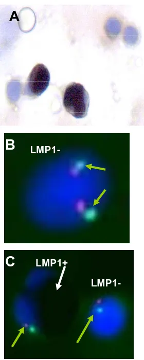
[image:4.595.58.284.344.723.2]A
B
C
LMP1+
LMP1-Figure 3. Immunohistochemistry using LMP1 antibodies and FISH analysis for chromosome 7 of the BM smears. (A) LMP1 positive and negative cells in a BM smear (400×); (B) LMP1 negative cell with two chromosome 7 signals; (C) Positive and negative cells with one signal of chro- mosome 7 (LSI D7S522 (7q31) spectrum orange/CEP spectrum green (VYSIS) probe) (1000×).
tively highly expressed [13,21-23]. Moreover, in immuno compromised patients abundant LMP1 expression can be observed in tonsillar B cells prior to onset of lymphopro- liferative disease [24].
Myelodysplastic syndromes (MDS) are characterized by ineffective hematopoiesis and a high propensity to transform to acute myeloid leukemia (AML). These are common hematological diseases in the elderly but are rarely observed in children [25]. The etiology and mo-lecular pathogenesis of MDS are still poorly understood [26]. Both gene mutations and cytogenetic changes play important roles in the pathogenesis of this disease. The main genetic diagnosis of MDS so far is by karyotyping [27]; monosomy 7 is one of the common chromosomal abnormalities in MDS. The outcome of monosomy 7
MDS in children is often rather poor [28] and the treat-ment in such cases is BM transplantation.
Cytogenetic, histologic, and virologic analyses of the present patient were consistent with MDS, a subtype refractory anemia associated with EBV infection.
Two patterns of monosomy 7 MDS in children are de-scribed in the literature. One is the sporadic monosomy 7 MDS characterized by a striking sex difference, with males affected 3 - 10 times more than females [4,29]; median age at disease onset is relatively young (3 years). The second is the familial monosomy 7 MDS with no sex preference and with later onset of the disease at a median age of 10 - 14 years [9,28,30]. The inheritance mode of both MDS patterns is thought to be poly-genic-mulifactorial.
The described patient is most probably a sporadic MDS case, as there was no evidence for familial monosomy 7 MDS. The patient’s age is close to the me-dian age for sporadic MDS. However, the patient is a female in contrast to the male prevalence in sporadic MDS. In polygenic multifactorial inheritance this strengthens the possibility of prominent genetic in-volvement. The patient’s high susceptibility to bacterial and viral infections may also strengthen the possibility of a genetic predisposion for leukemia disorder develop-ment.
The virology results showed a high titer of VCA IgM and IgG at 2 weeks after EBV infection symptoms onset (on hospital admittance) followed by an increase of EBNA IgG titer together with EBV DNA positive naso-pharingeal smear, as well as in blood samples, further strengthening the diagnosis of EBV acute infection. The presence of <25 - 100 copies/ml of EBV in blood sam-ples during the 55 weeks post-infection, may reflect the latent stage of the EBV in the patient.
Although CMV IgM and IgG were positive in the pa-tient’s blood, the high avidity points strongly towards reactivation response. Also, CMV is known to be less tumorogenic compared to EBV.
The patient received supportive care of transfusions of packed cells during the 24 weeks post-disease onset with no antileukemic therapy (Figure 2). The patient is a can-
didate for BM transplantation.
Cytogenetic and FISH analyses follow-up of mono- somy 7 percentage in bone marrow and peipheral blood samples showed, in general, a decrease in the percentage of cells carrying monosomy 7 during the 13 months after disease onset. This is an original observation.
Table 1 shows a discrepancy between the cytogenetics
[image:5.595.102.242.84.434.2]normal cells compared with the normal cells. We found a similar discrepancy when other FISH probes were used (7q13, 5q33, and centromere 8—personal experience, unpublished data). These results are in agreement with those of Harrison et al., [31] showing that in some cases
there is preference for the cell carrying the chromosomal abnormality to divide, compared to normal cells.
Immunohistochemistry of the BM smears using anti- bodies against EBV LMP1 antigen showed 9% LMP1 positive cells in all examined samples. FISH analysis of the LMP1 stained smears for monosomy 7 showed an increase in the percentage of the LMP1 positive cells carrying monosomy 7. We could not differentiate be- tween the specific and non-specific EBV LMP1 staining since the LMP1 antibodies used in this study cross-react with young myeloid cells [32]. Nevertheless, according to their morphology, LMP1 positive cells were neutron- phils. The significance of the increasing LMP1 positive cell percentage carrying monosomy 7 is unclear.
As far as we know there are no reports on pediatric MDS caused by EBV infection. However, there are some reports that point to the possibility that EBV plays an important role in MDS or leukemia development. Ma- honey et al. [33] described persistent EBV infection co-
inciding with the evolution of myelodysplasia in a 5-year-old child. Manabe et al. [34] showed an
associa-tion between reactivaassocia-tion of EBV infecassocia-tion and juvenile myelomonocytic leukemia (JMML) in 4 patients; Stoll- man et al. [35] described a child with persistent EBV
infection and a t(3,5)-positive myeloproliferative disease resembling JMML.
Moreover, in patients with chronic active EBV infec- tion, which can evolve to virus-associated hemophago- cytic syndrome [36], the peripheral blood T cells and natural killer cells showed monoclonal integration of the EBV genome [37], a hallmark of natural killer cell-as- sociated leukemia/lymphoma. Recently, Borze et al. [38]
showed that EBV miRNA ebv-miR-BART13 was upre- gulated in 19 cases of MDS. All the above reports sup- port the possibility that EBV plays an important role in MDS development.
4. CONCLUSION
The simultanous occurrence of both diseases is convinc-ing that they are related. Our results suggest that EBV can be either the cause of the MDS development or a crucial co-factor in its development. It is also possible that a pre-existing subclinical MDS became apparent at time of a primary EBV-infection due to additional mye-losuppression or was promoted by virus-induced immu-nomodulation. The possibility that EBV or other viruses contribute to MDS pathogenesis by stimulating a pre- existing clone or pre-existing genetic predisposition to develop myeloproliferative disorders warrants further
investigation.
REFERENCES
[1] Niemeyer, C.M. and Baumann, I. (2008) Myelodysplastic syndrome in children and adolescence. Seminars in He-
matology, 45, 60-70.
doi:10.1053/j.seminhematol.2007.10.006
[2] Mitelman, F. and Heim, S. (1992) Quantitative acute leu- kemia cytogenetics. Genes Chromosomes & Cancer, 5, 57- 66. doi:10.1002/gcc.2870050109
[3] Johansson, B., Mertens, F. and Mitelman, F. (1993) Cy- togenetic deletion maps of hematologic neoplasms; Cir- cumstantial evidence for tumor suppressor loci. Genes
Chromosomes & Cancer, 8, 205-218.
doi:10.1002/gcc.2870080402
[4] Luna-Fineman, S., Shannon, K.M. and Lange, B.J. (1995) Childhood monosomy 7: Epidemyology, biology and me- chanistic implications. Blood, 85, 1985-1999.
[5] Sieff, C., Chessells, J., Harvey, A., et al. (1981) Mono- somy 7 in childhood: A myeloproliferative disorder. Brit-
ish Journal of Haematology, 49, 235-249.
doi:10.1111/j.1365-2141.1981.tb07220.x
[6] Hasle, H., Arico, M., Basso, G., et al. (1999) Myelo- dysplastic syndrom, juvenile myelomonocytic leukemia, and acute myeloid leukemia associated with complete or partial monosomy 7. Leukemia, 13, 376-385.
doi:10.1038/sj.leu.2401342
[7] Kardos, G., Bumann, I., Passmore, S.I., et al. (2003) Re- fractory anemia in childhood: A retrospective analysis of 67 patients with particular reference to monosomy 7.
Blood, 102, 1997-2003. doi:10.1182/blood-2002-11-3444
[8] Johnson, E. and Cotter, F. (1997) Monosomy 7 and 7q- associated with myeloid malignancy. Blood Reviews, 11, 46-55. doi:10.1016/S0268-960X(97)90006-0
[9] Hall, G.W. (2001) Childhood myeloid leukemias. Best
Practice & Research Clinical Haematology, 14, 573-591.
doi:10.1053/beha.2001.0155
[10] Epstein, M.A., Achong, B.G. and Barr, S.Y.M. (1964) Virus particles in cultured lymphoblasts from Burkitt’s lymphoma. Lancet, 1, 702-703.
doi:10.1016/S0140-6736(64)91524-7
[11] Williams, H. and Crawford, D.H. (2006) Epstein-Barr virus: The impact of scientific advances on clinical prac- tice. Blood, 107, 862-869.
doi:10.1182/blood-2005-07-2702
[12] Middeldorp, J.M., Brink, A.A., van den Brule, A.J., et al. (2003) Pathogenic roles for Epstein-Bar virus (EBV) gene products in EBV-associated proliferative disorders.
Critical Reviews in Oncology Hematology, 45, 1-36.
doi:10.1016/S1040-8428(02)00078-1
[13] Geiger, T.R. and Martin, J.M. (2006) The Epstein-Barr virus-encoded LMP-1 oncoprotein negatively affects Tyk2 phosphorylation and interferon signaling in human B cells. Jounal of Virology, 80, 11638-11650.
doi:10.1128/JVI.01570-06
Epstien Barr virus infection mimicking juvenile chronic myelogenous leukemia: Immunologic and hemathologic studies. Blood, 61, 1098-1104.
[15] Kirby, M.A., Weitzman, S. and Freedman, N.H. (1990) Juvenile chronic myelogenous leukemia: Differentiation from infantile cytomegalovirus infection. Journal of Pe-
diatric Hematology Oncology, 12, 292-296.
doi:10.1097/00043426-199023000-00007
[16] Lorenzan, L., Lyons, H., Sawaf, H., et al. (2002) Human herpes virus 6 infection mimicking juvenile chronic mye- logenous leukemia in an infant. Journal of Pediatric He-
matology Oncology, 24, 136-141.
doi:10.1097/00043426-200202000-00016
[17]Shaffer, L.G., Slovak, M.L. and Campbell, L.J., Eds. (2009) An international system for human cytogenetic nomen- clature. S. Karger, Basel.
[18] Niesters, H.G., Van Esser, J., Fries, E., et al. (2000) Dev- elopment of a real-time quantitative assay for detection of Epstein-Barr virus. Journal of Clinical Microbiology, 38, 712-715.
[19] Parkin, D.M. (2003) The global health burden of infec- tion-associated cancers in the year 2002 (review). Inter-
national Journal of Cancer, 118, 3030-3040.
doi:10.1002/ijc.21731
[20] Cohen, J.I. (2000) Epstein-Barr virus infection. New Eng-
land Journal of Medicine, 343, 481-492.
doi:10.1056/NEJM200008173430707
[21] Leibowitz, D. (1998) Epstein-Bar virus and a cellular signaling pathway in lymphomas from immunosuppres- sed patients. New England Journal of Medicine, 338, 1413- 1421. doi:10.1056/NEJM199805143382003
[22] Dukers, D.F., Jaspars, L.H., Vos, W., et al. (2000) Quan- titative immunohistochemical analysis of cytokine pro- files in Epstein-Barr virus-positive and negative cases of Hodgkin’s disease. Journal of Pathology, 190, 143-149. doi:10.1002/(SICI)1096-9896(200002)190:2<143::AID-P ATH519>3.0.CO;2-5
[23] Khabir, A., Karray, H., Rodriguez, S., et al. (2005) EBV latent membrane protein 1 abundance correlates with pa- tient age but not with metastatic behavior in north African nasopharyngeal carcinomas. Virology Journal, 2, 39. doi:10.1186/1743-422X-2-39
[24] Mowry, S.E., Strocker, A.M., Chan, J., et al. (2008) Im- munohistochemical analysis and Epstein-Barr virus in the tonsils of transplant recipients and healthy controls. Ar-
chives of Otolaryngology-Head & Neck Surgery, 134,
936-939. doi:10.1001/archotol.134.9.936
[25] Hasle, H. and Niemeyer, C.M. (2002) Myelodysplastic syndrome and juvenile myelomonocytic leukemia in children. In: Bennett, J.M., Ed., The Myelodysplastic Syndromes: Pathology and Clinical Management, Marcel Dekker Inc., New York, 299-344.
[26] Platzbecker, M., Meredyth-Stewart, M. and Eninger, G. (2007) The pathogenesis of meylodysplatic syndromes (MDS). Cancer Treatment Reviews, 33, S53-S58.
doi:10.1016/j.ctrv.2007.07.021
[27] Bejar, R. and Ebert, B.L. (2010) The genetic basis of myelodysplatic syndromes. Hematology-Oncology Clin-
ics of North America, 24, 295-315.
doi:10.1016/j.hoc.2010.02.001
[28] Kwong, Y.L., Ng, M.H. and Ma, S.K. (2000) Familial acute myeloid leukemia with monosomy 7: Late onset and involvement of a multipotential progenitor cell. Can-
cer Genetics and Cytogenetics, 116, 170-173.
doi:10.1016/S0165-4608(99)00121-1
[29] Kwong, Y.L. and Chan, L.C. (1994) Involvement of eosi- nophils in acute myeloid leukemia with monosomy 7 demonstrated by in situ hybridization. British Journal of
Haematology, 88, 389-391.
doi:10.1111/j.1365-2141.1994.tb05035.x
[30] Shannon, K.M., Turhan, A.G., Rogers, P.C.J., et al. (1992) Evidence implicating heterozygous deletion of chromo- some 7 in the pathogenesis of familial leukemia associ- ated with monosomy 7. Genomics, 80, 332-336.
[31] Harrison, K.J., Massing, B., McKenna, C., et al. (1995) Molecular cytogenetic analysis of monosomy 7 in pediat- ric patients with myelodysplastic syndrome. American
Journal of Hematology, 48, 88-91.
doi:10.1002/ajh.2830480204
[32] Leong, A.S.Y., Cooper, K., Joel, F. and Leong, W.M. (1999) Manual of diagnostic antibodies for immunohis- tology. Oxford University Press, Oxford, 162.
[33] Mahoney Jr., D.H., McClain, K.L., Hanson, I.C., et al. (1989) Acquired immune deficiency, myelodysplasia and acute nonlymphocytic leukemia associated with mono- somy 7 and t(3;3) (q21;q26) in a child with langerhans cell histocytosis. American Journal of Pediatric Hema-
tology, 11, 153-157.
[34] Manabe, A., Yoshimasu, T., Ebihara, Y., et al. (2004) Viral infection in juvenile myelomonocytic leukemia: Preva- lence and clinical implications. Journal of Pediatric He-
matology Oncology, 26, 636-641.
doi:10.1097/01.mph.0000140653.50344.5c
[35] Stollmann, B., Fonatsch, C.H. and Havers, W. (1985) Per- sistent Epstein-Barr virus infection associated with mono- somy 7 or chromosome 3 abnormality in childhood mye- loproliferative disorders. British Journal of Haematology, 60, 183-196. doi:10.1111/j.1365-2141.1985.tb07399.x [36] Straus, S.E. (1998) The chronic mononucleosis syndrome.
Journal of Infectious Diseases, 157, 280-286.
[37] Kimura, H., Hohino, Y., Kanegane, H., et al. (2001) Clini- cal and virologic characteristics of chronic Epstein-Barr virus infection. Blood, 98, 280-286.
doi:10.1182/blood.V98.2.280
[38] Borze, I., Scheinin, I., Sitonen, S., et al. (2011) miRNA expression profiles in myelodysplastic syndromes reveal Epstein-Barr virus miR-BART 13 dysregulation. Leuke-
mia & Lymphoma, 52, 1567-1573.