ISSN Online: 2151-1942 ISSN Print: 2151-1934
Study of Minimal Residual Disease in Adults
with B-Lineage Acute Lymphoblastic Leukemia
by Flowcytometry
Rania A. Ghonaim
1, Tarek A. Elgohary
21Department of Clinical Pathology, Faculty of Medicine, Zagazig University, Zagazig, Egypt
2Department of Medical Oncology & Hematology, Faculty of Medicine, Zagazig University, Zagazig, Egypt
Abstract
Background: After achieving morphological remission, existence of few number of leukemic cells in the patient’s blood represents the minimal resi-dual disease (MRD) and its monitoring helps in evaluating early treatment response and future relapse. Patients and methods: Eighty seven newly di-agnosed (B-ALL) cases were enrolled in the present study in the time period from October 2013 to October 2016. A panel of 4 monoclonal antibodies (CD10FITC, CD19PE, CD34PercP and CD45APC) were defined at diagnosis and after morphological remission for tracing of minimal residual disease (MRD). Results: Eighty seven newly diagnosed B-ALL cases were included in the present study of which 73 (84%) showed positive expression to CD45 in combination with (CD10, CD19 and CD34) at diagnosis, which allow us to use this combination for further assessment of MRD after morphological re-mission. In our study 65% of patients had negative MRD (<0.01), while 35% of patients had positive MRD (≥0.01). The DFS and OS for patients with MRD−ve were significantly higher than those with MRD + ve (P = 0.01 & P = 0.04) respectively. Conclusion: MRD detection by flow cytometry using the combination of CD45 with CD10, CD19 & CD34 is an easy and reliable me-thod. Patients with positive MRD are at higher risk of relapse and have infe-rior overall survival rates compared to those with MRD−ve. Future studies focusing on treatment intensification for the group of patients with +ve MRD aiming to improve the treatment outcome are warranted.
Keywords
Acute Lymphoblastic Leukemia (ALL), Minimal Residual Disease (MRD), Flowcytometry (FCM), Complete Response (CR), Disease Free Survival (DFS), Overall Survival (OS)
How to cite this paper: Ghonaim, R.A. and Elgohary, T.A. (2017) Study of Minim-al ResiduMinim-al Disease in Adults with B-Li- neage Acute Lymphoblastic Leukaemia by Flowcytometry. Journal of Cancer Therapy, 8, 386-398.
https://doi.org/10.4236/jct.2017.84033
Received: March 9, 2017 Accepted: April 25, 2017 Published: April 28, 2017
Copyright © 2017 by authors and Scientific Research Publishing Inc. This work is licensed under the Creative Commons Attribution International License (CC BY 4.0).
http://creativecommons.org/licenses/by/4.0/
1. Introduction
Minimal residual disease describes the leukemic cells that remain in the blood and cannot be detected after morphological remission. Its monitoring has be-come routine clinical practice nearly in all children acute lymphoblastic leuke-mia (ALL) treatment and in many adult ALL patients [1].
According to the 2008-2011 National Cancer Registry Program, the incidence of lymphoid leukemia in patients aged 18 years or more was 33.7/100,000 men and 20.2/100,000 women [2].
Monitoring of minimal residual disease (MRD) in patients with ALL offers a way in assessing early treatment response and relapse detection. MRD mea-surement by flowcytometry depends on the detection of leukemia associated immunophenotypes, which can be used to differentiate them from normal he-matopoietic cells [3].
The main principle underlying all MRD assays is that the process of leukemo-genesis has resulted in both molecular and cellular changes that distinguish leu-kemic cells from their normal counterparts, so these leukemia-associated fea-tures are identified at diagnosis or at relapse and then used to monitor MRD. Consequently, techniques for MRD detection can be classified based on the type of cell marker used to identify the malignant cell clone. MRD measurements can detect the effect of the novel treatment and can be used as a surrogate end point. Identification of new markers in leukemia and the use of highly specific assays should facilitate routine monitoring of MRD and so help in understanding the cellular and biologic features of leukemic cells that resist chemotherapy in vivo
[4].
Selection of treatment intensity and duration can depend on results of MRD researches and estimation of the optimal time for hematopoietic stem cell trans-plantation [5].
2. Aim of the Study
The aim of this study is to determine the value of MRD monitoring by FCM in adult B-ALL patients after achieving morphological remission and its impact on overall & disease free survival.
3. Subjects and Methods
The present study was carried out at Medical Oncology & Hematology Depart-ment and Clinical Pathology DepartDepart-ment of Zagazig University Hospitals in the time period from October 2013 and October 2016.
3.1. Subject
54 years. All patients received hyper CVAD regimen.
All participants were informed adequately about the aim of the study and consented to donate samples for research purpose. Samples were obtained after informed consents and in accordance with the procedures approved by the hu-man ethics committee.
Eligibility criteria: • Denovo B-ALL • Phladelphia−ve • Age >15 years
• No significant chronic illness • No previous malignancy • No previous chemotherapy
Exclusion criteria:
• Relapsed or refractory ALL • T-ALL
• Phladelphia + ve
• Previously diagnosed with cancer and/or received chemotherapy
• Significant co-morbidity e.g. heart failure, renal insufficiency, chronic liver disease
3.2. Methods
All patients were subjected to thorough history taking, full clinical examination, complete blood count (Sysmex XT1800) with examination of a Leishman- stained smear, bone marrow (BM) aspiration for morphology and cytochemi-stry. Liver and kidney functions tests, uric acid level and lactate dehydrogenase (Dimension R-XL max), serum electrolytes (Cobas Integra 400 plus). Cerebros-pinal fluid (CSF) examination was also done in addition to cytogenetics for Phi- ladelphia chromosome. Chest radiographs, abdominal ultrasound and other ra-diological examinations.
from residual blasts in ALL cases after induction. The rational for MRD detec-tion is to use sequential gating.
In all cases, tight lympho-population gate was applied on SSC vs. FSC then CD45 vs. SSC and CD19 co-expressing CD34 population, then subsequently gating on CD34 and CD45. Leukemic events were defined at dot plot in a region with estimated number of events from statistics, distinguishing of leukemic cells from their normal counterparts. In cases of B-ALL cells, the normal equivalent cells are the B cells progenitors (hematogones), which normally reside in the bone marrow and can also be found in low proportion in peripheral blood, so the challenge is to differentiate these normal progenitors from the malignant cells [6].
[image:4.595.215.532.568.702.2]We used CD45 mean fluorescence intensity (MFI) to differentiate between the blasts, which are dim for CD45 and the lymphocytes, which are bright for CD45. Gates were performed to detect the position of the blasts from the time of diag-nosis, and we chose the site of CD45 expression which is dim or moderate and not high. Then on doing follow up after morphological remission comparison was done on the same chosen gate for blast detection away from position of pro-liferated lymphocytes (hematogones) with high expression of CD45as shown in
Figure 1.
Minimum target sensitivity for quantification of MRD was defined as (0.01%). Cut off point of MRD was <10−4 (0.01%). MRD low risk for patients with MRD <10−4 at all examined time points after induction and MRD high risk for patients with MRD >10−4 at any time-point [7] [8].
3.3. Treatment Plan
Four cycles of hyper-CVAD alternating with 4 cycles of high dose methotrexate and cytarabine were given every 21 days as follows:
Hyper-CVAD regimen:
Cyclophosphamide: 300 mg /m2 IV every 12 hours for 6 doses on days 1 - 3 Mesna: 600 mg IV over 24 hours on days 1 - 3 ending 6 hours after the last dose of cyclophosphamide
Vincristine: 2 mg IV on days 4 and 11
(a) (b)
Figure 1. Comparison between blast events & MFI of CD45 at day (0) and after morpho-
Doxorubicin: 50 mg/m2 IV on day 4
Dexamethasone: 40 mg IV on days 1 - 4 and 11 - 14
High dose methotrexate and cytarabine:
Methotrexate 200 mg/m2 IV over 2 hours, followed by 800 mg/m2 IV over 24 hours on day 1
Leucovorin: 15 mg IV every 6 hours for 8 doses, starting 24 hours after the completion of methotrexate infusion
Cytarabine: 2000 mg/m2 IV every 12 hours for 4 doses on days 2 - 3 Methyl prednisolone: 50 mg IV bid days 1 - 3
CNS prophylaxis: repeated with every chemotherapy cycle “depending on risk of CNS disease”
Methotrexate: 12 mg IT on day 2 Cytarabine: 100 mg IT on day 8
Supportive care
Ciprofloxacin: 500 mg PO bid Fluconazole: 200 mg IV OD Acyclovir: 200 mg PO bid
G-CSF: 10 ug/kg/day SC starting 24 hours after the end of chemotherapy
3.4. Statistical Analysis
The statistical package for the social sciences (SPSS software 19; SPSS Inc., Chi-cago, USA) was used for data analysis [9]. Survival analysis was done using the Kaplan Meier method and the Log Rank test was used to compare survival curves. Correlation between quantitative variables was done by the r-test. Signi-ficance level of 0.05 was used in all statistical tests.
4. Results
Eighty seven adult B-ALL patients were enrolled in the study according to the eligibility criteria, only 73 (84%) of them showed positive expression to CD45 in combination with (CD10, CD19 and CD34) and were included in our research. They were 54 males and 19 females. Median age was 29 years ranging from (15-54 years). Patients′ characteristics at diagnosis are shown in Table 1.
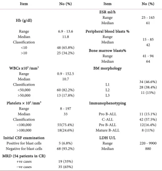
The median total leucocytic count (TLC) was 10.7 × 10³/mm with a range of 0.9 − 152.5 × 10³/mm. CNS leukemia infiltration was present in 6.8% (5/73) of patients. Laboratory characteristics of the patients are shown in Table 2.
Response to Induction Chemotherapy 54 (74%) of patients achieved morpho-logic remission, while 19 (26%) failed to achieve remission. Patients achieving morphologic remission were evaluated for MRD using flowcytometric analysis. MRD positivity was found in 19 cases (35%), while 35 patients (65%) were MRD−ve.
Table 3 clarifies the correlation between blast events (CD34+/CD45+) & MFI
Table 1. Clinical Patients characteristics.
Item No (%) Item No (%)
Age(year) Range
Median 15 - 54 29
Lymphedenopathy Present
Absent 23 (31.5) 50 (68.5) Sex
Female
Male 19 (26%) 54 (74%)
Splenomegaly Present
Absent 43 (59) 30 (41) Fever
Present
Absent 40 (54.8) 33 (45.2)
Hepatomegaly Present
Absent 20 (27.4) 53 (72.6) Pallor
Present
Absent 45 (61.6) 28 (38.4)
Purpura Present
Absent 14 (19.2) 59 (80.8)
CNS manifestation 4 (5.4%)
Response to chemotherapy CR
No CR 54 (74%) 19 (26%)
Table 2. Laboratory Patients characteristics.
Item No (%) Item No (%)
Hb (g/dl) Range Median Classification <10 >10
6.9 - 13.6 11.8
48 (65.8%) 25 (34.2%)
ESR ml/h Range
Median 25 - 145 61 Peripheral blood blasts %
Range
Median 15 - 85 42 Bone marrow blasts%
Range
Median 41 - 94 64 WBCs x103 /mm3
Range Median Classification
<50,000 >50,000
0.9 - 152.5 10.7 60 (82.2%) 13 (17.8%) BM morphology L1 L2 L3 34 (46.6%) 28 (38.4%) 11 (15%)
Platelets × 103 /mm3
Range Median Classification
<100,000 >100,000
8 - 197 33 55(75.4%) 18(24.6%) Immunophenotyping Pro B-ALL C-ALL Pre B-ALL Mature B-ALL 11 (15.1%) 42 (57.5%) 12(16.4%) 8 (11%) Initial CSF examination
Positive for blast cells
Negative for blast cells 68 (93.2%) 5 (6.8%)
LDH U/L Range
Median 220 - 9900 880 MRD (54 patients in CR)
+ve cases
−ve cases 19 (35%) 35 (65%)
Disease Free Survival:
[image:6.595.211.539.325.672.2]with MRD−ve (58%) compared to (14%) for patients with MRD +ve (Log Rank Test; P = 0.01) as shown in Table 4.
Overall Survival:
The 2-year OS for the studied patients was 46%. The median OS (95% CI) was 26.1 months (23.5 - 29.2). The 2-year OS was significantly higher in patients with MRD-ve (58%) compared to (22%) for those with MRD +ve (Log Rank Test; P = 0.04) as shown in Table 4.
Figure 1 represents the comparison between blast events & MFI of CD45 at
day (0) and after morphological remission for the same B-ALL patient. The dot-plot analysis of flowcytometry (a) shows that the region gate R5 represent CD34positive blasts with dim expression of CD45 (MFI 23.75) and region gate R6 represent Hematogones (CD34 positive with high expression of CD45) at day (0); (b) shows that there’s a marked decrease in blast number in region gate R5 which represent MRD, MFI of CD45 (9.9), MRD = 0.06.
5. Discussion
MRD is attributed to the few number of cells remaining in the patient’s blood during treatment or during the remission period and is the main reason for re-lapse in leukemic patients. Molecular methods are used nowadays as a golden standard, where FCM for MRD determination is getting developed day-by-day.
[image:7.595.208.541.505.568.2]Detection of MRD is very important in detecting the efficacy of treatment and the patient’s remission status and expecting the possibility of relapse. MRD di-agnosis also helps in assigning different treatment regime ranging from signifi-cant treatment reduction to mild or strong intensification. In addition to its great role in relapsed patients and that undergoing stem cell transplantation as a guide to treatment strategy.
Table 3. Correlation between blast events (CD34+/CD45+) & MFI of CD45.
MFI of blasts with CD45+
R P
Percent of blasts CD45+/CD34+ −0.128 >0.05
[image:7.595.209.541.614.730.2]P > 0.05 is not significant.
Table 4. Comparison between DFS & OS in both MRD+ve and MRD−ve Patients.
All Patients MRD−ve MRD+ve P
DFS (month) Mean (CI)
Median (CI) 21.7 (18.2 - 25.2) 22 (14.2 - 29.7) 25.6 (21.7 - 29.5) Not reached 13.1 (9.3 - 16.8) 12 (6.7 - 17.3) 0.01
OS (month) Mean (CI)
The outcome of ALL in adults is challenging as compared with that observed in children. The rate of disease relapse is much higher in adults. Although cure rates in children approach 90%, no more than 40% of adult patients remain free of leukemia after 5 years, and this rate is much lower in older patients [10].
The CR rate in our study was 74% compared to 88% in a study done in NCI
[11], however their study included patients with both B and T-ALL, while our patients were B-ALL as shown in Table 1.
Measurement of MRD might be used as an end point that significantly short-ens the follow up period. MRD has been shown to be prognostic essentially at every time point studied, though the most useful measurements appear to be relatively early in therapy, during or after induction and early in consolidation
[12].
There are cut-off points for MRD, where >0. 01% blasts in bone marrow at any time points during treatment had a significantly increase risk to relapse [13]. MRD ≥1% at the end of induction remission therapy and MRD ≥0.1% during continuation therapy are certainly at high risk of relapse. On the other hand MRD can help in identifying patients with favorable outcome [13].
Many cutoff values were applied by adult ALL study groups, which differ from one another in patients population and the MRD time point. NILG takes cutoff value of 1 × 10⁻⁴ at week 16 and absence of detectable MRD at week 22 [14]. PETHEMA takes a cut off value of 5 × 10⁻⁴ at weeks 16 to 18.7. Another Group for Research on Adult Acute Lymphoblastic Leukemia takes a cutoff value of 1 × 10⁻⁴ at week 6 for all ALL Philadelphia negative patients [15], while a cutoff 10−3 for high-risk patients [12]. However, all these studies highlight the prognostic value of MRD detection in adult patients with ALL. In our study we performed flow cytometric analysis at day (O) to be followed by MRD flowcytometric de-tection to patients in CR (Complete Response) after achieving morphological remission.
In our study, the results of minimal residual disease status of B-ALL patients after morphological remission showed that (65%) of patients had negative MRD (<0.01), 35% of patients had positive MRD (≥0.01) as shown in Table 2.
CD45 in combination with CD10, CD19 and CD34were positive in 84% of our patients at diagnosis, which allow us to use this combination for further assess-ment of MRD after morphological remission, Campana and Coustan Smith,
2003 [16], found the combination which includes (CD19, CD34, CD10, CD45)
to be informative in 30% - 50% of cases.
Lucio et al., (2001) [17] reported that CD45, CD19 and CD34 are informative in 22.2% of B-ALL, whereas Vidriales et al., (2003) [17] stated that CD45, CD10, CD19, and anti-TdT combination was applicable to 70% - 80% of B-ALL. Patkar
et al., (2012) [18] found that CD20, CD10, CD45 and CD19 combination was informative in 71.4% of B-ALL cases.
to 49% in NCI [11] and 43% at 3-years follow up reported by Larson et al.[21].
Our data are shown in Figures 2-5.
[image:9.595.210.540.191.439.2]It is accepted that cluster of MRD cells between 10 and 100 have to be identi-fied in a given sample to assure that MRD cells have been seen. This is for achieving sensitivity between 10−4 to 10−5, consequently 105 to 106 leukocytes have to be screened. In a study done by [22] they stated that the levels of MRD are proportional to the risk of relapse, MRD equal or greater than 1% at the end
Figure 2. Disease free survival for all patients.
Figure 3. Disease free survival according to MRD status.
MRD -ve
MRD +ve
[image:9.595.217.531.478.718.2]Figure 4. Overall survival for all patients.
Figure 5. Overall survival according to MRD status.
of induction remission therapy is accompanied with unfavorable outcome, so recommending transplantion in first remission for these patients. The Interna-tional Berline-Frankfurt-Munstere (I-BFM) Study Group found that patients with MRD levels of 0.1% or higher on both day 33 and day 78 of treatment had a relapse rate of 75%, prompting treatment intensification for this group of pa-tients is needed [23].
MRD -ve
MRD +ve
[image:10.595.217.518.370.603.2]Patients with early clearance of leukemic cells typically remain MRD-negative, and their prognosis is excellent with current treatment protocols [24]. Some of the studies stated that treatment deintensification should be considered for these patients, others were against this and stated that early MRD negativity might be a good prognostic feature only in the context of intensive therapy. Thus, if ther-apy is deintensified, the risk of relapse of MRD-negative patients might increase significantly, we agree with the second opinion, however deintensification might be especially useful when intensive therapy confers a high risk of serious toxici-ties or in older adult patients, where the potential benefits of treatment deinten-sification might outweigh the risk of relapse.
6. Conclusion
MRD detection by flow cytometry using the combination of CD45 with CD10, CD19 & CD34 is an easy and reliable method. Patients with positive MRD are at higher risk of relapse and have lower overall survival rates compared to those with MRD−ve. Future studies focusing on treatment intensification for the group of patients with +ve MRD aiming to improve the treatment outcome are warranted.
References
[1] Kern, W., Schoch, C., Haferlach, T., et al. (2005) Monitoring of Minimal Residual Disease in Acute Myeloid Leukemia. Critical Reviews in Oncology/Hematology, 56, 283-309. https://doi.org/10.1016/j.critrevonc.2004.06.004
[2] Ibrahim, A.S., Khaled, H.M., Mikhail, N.N., Baraka, H. and Kamel, H. (2014) Can-cer Incidence in Egypt: Results of the National Population-Based CanCan-cer Registry Program. Journal of Cancer Epidemiology, 2014, Article ID: 437971.
https://doi.org/10.1155/2014/437971
[3] Bruggemann, M. Raff, T. and Kneba, M. (2012) Has MRD Monitoring Superseded Other Prognostic Factors in ALL? Blood, 120, 4470-4481.
https://doi.org/10.1182/blood-2012-06-379040
[4] Schrappe, M. (2012) Minimal Residual Disease: Optimal Methods, Timing, and Clinical Relevance for an Individual Patient. Hematology, 2012, 137-142.
[5] Beldjord, K., Chevret, S., Asnafi, V., et al., Group for Research on Adult Acute Lymphoblastic Leukemia (GRAALL) (2014) Oncogenetics and Minimal Residual Disease Are Independent Outcome Predictors in Adult Patients with Acute Lym-phoblastic Leukemia. Blood, 123, 3739-3749.
https://doi.org/10.1182/blood-2014-01-547695
[6] Deborah, S., Adrianac-Foxwell, E., Govind, B., et al. (2010) Hematogones: A Review and Update. Leukemia & Lymphoma, 51, 10-19.
[7] Hoelzer, D., Gökbuget, N., Ottmann, O., Pui, C.H., Relling, M.V., Appelbaum, F.R.,
et al. (2002) Acute Lymphoblastic Leukemia. Hematology, 2002, 162-192.
https://doi.org/10.1182/asheducation-2002.1.162
[9] Norusis, M. (2009) SPSS 17.0 Advanced Statistical Procedures Companion. Prentice Hall, Inc., Upper Saddle-River, NJ.
[10] Hoffbrand, A.V. and Moss, P.A.H. (2011) Essential Hematology. 6th Edition, Blackwell Publishing Ltd., Oxford, UK.
[11] Samra, M.A., Mahmoud, H.K., Abdelhamid, T.M., Kamel, A.M., et al. (2013) The Prognostic Significance of Minimal Residual Disease in Adult Egyptian Patients with Precursor Acute Lymphoblastic Leukemia. Journal of the Egyptian National Cancer Institute, 25, 135-142. https://doi.org/10.1016/j.jnci.2013.05.004
[12] Dhédin, N., Huynh, A., Maury, S., et al., GRAALL Group (2015) Role of Allogeneic Stem Cell Transplantation in Adult Patients with Ph-Negative Acute Lymphoblastic Leukemia. Blood, 125, 2486-2496. https://doi.org/10.1182/blood-2014-09-599894
[13] Coustan-Smith, E., Sancho, J., Behm, F.G., et al. (2002) Prognostic Importance of Measuring Early Clearance of Leukemic Cells by Flow Cytometry in Childhood Acute Lymphoblastic Leukemia. Blood, 100, 52-58.
https://doi.org/10.1182/blood-2002-01-0006
[14] Bassan, R., Spinelli, O., Oldani, E., et al. (2009) Improved Risk Classification for Risk-Specific Therapy Based on the Molecular Study of Minimal Residual Disease (MRD) in Adult Acute Lymphoblastic Leukemia (ALL). Blood, 113, 4153-4162.
https://doi.org/10.1182/blood-2008-11-185132
[15] Campana, D. and Coustan-Smith, E. (2003) Minimal Residual Disease Studies by Flow Cytometry in Acute Leukemia. Acta Haematologica, 112, 8-15.
https://doi.org/10.1159/000077554
[16] Lucio, P., Gaipa, G., van Lochem, E.G., et al. (2001) BIOMED-I Concerted Action Report: Flow Cytometric Immunophenotyping of Precursor B-ALL with Standar-dized Triple-Staining. Leukemia, 15, 1185-1192.
https://doi.org/10.1038/sj.leu.2402150
[17] Vidriales, M.B., Perez, J.J., Lopez-Berges, M.C., Gutierrez, N., Ciudad, J., Lucio, P.,
et al. (2003) Minimal Residual Disease (MRD) in Adolescent (>14 Years) and Adult Acute Lymphoblastic Leukemias (ALL): Early Immunophenotypical Evaluation Has High Clinical Value. Blood, 101, 4695-4700.
https://doi.org/10.1182/blood-2002-08-2613
[18] Patkar Nikhil, A.A., Bargavi, B., Ahmed, R., et al. (2012) Standardizing Minimal Re-sidual Disease by Flow Cytometry for Precursor B Lineage Acute Lymphoblastic Leukemia in a Developing Country. Cytometry Part B (Clinical Cytometry), 82B, 252-258. https://doi.org/10.1002/cyto.b.21017
[19] Abdel Hamid, T.M., Mahmoud, K.H., Kamel, A.M., El Sharkawy, N., et al. (2005) Risk Adapted Chemotherapy Treatment for Adult Acute Lymphoblastic Leukemia.
The Egyptian Journal of Haematology, 30, 579.
[20] Mahmoud, H.K., Hamza, M.R., Gad El Mawla, N., Khaled, H.K., Elzawahry, H., Abdelhamid, T., et al. (1995) The Impact of Intensified Induction Protocol on Re-mission Rate and Duration in Adult Acute Lymphoblastic Leukemia. The Egyptian Journal of Haematology, 20, 225.
[21] Larson, R.A., Dodge, R.K., Linker, C.A., Stone, R.M., Powel, B.L., Lee, E.V., et al.
(1998) A Randomized Controlled Trial of Filgrastim during Remission Induction and Consolidation Chemotherapy for Adults with Acute Lymphoblastic Leukemia: CALGB Study 9111. Blood, 92, 1556-1564.
[22] Pui, C.H. and Evans, W.E. (2006) Treatment of Acute Lymphoblastic Leukemia.
The New England Journal of Medicine, 354, 166-178.
[23] Flohr, T., Schrauder, A., Cazzaniga, G., Panzer-Grumayer, R., van der Velden, V., Fischer, S., et al. (2008) Minimal Residual Disease-Directed Risk Stratification Us-ing Real-Time Quantitative PCR Analysis of Immunoglobulin and T-Cell Receptor Gene Rearrangements in the International Multicenter Trial AIEOP-BFM ALL 2000 for Childhood Acute Lymphoblastic Leukemia. Leukemia, 22, 771-782.
https://doi.org/10.1038/leu.2008.5
[24] National Institutes of Health (NIH) (2009) Minimal Residual Disease in Acute Lymphocytic Leukemia. Seminars in Hematology, 46, 100-106.
Submit or recommend next manuscript to SCIRP and we will provide best service for you:
Accepting pre-submission inquiries through Email, Facebook, LinkedIn, Twitter, etc. A wide selection of journals (inclusive of 9 subjects, more than 200 journals)
Providing 24-hour high-quality service User-friendly online submission system Fair and swift peer-review system
Efficient typesetting and proofreading procedure
Display of the result of downloads and visits, as well as the number of cited articles Maximum dissemination of your research work
Submit your manuscript at: http://papersubmission.scirp.org/