Oncoplastic Breast Surgery Using Spindle Shaped-Partial
Mastectomy for Early Breast Cancer in the Upper
Quadrant Area
*
Yuko Kijima#, Heiji Yoshinaka, Munetsugu Hirata, Yoshiaki Shinden, Sumiya Ishigami, Akihiro Nakajo, Hideo Arima, Takaaki Arigami, Hiroshi Okumura, Shoji Natsugoe
Department of Digestive Surgery, Breast and Thyroid Surgery, Kagoshima University Graduate School of Medical and Dental Sci-ences, Kagoshima, Japan.
Email: #ykijima@m3.kufm.kagoshima-u.ac.jp
Received February 25th, 2013; revised March 27th, 2013; accepted April 4th, 2013
Copyright © 2013 Yuko Kijima et al. This is an open access article distributed under the Creative Commons Attribution License, which permits unrestricted use, distribution, and reproduction in any medium, provided the original work is properly cited.
ABSTRACT
Background: Oncoplastic surgery is becoming more common, however, only several reports have been published in Japan. We report the results of simple oncoplastic surgery for Japanese patients with early breast cancer in the upper quadrant area. Methods: In seven patients with a past history of breast-feeding and ptotic breasts, we performed on-coplastic surgery involving partial mastectomy and the resection of excess skin and parenchymal tissue. Results: None of the patients received a contralateral operation to produce symmetrical breasts. The width of the resected excess skin tissue ranged from 20 to 50 mm, with the mean width being 30 mm, and its length ranged from 50 to 90, with the mean length being 77 mm. The width of the resected gland tissue ranged from 40 to 65 mm, with the mean width being 53 mm, and its length ranged from 70 to 100 mm, with the mean length being 97 mm. The cosmetic results were excellent. Conclusions: Oncoplastic surgery using spindle shaped-resection was successfully performed in patients with upper quadrant lesions, and the cosmetic results were excellent.
Keywords: Breast Cancer; Spindle-Shaped Resection; Breast-Conserving Surgery; Oncoplastic Surgery; Reduction Mammoplasty
1. Introduction
Oncoplastic Breast Surgery (OBS), which combine the concepts of oncologic and plastic surgery, are becoming more common, especially in Western countries [1,2]. There are many different oncoplastic surgical techniques, one of which involves careful planning of skin and enchymal excisions, reshaping of the gland after the par-enchymal excisions, and repositioning of the nipple are-ola complex (NAC) to the center of the breast mound with or without correction of the contralateral breast to achieve better symmetry [3-5]. We have reported that oncoplastic surgery combining partial mastectomy and recentralization of the NAC with/without a contralateral operation produced excellent results in Japanese patients
with ptotic breasts as well as Western women [6-8]. On the other hand, the resection of upper deformities fol-lowed by immediate volume replacement using a local flap or a distant autologous graft resulted in good out-comes [9,10].
We herein report our early experiences of oncoplastic surgery involving partial mastectomy and the resection of excess skin tissue without a contralateral operation in six Japanese patients with early breast cancer in the upper quadrant region of their ptotic breasts.
2. Patients
From June 2006 to May 2009, seven Japanese patients were diagnosed with early breast cancer and received successful breast conserving surgery without any preop-erative systemic therapy. The indications for spindle- shaped resection of breast tissue and excess skin tissue were as follows: 1) the patient had ptotic (the nipple level was beneath the inframammary line) or large breasts; 2) *Conflict of interest statement: We declare that there are no financial
relationships or other interests with regard to this manuscript that might be construed as constituting a conflict of interest for any author. Informed consent: Written informed consent from each patient has been received before submission.
the cancer lesion was restricted to the upper-outer or up-per-inner quadrant; and 3) informed consent was ob-tained preoperatively after an explanation of the surgical procedure. A digital camera with a resolution of 14.1 megapixels was used, and a blue panel was used as the background. Photographs were taken in four positions with the patient standing on floor marks: facing the cam-era with their arms down, facing the camcam-era with their arms up, from the left side with their arms up, and from the right side with their arms up.
None of the seven patients who agreed to undergo spindle-shaped partial mastectomy combined with excess skin removal, received preoperative systemic chemo- or endocrine therapy. The mean patient age was 72.4 years (range: 57 - 84). All patients were postmenopausal and had experiences of breast feeding 2 to 4 children, with the mean number of breastfed children being 2.6. The mean size of the tumor was 11.9 mm (range: 7 - 27) (Ta-ble 1). The results of tumor histology are shown in Ta(Ta-ble 1. Axillary lymph adenectomy was performed for one patient with a clinical diagnosis of T2N0M0. Sentinel lymph node (SLN) biopsy using the radioisotope (RI) method and dye method was performed in the remaining 6 patients, who were preoperatively diagnosed with T1N0M0 tumors. In these six patients, the SLN were intraoperatively revealed to be negative, so axillary con-
servation was performed (Table 2).
3. Surgical Procedure
3.1. Design
The patients were seen by the breast surgeon two days before the surgery so that he could plan the operation, make drawings, and explain the different surgical options to the patient, e.g., other oncoplastic surgical techniques such as immediate volume replacement using a free graft or local flap, which were described previously. For spin-dle-shaped partial mastectomy and resection of excess skin tissue, the incision lines were drawn with the patient in a standing position after marking the resection area together with a surgical margin of at least 20 mm with the patient in a supine position (Figure 1).
3.2. Sentinel Lymph Node Biopsy and Axillary Lymphadenectomy
[image:2.595.58.538.434.552.2]In one patient who received axillary lymphadenectomy and two of six patients who underwent SLN biopsy, an-other incision was made in the axillary area to allow SLN biopsy or axillary lymphadenectomy. In the remaining four patients, the SLN were biopsied via the same inci-sion as was used for the partial mastectomy.
Table 1. Patients’ clinical data.
Case Age Laterality Location Height Weight %IBW No. of children Ptosis Systemic disease Distance to nipple Tumor size (mm)
1 84 Right Upper-inner 141 48 113 4 Yes None 35 27
2 76 Left Upper-inner 152 61 108 4 Yes None 80 12
3 63 Left Upper-inner 160 80 149 2 Yes Diabetes 60 7
4 71 Left Upper-inner 144 52 112 2 Yes Arrhythmia 50 12
5 57 Right Upper-inner 158 70 108 3 Yes None 60 8
6 80 Right Upper-outer 157 59 114 2 Yes None 30 9
7 76 Left Upper-outer 151 51 101 1 No None 90 8
Table 2. Surgical findings.
Case
Surgical margin (to lateral side)
mm
Surgical margin (to nipple) mm
Skin size (width) mm Skin size (length) mm Size of resected gland (width) mm Size of resected gland (length) mm Ax* Operative period (min) Bleeding (ml) Cosmesis Score (JBCS) Cosmesis Score (ABNSW)
1 25 40 35 85 80 110 Yes 90 15 10 11
2 20 50 20 50 65 110 SLN** 80 5 11 13
3 20 60 30 90 40 100 SLN** 149 15 10 12
4 20 20 25 80 50 100 SLN** 131 10 11 13
5 20 40 25 70 50 70 SLN** 106 5 11 14
6 25 25 30 80 60 100 SLN** 162 20 11 12
7 20 40 50 90 50 100 SLN** 92 20 12 15
*
[image:2.595.61.537.523.717.2]3.3. Spindle Shaped-Partial Mastectomy
On the day of surgery, both breasts were placed into the operative field to allow evaluation of their symmetry during the operation. The partial mastectomy consisted of the removal of tissue from the breast containing the tumor together with a tumor-free margin of at least 2 cm, including a spindle-shaped area of skin above the tumor and the pectoral fascia below it (Figures 2(a)-(c)).
During the operation, the surgical margins were his-tologically examined to ensure that they did not include the cancer lesion. It took 30 - 50 minutes to obtain the results from the pathologist, and this time is included in the total operative period shown in Table 1. For both
tumor types; i.e., the upper-outer and upper-inner quad-
rant tumors, SLN biopsy was performed using the same incision as was used for the partial mastectomy.
3.4. Reconstruction of Defects
After washing them with saline, the inner and lateral glandular flaps were lined up and sutured together to cover the excision defect (Figures 2(d)-(f)). A closed suction drainage line was placed onto the surface of the pectoralis major muscle and/or the axillary defect in each case.
3.5. Adjuvant Therapy
In all patients, pathological margins of over 10 mm were maintained from the edge of the resected area to the can-cer lesion. The treated lesions included both invasive and intraductal lesions. No patient displayed metastatic dis-ease or received postoperative radiotherapy to the rem-nant gland. Six of them received aromatize inhibitor (1
mg/day of anastrozol or 2.5 mg/day of letrozol) as an adjuvant hormone therapy.
4. Cosmetic Evaluation
The cosmetic assessment after the breast-conserving therapy was performed according to the method reported by Sawai’s group, which was supported by the Japanese Breast Cancer Society [11]. This assessment contains eight items: 1) breast size; 2) breast shape; 3) wound scar; 4) softness of the breast; 5) shape and size of the nip-ple-areola complex; 6) color of the nipnip-ple-areola com-plex; 7) level of the nipple (difference in distance from the suprasternal notch between the bilateral nipples); and 8) the lowest point of the breast (difference between the bilateral breasts). The cosmetic outcome was evaluated as excellent when the total score was 12 points, good when it was 9 to 11, fair when it was 5 to 8, and poor when it was 0 to 4.
Breast shape was also subjectively assessed by two of the authors and scored using the ABNSW system report-
ed by Yamashita et al. [12]. The ABNSW contains five
items: asymmetry (A), breast shape (B), nipple deforma-tion (N), skin condideforma-tion (S), and wound scar (W). The score in each category is graded as follows: 3—excellent: at first sight, there was no visible difference between the breasts; 2—good: there were few differences between the bilateral breasts and these were only apparent from close observation; 1—fair: there were marked differences be-tween the bilateral breasts from a distance; 0—poor: there were severe, ugly differences between the bilateral breasts. We scored all five items from 0 to 3 to produce the total score. The cosmetic outcome was evaluated as excellent when the total score was 15 points, good
[image:3.595.71.527.510.669.2](a) (b) (c)
(a) (b) (c)
(d) (e) (f)
Figure 2. (a) Preoperative marking. The incision line and surgical margin of the gland were drawn as a black line and dotted line, respectively; (b) A spindle shaped area of skin was also removed; (c) A spindle shaped area of columnar tissue was re-sected together with the fascia of the pectoral major muscle; (d) Medial and lateral glandular flaps were sutured using 3-0 vicryl sutures; (e) and (f) Subcutaneous 4-0 PDS sutures were also added. Mj: Pectoral major muscle.
when it was 11 to 14, fair when it was 6 to 10, and poor when it was 0 to 5.
5. Results
The patients’ clinical data are shown in Table 1. No ad-ditional resections due to a positive margin were neces-sary. The total operative period ranged from 92 to 162 minutes, with the mean period being 116 minutes. The suction drainage period required for each breast ranged from 3 to 5 days postoperatively.
The postoperative observation period ranged from 5 to 60 months (median: 20 months). There were no compli-cations in any of the treated breasts, and no local or dis-tant recurrence was seen in any case (Figures 3 and 4). In the objective assessment (Japanese Breast Cancer Society), the total cosmetic score ranged from 10 to 12;
the score was 12 in one patient, 11 in four, and 10 in two patients. In the subjective assessment (ABNSW), the total cosmetic scores ranged from 12 to 15; the score was 15 in one patient, 14 in one, 13 in two, 12 in two, and 11 in one patient. The cosmetic outcomes were excellent or good in both cosmetic assessments.
6. Discussion
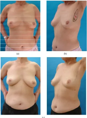
[image:4.595.69.523.90.478.2]
(a) (b)
Figure 3. (a) The resected spindle-shaped area of columnar tissue and excess skin; (b) One-year postoperative findings (Case 1).
(a) (b)
[image:5.595.145.453.95.279.2]
(c)
[image:5.595.154.442.307.697.2]removing so much breast tissue that it will leave a de-formed breast or a large discrepancy compared with the contralateral breast. Quadrantectomy increases the risk of a poor aesthetic result if no partial breast reconstruction is performed [18-20]. The impact of tumor surgery on cosmesis in BCT has also been emphasized by leading surgeons: surgical technique may influence the aesthetic outcome to a greater extent than radiation effects [21].
Postquadrantectomy deformities include localized de-fects in skin and glandular tissue, distortion and/or dislo-cation of the areola, and retraction of the breast tissue. Perhaps the most prominent and frequent failure in achieving a good aesthetic outcome is due to a lack of breast symmetry [22]. Surgical techniques used to ad-dress the conflicts between oncological and cosmetic results, including both total and partial mastectomy, can be classed under the general term of oncoplastic surgery, which is a new surgical approach that allows wide exci-sion but prevents breast deformities by reconstructing large resection defects immediately [13]. Several on-coplastic surgical procedures have been discussed in pre-vious reports. Hoffmann classified all breast surgeries into 12 subgroups according to a two-type, six-tier clas-sification system comprising 12 main categories [23]. In breast conserving surgery, ranging from simple excision up to quadrantectomy, defect repair without tissue mobi-lization is regarded as grade 1 complexity, and complex oncoplastic or reconstructive breast cancer procedures involving pedicled or free distant flap reconstruction and, where indicated, microvascular anastomosis for defect coverage are regarded as grade 6. Our procedure, a spin-dle shaped partial mastectomy in which excess gland and skin tissue are removed, is regarded to be of grade 1 complexity according to their classification. In fact, it is easier than other oncoplastic breast surgical techniques that we described previously, such as volume replace-ment or reduction type breast surgery.
Masetti et al. outlined several important aspects of
oncoplastic breast surgery procedures: first, careful plan-ning of skin incisions and parenchymal excisions must be made, following the templates used for reduction mam-maplasty and mastopexy; second, the gland must be re-shaped after the parenchymal excision; third, the nip-ple-areola complex must be repositioned in the center of the breast mound; and fourth, the symmetry of the con-tralateral breast must be corrected. Another researcher highlighted four integral features of oncoplastic surgery [3]. All of these require planning with respect to the placement and closure of wounds and careful assessment of the oncological need and aesthetic aim [24].
We reported good results for immediate volume re-placement surgery using a free dermal fat graft for the treatment of upper-inner lesions [10,25,26]. This is thought
to be sufficient for slim ladies with non-ptotic, small breasts, e.g., average Japanese ladies. It is easy to per-form; however, there are some disadvantages to this pro-cedure, for example, it is difficult to maintain softness, and an additional scar is made at the donor site. In such cases with breast cancers on upper quadrant lesion on ptotic breasts, OBS combining with partial mastectomy and reduction type mammoplasty brought excellent cos-metic results, although surgical techniques might be more complicated than former ones [27].
We performed oncoplastic surgery involving spindle- shaped partial mastectomy and excess skin removal in several older ladies in this series, and all procedures were successful. We produced satisfactory cosmetic results, and the observation period was thought to be sufficient. One drawback of this technique is the length of the scar it leaves. In particular, hypertrophic scars in the upper- inner quadrant area will reduce cosmesis. In this series, the patients’ satisfaction was high because the symmetry of the bilateral breasts was well maintained, especially in the position of NAC, and the softness of the whole breast was also retained by avoiding tissue mobilization to re-pair the breast defects.
The number of cases in this paper was not so large, and the follow-up period was short; however, we have revealed that oncoplastic surgery using spindle shaped- partial mastectomy combined with the removal of excess skin for patients with upper quadrant lesions in ptotic or large breasts produced excellent cosmetic results via a simple technique. This simple oncoplastic technique is an excellent procedure that yields very satisfactory cosmetic results, and it should be considered a suitable therapeutic option.
7. Conclusion
OBS involving spindle-shaped partial mastectomy com-bined with excess skin removal was successfully per-formed and is expected to become more popular for the treatment of aging patients.
REFERENCES
[1] W. P. Audretsch, M. Rezai, C. Kolotas, N. Zamboglou, T. Schnabel and H. Bojar, “Onco-Plastic Surgery: ‘Target’ Volume Reduction (BCT-Mastopexy), Lumpectomy Re-construction (BCT-ReRe-construction) and Flap-Supported Operability in Breast Cancer,” Proceeding 2nd European Congress on Senology, Vienna, 2-6 October 1994, pp. 139-157.
Chi-esa and A. Antinori, “Oncoplastic Techniques in the Con-servative Surgical Treatment of Breast Cancer,” Breast cancer, Vol. 7, No. 4, 2000, pp. 276-280.
doi:10.1007/BF02966389
[4] Y. Kijima, H. Yoshinaka, Y. Funasako, S. Natsugoe and T. Aikou, “Oncoplastic Surgery after Mammary Reduc-tion and Mastopexy for Bilateral Breast Cancer Lesions: Reports of a Case,” Surgery Today, Vol. 38, No. 4, 2008, pp. 335-339.doi:10.1007/s00595-007-3628-z
[5] H. Zaha, O. Hakazu, M. Watanabe and M. Higa, “Breast- Conserving Surgery Using Reduction Mammoplasty,”
Japanese Journal of Breast Cancer, Vol. 23, 2008, pp. 211-215.
[6] Y. Kijima, H. Yoshinaka, M. Hirata, T. Mizoguchi, S. Ishigami, A. Nakajo, H. Arima, S. Ueno and S. Natsugoe, “Oncoplastic Surgery in a Japanese Patient with Breast Cancer in the Lower Inner Quadrant Area: Partial Mas-tectomy Using Horizontal Reduction Mammoplasty,”
Breast Cancer, 2010.
[7] Y. Kijima, H. Yoshinaka, M. Hirata, K. Kaneko, M. Hi-rata, T. Mizoguchi, S. Ishigami, H. Arima, A. Nakajo, S. Ueno and S. Natsugoe, “Oncoplastic Surgery Combining Partial Mastectomy with Breast Reconstruction Using the Free Nipple-Areola Graft Technique for a Japanese Pa-tient with DCIS in a Ptotic Breast: A Case Report,” Sur-gery Today, Vol. 41, No. 3, 2011, pp. 390-395.
doi:10.1007/s00595-010-4294-0
[8] Y. Kijima, H. Yoshinaka, H. Munetsugu, T. Mizoguchi, S. Ishigami, A. Nakajo, H. Arima, S. Ueno and S. Natsugoe, “Oncoplastic Surgery for Japanese Patients with Breast Cancer of the Lower Pole,” Surgery Today, Vol. 41, No. 10, 2011, pp. 1461-1465.doi:10.1007/s00595-011-4490-6 [9] Y. Kijima, H. Yoshinaka, Y. Funasako, K. Kaneko, M.
Hirata, S. Ishigami and S. Natsugoe, “Immediate Recon-struction Using Thoracodorsal Adipofascial Flap after Partial Mastectomy,” Breast, Vol. 18, No. 2, 2009, pp. 126-129.doi:10.1016/j.breast.2009.02.006
[10] Y. Kijima, H. Yoshinaka, T. Owaki and T. Aikou, “Early Experience of Immediate Reconstruction Using Autolo-gous Free Dermal Fat Graft after Breast Conservational Surgery,” Journal of Plastic, Reconstructive & Aesthetic Surgery, Vol. 60, No. 5, 2007, pp. 495-502.
doi:10.1016/j.bjps.2006.06.004
[11] Japanese Breast Cancer Society, “Cosmetic Assessment after Breast Conserving Surgery,” The 12th Annual Meet-ing of the Japanese Breast Cancer Society, 2004, pp. 107-109.
[12] K. Yamashita, “Cosmetic Assessment,” Journal of Japan Society for Endoscopic Surgery, Vol. 10, No. 2, 2004, pp. 165-170.
[13] K. B. Clough, J. Cuminet, A. Fitoussi, C. Nos and V. Mosseri, “Cosmetic Sequelae after Conservative Treat-ment for Breast Cancer: Classification and Results of Sur-gical Correction,” Annals of Plastic Surgery, Vol. 41, No. 5, 1998, pp. 471-481.
doi:10.1097/00000637-199811000-00004
[14] J. Bostwick III, C. Paletta, C. R. Hartramph, “Conserva-tive Treatment for Breast Cancer: Complications
Requir-ing Reconstructive Surgery,” Annals of Surgery, Vol. 203, No. 5, 1986, pp. 481-490.
doi:10.1097/00000658-198605000-00006
[15] J. Y. Petit and M. Rietjens, “Deormities Following Tu-morectomy and Partial Mastectomy,” In: B Noon, Ed.,
Plastic and Reconstructive Surgery of the Breast, Marcel Decker, Philadelphia, 1991.
[16] K. B. Clough, C. Nos, R. J. Salmon, M. Soussaline and J. C. Durand, “Conservative Treatment of Breast Cancers by Mammaplasty and Irradiation: A New Approach to Lower Quadrant Tumors,” Plastic & Reconstructive Sur-gery, Vol. 96, No. 2, 1995, pp. 363-370.
doi:10.1097/00006534-199508000-00015
[17] M. Noguchi, Y. Saito, Y. Mizukami, A. Nonomura, N. Ohta, N. Koyasaki, et al., “Breast Deformity, Its Correc-tion, and Assessment of Breast Conserving Surgery,”
Breast Cancer Research and Treatment, Vol. 18, No. 2, 1991, pp. 111-118.doi:10.1007/BF01980973
[18] P. Berrino, E. Campora and P. Santi, “Postquadrantec-tomy Breast Deformities: Classification and Techniques of Surgical Correction,” Plastic & Reconstructive Surgery, Vol. 79, No. 4, 1987, pp. 567-572.
doi:10.1097/00006534-198704000-00010
[19] D. Clarke, A. Martinez and R. S. Cox, “Analysis of Cos-metic Results and Complications in Patients with Stage I and II Breast Cancer Treated with Biopsy and Irradia-tion,” International Journal of Radiation Oncology, Bi-ology, Physics, Vol. 9, No. 12, 1983, pp. 1807-1813.
doi:10.1016/0360-3016(83)90348-6
[20] J. R. Harris, M. B. Levene, G. Svensson and S. Hellman, “Analysis of Cosmetic Results Following Primary Radia-tion Therapy for Stage I and II Carcinoma of the Breast,”
International Journal of Radiation Oncology Biology Physics, Vol. 5, No. 2, 1979, pp. 257-261.
doi:10.1016/0360-3016(79)90729-6
[21] W. E. Matory Jr., M. Wertheimer, T. J. Fitzgerald, R. L. Walton, S. Love and W. E. Matory, “Aesthetic Results Following Partial Mastectomy and Radiation Therapy,”
Plastic & Reconstructive Surgery, Vol. 85, No. 5, 1990, pp. 739-746.doi:10.1097/00006534-199005000-00014 [22] A. Grisotti, “Immediate Reconstruction after Partial
Mas-tectomy,” Operative Techniques in Plastic and Recon-structive Surgery, Vol. 1, No. 1, 1994, pp. 1-12.
doi:10.1016/S1071-0949(10)80013-7
[23] J. Hoffmann and D. Wallwiener, “Classifying Breast Cancer Surgery: A Novel, Complexity-Based System for Oncological, Oncoplastic and Reconstructive Procedures, and Proof of Principle by Analysis of 1225 Operations in 1166 Patients,” BMC Cancer, Vol. 9, 2009, p. 108.
doi:10.1186/1471-2407-9-108
[24] A. D. Baildam, “Oncoplastic Surgery of the Breast,” Bri- tish Journal of Surgery, Vol. 89, No. 5, 2002, pp. 532-533.
doi:10.1046/j.1365-2168.2002.02077.x
Cancer Lesion,” Surgery Today, Vol. 41, No. 4, 2011, pp. 477-489.doi:10.1007/s00595-010-4307-z
[26] Y. Kijima, H. Yoshinaka, M. Hirata, Y. Umekita, M. Sohda, C. Koriyama, et al., “Clinical and Pathological Evaluation of Free Dermal Fat Graft after Breast Conser-vative Surgery and Immediate Breast Reconstruction,”
Surgery, Vol. 151, No. 3, 2012, pp. 444-455.
doi:10.1016/j.surg.2011.07.031