THE USE OF KEY
PERFORMANCE INDICATORS
FOR EXTERNAL QUALITY
CONTROL IN HISTOLOGICAL
DISSECTION
Matthew Paul Griffiths
A thesis submitted in partial fulfilment of the requirements of the University of the West of England, Bristol for the degree of Professional Doctorate in Biomedical Science
FACULTY OF HEALTH AND APPLIED SCIENCES, UINVERSITY OF THE WEST OF ENGLAND DEPARTMENT OF HISTOPATHOLOGY, DERBY ROYAL HOSPITAL
i
Contents
List of Figures ... v
List of Tables ... vii
Acknowledgements ... 1
Abstract ... 2
Introduction ... 4
Research Aims and Objectives ... 6
Thesis Overview ... 6
Chapter One ... 6
Chapter Two ... 7
Chapter Three ... 7
Chapter Four ... 7
Chapter Five ... 8
Chapter Six ... 8
Chapter One - Literature Review ... 9
Histological Dissection ... 9
Evidence Based Medicine ...10
Devolution of Responsibility ...11
Quality in Pathology ...13
Key Performance Indicators ...19
Standardisation of Dissection ...21
ii
Change Management ...33
Chapter Two – Study 1 – Review of Recent Practice ...39
Introduction ...40
Method ...41
Results ...44
t – test ...45
Discussion ...53
Chapter Three – Study 2 – The Training Intervention ...57
Introduction ...58
Study Two Part One – Creation of the checklists and collecting baseline data .60 Study Two Part Two – The introduction of the checklist ...62
Study Two Part Three – The training event plus checklist ...63
Study Two Part Four – The training event only ...66
Study Two Part Five – The use of diagrams only ...67
Study Two Part Six – The use of the training event with diagrams ...68
Study Two Part Seven – The use of the checklists only ...69
Study Two Part Eight – Return to the training event with checklists ...70
Results of Study Two ...71
Study Two Part One – Creation of the checklists and collecting baseline data .72 Study Two Part Two – Checklist Introduction ...79
Study Two Part Three – Training Event and Checklists) ...85
iii
Study Two Part Five – Guide Diagrams ...97
Study Two Part Six – Training Event and Guide Diagrams ...102
Study Two Part Seven – Checklists only ...107
Study Two Part Eight – Training Event and Checklists ...112
Changes over time, by tissue type ...117
Changes over time for all specimen types ...130
Chapter Four – Study 3 – Participant Interviews ...137
Introduction ...138
Participants ...139
Ethics ...140
Method ...140
Thematic Analysis of Participant Interviews ...141
Knowledge – “it’s not part of our culture” ...144
Standardisation – “a little bit of not everyone not doing the same thing” ...150
Feedback – “You’re working blindly” ...153
Microscopy – “The amount I learnt is amazing!” ...157
Summary ...158
Chapter Five – Discussion ...162
Summary ...162
The results ...162
The checklists & training event ...166
iv
Attitudes, responsibility & change management ...179
New knowledge ...183
Limitations ...185
Further work ...186
Chapter Six – Conclusions ...188
References ...191
Appendix One – Tissue Pathways and Minimum Datasets ...203
Appendix Two – IBMS & RCPath Dissection Categories ...204
Appendix Three – Baseline Data Collection Sheets ...206
Appendix Four – Dissection Checklists ...210
Appendix Five – Dissection Guide Diagrams ...214
v
List of Figures
Figure 1 - Graph showing stage of colorectal tumour at presentation (Duke’s staging) ... 26
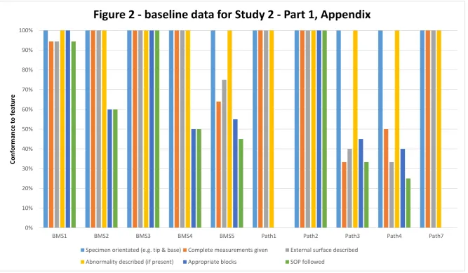
Figure 2 - Baseline Data for Study 2 Part 1, Appendix ... 75
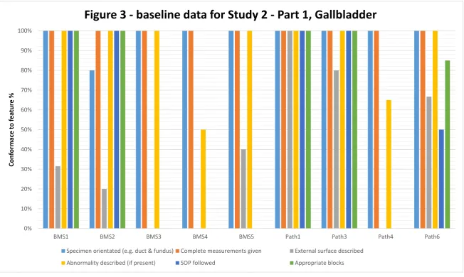
Figure 3 - Baseline Data for Study 2 Part 1, Gallbladder ... 76
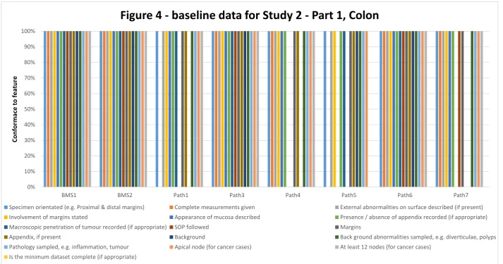
Figure 4 - Baseline Data for Study 2 Part 1, Colon ... 77
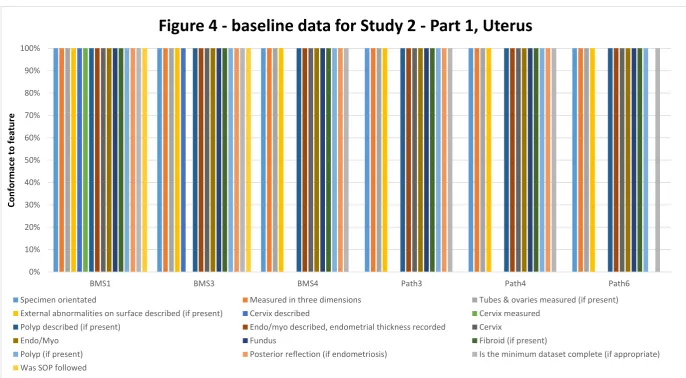
Figure 5 - Baseline Data for Study 2 Part 1, Uterus ... 78
Figure 6 - First Use of Checklist, Study 2 Part 2 – Appendix ... 81
Figure 7 - First Use of Checklist, Study 2 Part 2 – Gallbladder ... 82
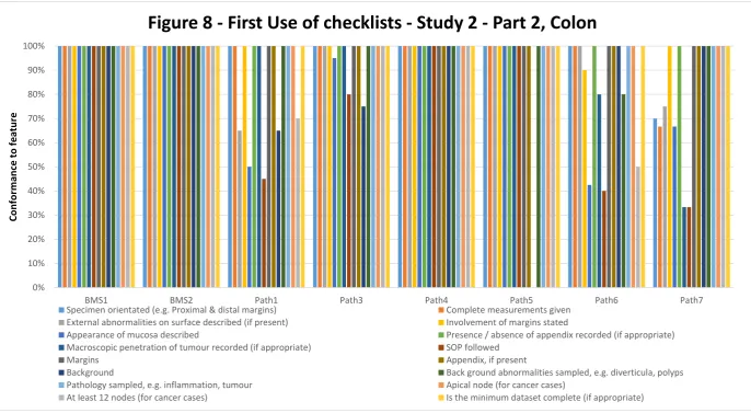
Figure 8 - First Use of Checklist, Study 2 Part 2 – Colon ... 83
Figure 9 - First Use of Checklist, Study 2 Part 2 – Uterus ... 84
Figure 10 - Training Event - Study 2 Part 3, Appendix ... 87
Figure 11 - Training Event - Study 2 Part 3, Gallbladder ... 88
Figure 12 - Training Event - Study 2 Part 3, Colon ... 89
Figure 13 - Training Event - Study 2 Part 3, Uterus ... 90
Figure 14 - Training Event Only - Study 2 Part 4, Appendix ... 93
Figure 15 - Training Event Only - Study 2 Part 4, Gallbladder ... 94
Figure 16 - Training Event Only - Study 2 Part 4, Colon ... 95
Figure 17 - Training Event Only - Study 2 Part 4, Uterus ... 96
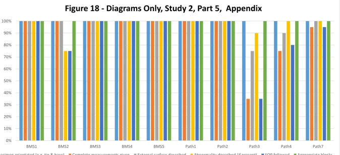
Figure 18 - Diagrams Only - Study 2 Part 5, Appendix ... 98
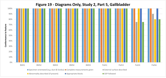
Figure 19 - Diagrams Only - Study 2 Part 5, Gallbladder ... 99
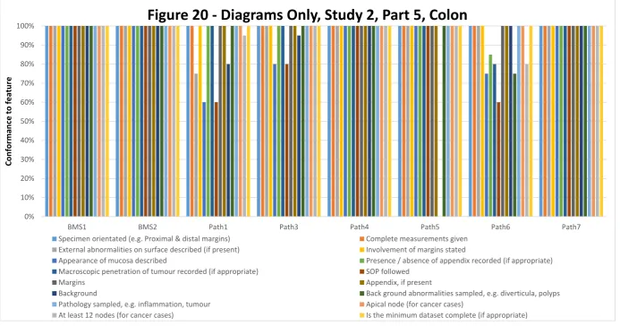
Figure 20 - Diagrams Only - Study 2 Part 5, Colon ... 100
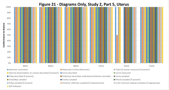
Figure 21 - Diagrams Only - Study 2 Part 5, Uterus ... 101
Figure 22- Training Event and Diagrams - Study 2 Part 6, Appendix ... 103
Figure 23 - Training Event and Diagrams - Study 2 Part 6, Gallbladder ... 104
Figure 24 - Training Event and Diagrams - Study 2 Part 6, Colon ... 105
vi
Figure 26 - Training Event and Diagrams - Study 2 Part 7, Appendix ... 108
Figure 27 - Training Event and Diagrams - Study 2 Part 7, Gallbladder ... 109
Figure 28 - Training Event and Diagrams - Study 2 Part 7, Colon ... 110
Figure 29 - Training Event and Diagrams - Study 2 Part 7, Uterus ... 111
Figure 30 - Training Event and Checklists – Study2 Part 8, Appendix ... 113
Figure 31 - Training Event and Checklists - Study 2 Part 8, Gallbladder ... 114
Figure 32 - Training Event and Checklists – Study 2 Part 8, Colon ... 115
Figure 33 - Training Event and Checklists - Study 2 Part 8, Uterus ... 116
Figure 34 – Average conformance to the SOP over time for appendix specimens. ... 117
Figure 35 - Average conformance to the SOP over time for gallbladder specimens ... 120
Figure 36 - Average conformance to the SOP over time for colorectal specimens. ... 123
Figure 37 - Average conformance to the SOP over time for uterus specimens. ... 127
Figure 38 – Overall conformance to SOP for each dissector ... 131
vii
List of Tables
Table 1 - Recommendations from Barnes ... 16
Table 2 - Performance assessment areas ... 30
Table 3 - Recent practice in appendix dissection ... 46
Table 4 - Recent practice in gallbladder dissection ... 48
Table 5 - Recent practice in colon dissection ... 50
Table 6 - Recent practice in uterus dissection ... 52
1
Acknowledgements
I would like to thank Dr Rachel Gilibrand for her support and guidance throughout this research doctorate. Her wide ranging knowledge and sense of humour helped motivate my failing spirits on more than one occasion. She picked up an unfamiliar subject area and gave a wonderfully fresh perspective to the most frequently neglected aspect of laboratory medicine, the people in the laboratory.
2
Abstract
The recent reports into standards in the NHS (Francis, 2013) and quality in pathology (Barnes, 2014) have focused scrutiny on the way in which we work in pathology and how we can provide assurance that this is of a sufficiently high standard. There are a number of external quality assurance schemes covering pathology generally and histopathology specifically (UKNEQAS), however, there is no scheme of any kind covering the process of histological surgical dissection. This is an area undergoing development currently, as it changes from an area which is the sole preserve of medically qualified pathologists, to an area utilising a number of highly trained biomedical scientists.
The concept of biomedical scientist led dissection has been around for some years, being codified into a document of best practice in 2005 (IBMS, 2005). This document placed the responsibility for the work of the BMS in the hands of the pathologist, without structured oversight or a quality assurance programme. Ten years on and specimen dissection, including the developing area of BMS led dissection, remains without any formal structured form of quality assurance.
4
Introduction
Few could fail to be aware of how badly Mid Staffordshire NHS Trust failed their patients. The Francis Report (Francis, 2013) examined in detail the practices of the Trust and found that the patient was not placed at the heart of the service, that targets were prioritised over good practice and that there were no effective sanctions to deal with poor performance. This was more than a decade after the Kennedy Report (2001), which looked into unnecessary deaths during paediatric heart surgery in Bristol. Kennedy noted that the mortality rate was double that in similar centres elsewhere. He also noted that the patients here were again not placed at the heart of the service. In his conclusions he stated that learning from errors is vital, and that openness and transparency are considered vital. Beyond this, he went on to consider the attitude of healthcare professionals to patients.
Kennedy speaks about the importance of a partnership between healthcare professionals and patients, one including respect and honesty. He is keen to move away from the traditional attitudes of “doctor in charge” and encourages an open collaborative approach. This is keenly echoed in the Francis Report, and, Secretary of State for Health, Jeremy Hunt’s statement in response to the report clearly criticised the attitudes encountered. The Secretary of State went on to call for accountability and transparency (Hunt, 2013).
5 detailed concerns regarding the methods used by an external agency appointed to investigate the issue; their apparent identification of “weak positivity” of oestrogen receptor (ER) staining, which they claimed had erroneously been reported as negative, were in fact false positives. The original report of ER negative was correct. This gave rise to another investigation, this time by Dr Barnes. The Barnes Report (2014) covered quality control in pathology and was quite exhaustive in nature. Barnes noted the reports from Francis and Sherwood Forrest, and echoed a number of their findings, particularly calling for open, transparent individual quality data and standardisation of practice in pathology. Currently there is some quality data available for pathology, however, it is obscure in nature and nonspecific. The national EQA scheme in histopathology is run by UKNEQAS (ukneqas.org.uk), the results generated by this scheme are fed back to the individual laboratory along with regional average rates. It is not possible to draw direct parallels between laboratories, nor is it possible for non-members of the scheme to view the data. Where a laboratory fails to meet the minimum standard, a letter is sent to the nominated “technical head” (UKNEQAS, 2015). This clearly does not fit within the framework of openness, accountability and transparency.
6
Research Aims and Objectives
This research aims to investigate the feasibility of developing Key Performance Indicators that can be applied to the process of histopathological surgical dissection. The aim of this is to enable the use of KPI data to demonstrate variation in practice by and between individuals; demonstration of this variation then allows a form of remedial and preventative action to be introduced, to reduce variation and increase standards.
The objectives of the research are to:
Create a set of histopathological dissection KPIs based on the best available professional and scientific guidance
Collect performance data in relation to the KPIs from a number of dissectors Develop a mechanism for correcting errors or weak / poor practice
Devise a team led system of training, education and feedback, based on the KPI data
Thesis Overview
Chapter One
7
Chapter Two
Chapter two presents the findings of the first study which reviewed current practice in the laboratory. The reports were reviewed, examining a number of criteria, selected as KPIs. These criteria were selected as examples, exploring the concept of KPI reporting in surgical dissection, and the difference in practice between pathologists and BMS. The KPI were selected, based on the recommendations from the RCPath for macroscopic description and sampling. This is used to establish a picture of current practice for pathologists and BMS. The difference between the two groups are considered and the significance of the difference is investigated. Discovery of the variance in practice between individuals and groups allowed a consideration of how quality might be assured by monitoring key performance indicators. It goes on to provide an initial consideration of the results and how to develop the investigation further.
Chapter Three
Chapter three presents study two, the development and delivery of a training intervention employing a face-to-face training event, the use of a practice checklist and diagrammatic representations of dissection. Study two was an eight part trial which first created the checklists and collected baseline data then introduced the training event, checklists and guide diagrams in different combinations to assess the individual effectiveness of each. The results are presented for each stage in the appendices and then brought together and presented by different dissection types at the end of this chapter.
Chapter Four
8
Chapter Five
Chapter five is a detailed discussion of the investigations in context. Here the variations are considered in their context and the significance of the variations in practice are discussed. The effect of the interventions is also discussed along with reflection upon individual practice and the methods that might be used to improved performance.
Chapter Six
Chapter six presents the conclusions of this work, where the new information gained in the study is presented in the context of a changing medical, social and political environment. The work is placed into the context of the changing NHS, with drives for identifiable, transparent quality performance data, with sanctions and rewards for poor or good performance.
9
Chapter One - Literature Review
Histological Dissection
The specimens received by the histology laboratory range from a single fragment of tissue, such as an endoscopic biopsy, to a large multi organ resection, such as a pelvic exenteration; these specimens must then be handled in such a way as to enable microscopic analysis.
There are many terms employed in laboratories to cover the process of transforming specimens of all types into samples suitable for chemical treatment and subsequent sectioning. In the experience of the author, “Cut up”, “Trimming” and “Dissection” are three of the more common. The term “dissection” will be used here, as this is the terminology used by the Royal College of Pathologists (RCPath) and the Institute of Biomedical Science (IBMS) in their co-authored documents (IBMS, 2009; IBMS, 2010a; IBMS, 2010b).
10
Evidence Based Medicine
Standardisation in medical practice has been increasingly linked to improved patient outcomes (ACOG, 2012; Dhingra, 2010; Coombs, 2009; Ferran, 2008) and is an integral part of the current drive for Evidence Based Medicine (EBM). EBM may be thought to have originated with Dr Archie Cochrane with his book Effectiveness and Efficiency: Random Reflections on Health Services in 1972 (Shah, 2009). EBM seeks to base the practice of medicine on the best available evidence, rather than the individual preferences of the practitioner. As research indicates better methods, the guidance is updated. As such, the practice of medicine should follow the available guidance, based on the current evidence, and all practitioners should be working in very much the same way. The logical extension of this is that variation in practice, either within or between individuals, should be eliminated where possible.
11 The concept of EBM, or rather evidence based practice (EPB), is not new to pathology. Within pathology the staging, diagnosis and reporting practices are based on evidence; even guidance on appropriate staffing is given, which is also evidence based (Lowe, 2014; Thorpe et al., 2012; Ellis et al., 2005). The practice of histopathological dissection is based on variable amounts of evidence and in considered in detail within the “Standardisation of Dissection” section.
Devolution of Responsibility
Another emerging trend within pathology, and within the wider medical community, is the devolving of tasks and responsibilities traditionally undertaken by medically qualified doctors to appropriately qualified paramedical professionals. This has occurred extensively within nursing, with a great deal of traditional nursing roles being undertaken by Healthcare Assistants (Bosley, 2008; Daykin & Clark, 2000). This is also occurring in pathology, whereby the process of surgical dissection, which was traditionally the preserve of medically qualified pathologists, has been passed to suitably trained biomedical scientists. The concept of pathological dissection being performed by non-medically qualified staff (specifically, qualified Biomedical Scientists) has been present for some time. There has been considerable variation in range and scope of practice, which until recently has been uncoordinated and left to local agreement. In recent years there have been steps to formalise and standardise the range of specimens and personnel that are authorised to work with them (IBMS, 2009; IBMS, 2010a; IBMS, 2010b; RCPath, 2004). However, there are no protocols in place to ensure the standard of this work; the quality must be monitored locally, there are some suggestions made on how to perform this (IBMS, 2009, RCPath, 2007), but no structured or formal systems have yet been developed.
12 led dissection, with 98.9% of respondents being either fully in favour of BMS dissection or in favour with some reservations. There remains some controversy and arguments regarding exactly how BMS led dissection should progress and be monitored, but the prevailing attitudes expressed are positive.
13 The importance of noting these historical circumstances and their subsequent changes is to place the devolution of responsibility and attitudes to assessment in a proper context. Being a medical doctor has long held a social weight, a certain cachet; this came with certain responsibilities as a part of the social contract. The fabric of this social contract, the trust with which the public hold their doctors, has been successively damaged over the years, in no small part by these scandals. The consequence of this is a move to what Dixon-Woods et al. (2011) call a “confidence” system, whereby individuals (the patients) put their faith in the system of assurance and regulation around the individual, rather than in the individual themselves. This means that we must be able to demonstrate our quality within that system in order to promote this confidence in us. With the current political environment and the drive for ever increasing competition, any commissioning group will also be looking for evidence for putting their confidence in any particular hospital, department, or individual. Demonstrating appropriate team behaviours, attitudes and quality is going to be a necessity.
Quality in Pathology
14 One of the methods used to ensure quality is External Quality Assurance (EQA). The value of EQA has been proven repeatedly (Hastings and Howell, 2010, Sciacovelli et al., 2010), over many years (Barr & Williams, 1982). EQA is a method whereby an external agency provides independent feedback on performance, against an agreed standard. Within UK pathology the main sources of EQA are UKNEQAS and the Royal College of Pathologists, where substandard performance is identified, this is reported back to the laboratory. EQA has been used across all areas of histopathology, with the sole exception of pathological dissection. There are schemes covering the microscopic analysis of tissue sections (NHSBSP, 2003; RCPath, 2007), routine and special staining (UKNEQAS, 2011a), immunocytochemistry and in situ hybridisation (UKNEQAS, 2011b). Duthie et al. (2004) specifically highlight the lack of published literature regarding the quality of specimen handling. When considering how quality assurance of histopathological dissection might be performed, whether BMS or pathologist led, it is significant that dissection involves compromising and damaging the sample. The training process suggested by the IBMS (2009) suggests preview prior to dissection, moving on to review after dissection, progressing to dissection with regular audit. The concept of regular audit is, however, not further defined; nor are any recommendations made regarding EQA. Additionally, one of the drivers for BMS led dissection is the time saved by consultants, it is not practicable for each specimen to be viewed before, during and/or after dissection, beyond an initial training stage. This gap in knowledge and oversight is something we seek to address here. Whilst EQA clearly has much to offer, it lacks a robust framework in which poor performance of individuals can be identified, or whereby poor performance can be managed.
15 indeed criticisms were made of the external agency appointed to investigate the alleged discrepancies, they identified a number of further issues within the EQA system (RCPath, 2013).
The review undertaken was extensive, involving rescreening a great many slides and determining the Oestrogen Receptor (ER) status of breast tumours. This highlighted a number of areas of variation, both within this centre and between centres. However, the main problem noted was related to the EQA scheme. As Sherwood Forest Trust was handling a small number of cases, a small amount of variation was sufficient to create the appearance that this trust was an outlier. The subsequent investigation into the matter was further compounded the issue by the use of the antibody clone 6F11. 6F11 is now know to show false positive staining (Ibrahim, 2012; Rakha, 2012). This is something which was picked up by the EQA scheme, however, there is clearly a significant time lag in identifying and reporting these issues. The Sherwood Forest Report also highlighted a number of systematic faults with the statistical aspects of the EQA service. It also recommended that statistical monitoring of individual performance be introduced, using radiology or cervical screening methods as a starting point (RCPath, 2013).
As a consequence of this investigation, and the background of the Francis report, the NHS Medical Director, Sir Bruce Keogh, appointed the National Clinical Director for Pathology, Dr Ian Barnes, to review pathology quality assurance. Barnes (2014) states “the current systems of quality assurance in pathology are no longer able to meet the needs of modern healthcare” and “we cannot say the best interests of the patients are being served”. This could not be stated more clearly. He based this assertion on a number of factors, including the lack of key assurance indicators (KAIs) and the inability of the pathology service to provide evidence of quality to the Care Quality Commission (CQC).
16 make to patient experience; held to account when they fail to offer the level of service patients expect.
Barnes noted that there were a number of failings in pathology practices, particularly in relation to KAI and demonstrating quality data (Table 1 below).
Table 1 - Recommendations from Barnes, 2014
1.7.1 The broad assurance framework has a lack of KAIs to evidence quality and safety of pathology services, and to enable effective contract management both within organisations and by commissioners.
1.7.2 Pathology assurance and governance is not consistently embedded in provider governance and assurance frameworks.
1.7.3 Pathology is unable to provide evidence to the Care Quality Commission (CQC)/Hospital Inspectorate of the overall quality of pathology services.
1.7.4 Pathology needs to respond to changing and additional requirements from commissioners and the public for information and assurance around consistency of provision and reporting.
1.7.5 The impact of new technology and processes (genomics, point of care testing (POCT), digitalisation, molecular techniques, informatics) on delivering pathology services, and the impact on a rapidly changing workforce, require a strengthened quality assurance framework.
1.7.6 There is too much variation between pathology services, and a lack of harmonisation and standards, which is unacceptable to patients and users. 1.7.7 The current system was fit for the purpose for which it was designed, but it is not
fit for the future, nor does it meet the emerging requirement for transparency and well-evidenced quality assurance. Therefore, the Review and the recommendations it makes will attempt to bring these features of the system into sharper focus, strengthening existing structures to ensure these gaps are filled.
One of the recommendations made by Barnes is:
17 professional bodies, led by RCPath, to review these issues and report back within twelve months on their findings.”
This clearly echoes the comments made in the Sherwood Forest investigation (RCPath, 2013), showing that these calls for individualised quality data are growing.
The RCPath, IBMS and Association of Clinical Biochemists (ACB) welcomed the publication of the report (Jayaram, 2014). They acknowledge that the report presents an opportunity for professionals in pathology to form a rationally based quality system. This work attempts to address some of the gaps identified by Barnes, by identifying KAIs / KPIs, demonstrating their evidence base, and investigating how poor performance or change in expectation can be addressed.
Foy et al. (2002) discuss how to best implement changes in professional practice. They performed an audit and regression analysis of the work done in the Gynaecology Audit Project in Scotland, and categorised the existing recommendations as having any of the following features:
Addresses common issue Precisely described Compatible with values
Key feature to a set of wider recommendations Based upon sound evidence
Fits patient expectations Observable benefits
Requires organizational change Requires changed routines High profile
Complex Trialable
18 correlation between these and noted that there were four features that most correlated with change. Two showed a positive correlation and two showed a negative correlation. Those recommendations compatible with the views and values already held by practitioners relating to the performance of their duties and those recommendations that were termed “key features”, which is to say they formed part of a larger set of recommendations key to wider goals were seen to show a positive correlation to recommendation uptake. A negative correlation was seen to those recommendations that required organisational change or a change in routine. Foy also noted that although only 3% of recommendations could be classified as being trialable, that is, they could be adopted as a temporary measure and discarded if needed, these showed a higher level of uptake.
As a change in guidelines is likely to involve change in routine practice, and may well relate to practices where there is some disagreement over what is appropriate practice, this presents something of an obstacle to change. Since Foy noted measures that could be adopted on a temporary basis showed a greater and more sustained uptake, this is something that has been considered in the development of this study.
19 unnecessary interventions were being made. They worked with a number of practitioners, explaining the research and the evidence for the recommendations, this enabled the practitioners to form their own opinion regarding the evidence and the recommendations, rather than simply responding to instruction, or ignoring the guidelines. This enabled the practitioners to preserve their autonomy and develop their knowledge and skills in light of new evidence. Ho et al. combined this with recommendations that could be adopted on a trial basis, as with the conclusions from Foy (2010), this showed the greatest change in practice. These papers have been considered in relation to the work currently being undertaken, whereby the training interventions are to involve as many people as possible, and are to be guided, interactive and initiated on a trial basis. This is considered in more depth later in the literature review, under the heading of “Change Management” on page 33.
Key Performance Indicators
20 to the findings of the Francis and Sherwood Forrest reports, examining in great depth the quality assurance (QA) processes in pathology. He concludes that QA has worked well up to now, but in the expanded and more complex role pathology plays, QA in pathology needs to be fundamentally re-assessed as it is no longer fit for purpose. This is discussed in more depth later, however, one of the recommendations he makes is developing Key Performance Indicators (KPIs).
21 feedback on these scores to the surgeons led to an improvement in the quality of the submitted specimen during the period of their study.
KPIs are not, of course, unique to healthcare, Gonzalez-Gil (2014) details the use of KPI in managing the energy use of a railway system, and Cai (2009) discussed their use in improving supply chain management. The unifying principle of KPIs and the inherent benefit of their incorporation into practice is that they are based on the best available evidence, and are amended in the light of new research. Here we identify KPIs relating to histological dissection and demonstrate how they can be used to demonstrate quality, how to improve quality and how to monitor quality.
Standardisation of Dissection
22 12–20) identified more lymph nodes than consultant pathologists (median 10, range 7–13), confirming the findings of Thomas (2006).
When considering the differences between BMS led dissection and pathologist led dissection one must be aware of the different training backgrounds. Pathologists are medically trained, whilst BMSs have followed a scientific pathway, which clearly leads to a very different sphere of knowledge. Simmons (2011) investigated attitudes to BMS led dissection, they surveyed over 1300 consultant histopathologists, 25% of respondents expressed concern at the potential lack of knowledge and increased supervision required by the BMS. One of the concerns held by consultant pathologists, reported by Simmons was whether the BMS is aware of important and unusual clinical features, and when specimens might be best dealt with by a non-standard approach. This is a judicious concern, Chandra et al. (2010) published a review of best practice in bladder pathology specimens; they indicate the importance of being aware of the significant aspects of clinical information and the previous history. However, this is specifically addressed in the IBMS document (2005) relating to good practice in dissection – they explicitly recommend that BMS dissectors should seek advice on unusual or complex cases. The IBMS good practice document should be read in conjunction with the protocols recommended in the RCPath minimum datasets and tissue pathways (RCPath, 2014a; RCPath, 2009). These are evidence based, Flemming & Griffiths (2005) detail in their best practice article much of the reasoning behind the protocols and minimum dataset points. Whilst this is essential information, the knowledge, awareness and comprehension of these points is not monitored in either BMS or pathologists once they have passed their exams as currently there is no requirement for a periodic or on-going skills test or CPD in dissection.
23 or of an ongoing nature. The paper is an excellent reference indicating how various colorectal resection specimens should be handled according to current state, although they make no recommendations for further development or how to asses or ensure quality. However, they raise an interesting point regarding the RCPath minimum requirements. For example, the minimum dataset requires at least 12 lymph nodes to be sampled – they suggest that for some dissectors this minimum number becomes a target, this is something which has been echoed during personal communication with other pathologists. There is no incentive to obtain more than the baseline minimum number of lymph nodes, or indeed to do anything more than meet any minimum standard. The authors indicate that despite audits on lymph node numbers retrieved, the numbers of retrieved nodes remains low in some centres. Within Shepherd’s laboratory there is an informal “leader board” with the number of nodes retrieved and the dissector noted, this, they state, has created a culture of ensuring high lymph node counts. Ludeman & Shepherd published their paper nine years ago, some eight years after the initial RCPath recommendation that a minimum of 12 nodes be obtained (Quirke and Williams, 1998) and with no on-going standardised framework to monitor this or to provide training / feedback, the authors consider this is unlikely to change. There is extensive evidence and evidence based guidance regarding lymph node retrieval, clearly though, the actual approach to lymph node retrieval is not meeting this guidance. Verrill (2004) highlighted the importance of sampling lymph nodes fully, detailing the prognostic relevance of tumour involvement in lymph nodes. Thus ensuring that minimum standards are consistently reached is critical.
25 from the Barnes report (detailed later), this clearly highlights a need for KPIs to be collated and actioned.
The literature supporting the notion of standardisation of dissection practise in this thesis is presented primarily from the perspective of lymph node sampling as the decision made on the number of lymph nodes required by the RCPath is possibly the most evidence based. The notion of stipulating a minimum number of lymph nodes began when Scott and Grace (1989) demonstrated a method of ensuring all the lymph nodes were recovered and examined. Scott and Grace collected data from 103 colorectal carcinoma specimens, traditional dissection methods yielded a mean 6.2 nodes whereas further sampling gained a further 12.4 nodes. These additional nodes showed tumour positivity in 5% of the cases which were initially reported as lymph node negative. Their data showed that when significantly more nodes, and more involved nodes, were found for each resection this resulted in upstaging of the tumours in 5% of the cases. The data analysis resulted in the recommendation that a minimum of 13 lymph nodes be sampled, forming a foundation for the later minimum number (12) given by the RCPath. Kim et al. (2007) determined that there was minimal added value to the extensive search for nodes, once 12 nodes had been found. Their conclusions suggested taking additional samples if no positive nodes were found, but if at least 12 nodes had already been found and at least one was positive, no further sampling was required. However, this recommendation of further sampling in the absence any positive nodes failed to make it into the RCPath guidelines (Quirke and Williams, 1998).
26 (RCPath, 2014a). The numerical data standards (number of lymph nodes) are easily assessed; other standards, such as adequacy of tumour sampling, are less easily judged. The distribution of tumour staging for different types of tumour is not available in any published format, an omission noted in the RCPath minimum dataset (RCPath, 2009). However, some data has been obtained in this area though freedom of information (FOI) requests and local data (Figure 1, below). This is currently unpublished, but does indicate that there is a broad concordance of stage at presentation.
Figure 1 - Graph showing stage of colorectal tumour at presentation (Duke’s staging). 2009 data, from 18 different cancer networks – data obtained via Freedom of Information Requests from the North Yorkshire Cancer Network.
27 Using a rolling mean of tumour staging of resections begins to provide a dissection practice profile. Adding further evidence based data points to this would allow a clearer picture of individual practice to be seen. Another measure detailed in the RCPath minimum dataset is the frequency of extramural venous invasion; again, no published data is available in regard to this. A comparative analysis between individuals would allow some discussion as to quality of practice. There are a number of studies which indicate the importance of accurate assessment of vascular invasion, and its prognostic significance. Stefansson et al. (2004) examined 237 endometrial cancers and were able to report that vascular invasion is a strong prognostic factor, independent to the stage or grade of the tumour. Westenend et al. (2004) investigated 50 cases of breast cancer, they reported that tumour size and vascular invasion were the most important prognostic factors. Whilst there is a recommendation that pathologists audit the number of cases which they report as showing vascular invasion, there is no suggestion that dissectors should do similar. Personal experience and anecdotal evidence shows that poor practice can lead to the appearance of vascular invasion; therefore, this is another possible area for comparison.
28 Shaw (2008) reports that less than 12 lymph nodes were found in 54% of the 1,194 cases they examined. Clearly, having guidelines in place does not mean that people will follow them or reach the standards detailed in them. There are a number of ways in which poor performance, or a change in expectations, can be managed. Mesmer-Magnus and Viswesvaran (2010) detail an extensive review of training methodologies, concluding that what they refer to as the “Why?” method is a particularly effective method. This methods causes the individual to move their attention from the details to the underlying process and back in a fluid manner. They detail a method of training primarily aimed at untrained individuals in which the trainee is encouraged to ask why certain things are done, or not done, in order to fully understand the process and their part in it. This is certainly something that is targeted to those in training; however, those who are already trained, but who take part in on-going training, CPD or reflective learning would also stand to benefit.
30 objective assessment is the “holy grail” of assessment. Cross notes a number of areas where performance needs to be assessed (Table 2, below).
Table 2 - Performance assessment areas (from Cross, 2005)
Diagnosis by light microscopy Not applicable to BMS, pathologists assessed by review, examination and EQA.
Diagnosis by other modalities This would include macroscopy, specimen dissection, and has no form of assessment beyond gateway examinations.
Knowledge base Assessed well by the appropriate pathological qualifications for both the BMS and pathologist.
Report generation and checking These are reviewed by the reporting consultant, and by the requesting clinician. For the BMS the report is limited to a report of the macroscopic appearance, which may show characteristic features giving an indication of the diagnosis.
Communication with colleagues Historically undervalued, but these are now being assessed as part of performance reviews in many centres.
Learning Theory
32 A fourth area which Torre et al. (2006) discuss is constructivist learning. Constructivist learning is facilitated by the educator, who fosters critical reflection and helps to develop meaning from experience. The IBMS strongly promotes reflective learning in their dissection portfolios (IBMS, 2009; IBMS, 2010a; IBMS 2010b) and their CPD scheme (IBMS, 2014b). Colliver (2002) publishes what amounts to a wholesale dismissal of constructivism as anything other than a philosophic stand point. He concludes that constructivism “offer[s] little of value to medical education”. However, Cobb (2002) rebuts all of Colliver’s points, expanding on the meaning and application of constructivism and highlights Colliver’s lack of questioning of assumptions. Cobb concludes that “constructivism might be of greatest relevance to educators”, this appears to be a more widespread opinion.
Torre et al. (2006; 2007) provide an explanation of how reflective practice may be performed in a medical context, in a way which is easily applicable to specimen dissection, and details what may be gained from such an approach. They also note that their method of documenting their reflective learning leads well into group work. Bennett et al. (2002) provide a review of small group teaching, where they note that explaining material to another party is one of the most effective means of cognitive elaboration. Their review is comprehensive, covering aspects such as the composition of the group in terms of gender and ethnic mix (which appears to make little difference) ability (mixed ability shows most improvement for most people) and group size (6 learners and a facilitator appears to be the optimum number).
33 The research above clearly shows that there are many evidence based standards of practice, and that many people are failing to meet these standards. Having identified the veracity of the evidence base for and need for these standards, and having identified the failure to meet these standards, how do we then address this? The evidence in the area of learning theory provides us with an insight into how we might best approach the educational deficits in our practitioners. However, what is needed is not just education, there needs to be a change in practice. Creating and sustaining a change is a multifactorial process, which comes under the heading of change management.
Change Management
34 suggested change and listening to the concerns of those involved was critical. The most successful method of change reported by Ho et al. (2010) was in cases where the staff had been part of research projects that investigated the change proposed. Active engagement of staff, allowing them to evaluate the evidence were key aspects. This is echoed by the comments made around the research of Pronovost (2006) detailed later in the literature review. The training intervention which is to follow this literature review will be structured with these concerns in mind, drawing on research in medical practice, change management, and behaviour management.
Lim et al. (2012) discuss an interesting study based in the area of mental health workers and evidence based practice (EBP). They ran a series of workshops during which they examined a number of methods and practices, went through with participants (n=238) whether or not these were evidence based and explained the implications of these aspects. The participants were found to have a more positive attitude towards EBP at the end of the workshops. With this and the preceding paragraph in mind, the proposed method will be developed in conversation and negotiation with active staff members. Attempting to impose a quality system on reluctant subjects is unlikely to result in long term success.
35 regarding communication or addressing performance issues, these should be addressed regardless of any training or feedback event, however these issues are frequently pushed aside as it is often easier to work around these issues than to address them. Fostering this supportive and collaborative environment will go a long way towards addressing these issues.
There are key lessons to be learnt from Chisholm, if we are to engage in changing attitudes and behaviours. The most important thing she reports is that people are more likely to engage in such discussion if they have had some form of formal training in the theory behind behaviour change techniques. Drawing on this may help with the current work, finding key personnel and enabling them to undergo such training could help with engagement and dissemination of findings and recommendations. If we are able to demonstrate a robust quality monitoring and feedback system, we shall be reliant on others to deliver it elsewhere. As such, we must also provide some form of instruction on how to implement these changes.
One intervention making use of KPIs and dealing with behaviour change demonstrated startling effectiveness. Pronovost et al. (2006) set out to determine whether central line related blood stream infections in Intensive Care Units (ICU) could be reduced. They developed a Team Checklist, centred on five evidence based infection control procedures. This was a multicentre trial, where hospital participation was optional. When a hospital opted in, the use of the checklist within the ICU was mandatory for all staff involved in the placing of central lines. A number of training and educational interventions were planned, training staff in the science of the safety measures. If the individual placing the central line was not following the approved procedure, any team member was authorised to stop the procedure (in non-emergency situations).
36 credited with a reduction in the rate of catheter-related bloodstream infection of up 66% sustained over the course of the 18 month study.
Checklists have been widely used in safety critical environments, notably aviation – the first published instance being in Life magazine (Anon, 1942). Since then there have been a number of attempts to bring checklists as a safety feature into the realm of healthcare. These continue to the current day, Ornato and Peberdy (2014) note the need for healthcare practitioners to respond in a consistent, high quality manner. Whilst they are talking in relation to emergency situations, this is clearly relatable to routine practice. They note the 2000 fold reduction in fatalities per million flight miles since 1929, and attribute this to the insistence on standardised procedures regardless of who is on the team. Kao and Thomas (2008) also strive to apply lessons from aviation into healthcare, specifically surgery. They note an intervention that reduced the incidence of wrong site surgery to 0, they also refer to a number of studies that state such interventions improve the safety climate and clinical outcomes. Pugel (2015) notes the number of errors in the operating room, and relates that communication errors are a common form of error. Dhingra (2010) backs this up, stating that 70% of serious events reported by the Joint Commission are due to communication errors. Pugel (2015) also discusses some of the
drawbacks to checklists. Pugel notes that previous safety checks may be dropped and that the checklist can interrupt the performance of other safety tasks. She also states that without a firm commitment to the checklist it may become a routine activity of checking off boxes without actually driving behaviour change or improvement and that checklists may give staff a false sense of security that issues have truly been resolved when in fact they have not
37 11% to zero. The reporting around this focused extensively on the checklist (Gawande, 2007; Lawrence, 2009; Seifert, 2009; Kingsbury, 2008). Whilst each of these reports makes some effort to explain the intervention around the checklist, it is clear that the checklist is given centre stage. This was noted by the authors, who published an update, giving greater emphasis on the surrounding parts of the intervention (Bosk et al., 2009).
Whilst the checklist was not the only factor in the intervention detailed by Pronovost et al., it is the central point around which the other factors revolve. Checklists have received considerable interest in medicine since their paper. After discussing the applications of aviation lessons to surgery, Kao and Thomas (2008) go on to discuss some of the differences. They note that when compared to pilots, surgeons tended to downplay the effect of stress and fatigue on decision making. Senior surgeons tended to prefer a rigid hierarchical structure, where junior members of the team do as instructed without question. Ornato and Peberdy (2013) support this conclusion, highlighting a difference in attitude between pilots and surgeons. They note that pilots recognise that permission to fly is a privilege, not a right and that this permission can be easily withdrawn. They also note that with pilots their own lives and safety are dependent upon the function of the team, whereas in surgery the only life at risk is likely to be that of the patient and the surgeon will take the majority of responsibility for the success, or not, of the surgery.
Sexton et al. (2000) reported that out of consultant surgeons, consultant anaesthetists and pilots there was a wide difference in who would acknowledge the effect of fatigue on performance (30%, 53% and 74%, respectively). Additionally, only 55% of consultant surgeons recommended a flat hierarchy in contrast to 94% of cockpit staff. Kao and Thomas (2008) suggest that approximately 25% of errors may not be admitted in morbidity and mortality.
38 attitude of the pilots, who accept the need to satisfy requirements to prove their competence whereas many of the surgeons resent and resist attempts to introduce similar requirements. Within the Keystone intervention reported by Pronovost et al. (2006) and further expounded upon by Bosk et al. (2009), there was a large social aspect to the intervention, with the implementation phase lasting 9 months. Additionally, a member of the senior executive within each hospital had to agree to work with their ICU; a senior physician and senior nurse were also identified, along with team leaders who were tasked with explaining the principles of safety, the specifics of the investigation and the evidence base. The explanation of the evidence base is something that Ho et al. (2010) identified as a key factor in driving behaviour change.
Clearly this was an investigation far greater in scope than the introduction of a checklist, indeed it was not one single checklist. There were in fact over 100 different checklists. Each ICU was encouraged to take the template checklist and adapt it according to their own needs, but keeping within the needs of the investigation and the demands of the evidence (Bosk et al., 2009).
39
Chapter Two – Study 1 – Review of Recent Practice
Study 2 – The Training Intervention
Part One – The creation of the checklists and collecting baseline data
Part Two – Checklist Introduction (BMS N=5, Pathologist, N=7)
Part Three – Training Event and Checklists (BMS N=5, Pathologist, N=7)
Part Four – Training Event only (BMS N=5, Pathologist, N=7)
Part Five – Guide Diagrams (BMS N=5, Pathologist, N=7)
Part Six – Training Event and Guide Diagrams (BMS N=5, Pathologist, N=5)
Part Seven – Checklists only ((BMS N=5, Pathologist, N=0)
Part Eight – Training Event and Checklists (BMS N=6, Pathologist, N=8)
Study 1 – Archival Review
100 appendix, 100 gallbladder, 100 colon, 100 uterus reports reviewed
Unknown number of participants
Study 3 – Participant Interviews
The dissectors were interviewed
40
Introduction
In order to investigate the level of variation in practice within the Royal Derby Hospital Histopathology Department, a retrospective review of practice was undertaken. A number of points were addressed, noted below in the procedure section, and these were completed against authorised reports from the preceding year.
41
Method
Design
An audit into the dissection processes performed in 2009 – 2010 was performed.
Participants
Individual participants were not personally identified at this stage, however, there were 18 practicing dissectors at this time (10 pathologists and 8 BMS). As the individual reports were examined, the dissector of that specimen was noted as either BMS or pathologist only.
Materials
42 Appendix Three – Baseline Data Collection Sheets, page 206), to capture data specific to the specimen type being considered. The specimens examined were appendix, gallbladder, uterus and colon. These were chosen as they represent an increasing level of complexity, whilst being within the scope of BMS dissection. Appendix and gallbladder are relatively straightforward specimens, with a limited range of presentations. The uterus and colon are of varying complexity and have a wide range of presentations, making dissection a more complex and multilevel activity.
Procedure
Ethical approval was granted by the University of the West of England Ethics Committee (UWEEC). The National Research Ethics Committee (NRES) determined that this investigation did not require ethical approval under their scheme. The Derby Teaching Hospitals NHS Foundation Trust (formerly the Derby Hospitals Foundation Trust) were satisfied with the ethical approval granted by UWEEC and the statement from NRES and gave their approval for this investigation to proceed, having registered the investigation with the trust R&D department. This covered study 1 and 2; study 3 required separate approval which is detailed in Chapter 4.
The pathology Laboratory Information Management System (LIMS) archive was searched, identifying 100 appendix, gallbladder, uterus and colon specimens from 2009 – 2010. The pathology report for each was checked against the data collection sheet and the sheets were structured as follows.
All specimens
Time from receipt to dissection (working days)
Need for extra investigations (e.g. levels, Immunocytochemistry) The status of dissector (e.g. Pathologist or BMS)
The number of blocks
43 The need for and number of extra blocks
A record of any errors in the macroscopic description
Whether the manner of dissection and macroscopic description allows the minimum dataset to be completed.
If there is agreement between the macroscopic description and the microscopic report.
Additionally, the macroscopic description was reviewed and more specific data was logged for each specimen type. These data points are all mandated by the Standard Operating Procedure (SOP), some are specifically required by the relevant minimum dataset. As such, if a feature is present it should be recorded; there is no requirement to record the absence of such features. The criteria marked in bold should return at 100% for all cases, as this mandatory information required by the SOP for that specimen. There is no expectation that the other criteria should return a specific value, simply that as all the dissectors are drawing their specimens from the same pool, theoretically working in the same manner, a similar value should be seen for each criteria for each group.
Appendix o Length
o Diameter
o Serosa, exudate, perforation, mucin, fibrosis, faecolith, pus o Appearance suggestive of carcinoid / neuroendocrine tumour o Normal / unremarkable appendix
Gall Bladder
o Length x width x breadth (or diameter)
o Whether the specimen was received open or unopened
o Serosal surface, the contents of the lumen, the presence or absence of choleliths (gall stones) and their type, the presence of an exudate, the presence of a perforation
o Fibrosis, mucin, the thickness of the gall bladder wall, mucosal polyps, mucosal cholesterolosis, empyema, Rokitansky-Aschoff sinuses, cystic duct lymph node, the mucosa
44 Hysterectomies
o The presence of fibroids, the appearance of the serosal surface o The thickness of the endometrium
o The appearance of the myometrium
o The presence of adenomyosis, the presence of endometriosis Colorectal
o Full measurements
o Background mucosa described
o Polyps, diverticulae, ulceration, fissuring, loss of mucosal fold pattern o Thickening / thinning of wall, oedema, congestion, exudate, perforation A sample of the data sheets were reviewed by an independent third party to check for accuracy. The data was transcribed on to an excel workbook, and the sheets shredded. Confidentially and data protection was maintained by following NHS Information Security Policy (NHS England, 2014a). All electronic records were kept within the NHS computer system within a password protected system, any paper copies were kept within a locked cabinet within a locked office, prior to shredding. No identifying characteristics for either staff or patients were carried over on to the data collection form, only a record of BMS or pathologist was made.
Results
45 present. Therefore the criteria in bold should be at 100% for both groups, and the other criteria should be at a similar level for both groups.
As was noted extensively in the literature review, variation in practice reflects poor patient outcomes (ACOG, 2012; Dhingra, 2010; Coombs, 2009; Ferran, 2008). As such, methods of identifying this variation, assessing the root cause, and providing an opportunity to reduce this variation, must be developed. This initial review provides an opportunity to assess practice and identify what, if any, variation is occurring.
t – test
46
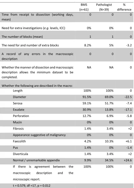
Table 3 - Recent practice in appendix dissection
BMS (n=61) Pathologist (N=39) % difference Time from receipt to dissection (working days,
mean)
0 0 0
Need for extra investigations (e.g. levels, ICC) 0% 0% 0
The number of blocks (mean) 1 1 0
The need for and number of extra blocks 8.2% 5% -3.2
A record of any errors in the macroscopic description
0 0 0
Whether the manner of dissection and macroscopic description allows the minimum dataset to be completed.
NA NA 0
Whether the following are described in the macro:
Length 100% 100% 0
Diameter 91.5% 69.0% -22.5
Serosa 59.1% 51.7% -7.4
Exudate 30.9% 13.8% -17.1
Perforation 12.7% 6.9% -5.8
Mucin 0% 0% 0
Fibrosis 1.4% 3.4% +2
Appearance suggestive of malignancy 0% 0% 0
Faecolith 4.2% 10.3% +6.1
Pus 1.4% 0% -1.4
Diverticula 1.4% 3.4% +2
Normal / unremarkable appendix 9.9% 34.5% +24.6
If there is agreement between the macroscopic description and the microscopic report.
100% 100% 0
t = 0.579, df =17, p = 0.012
47 discrepancies are identified, most notable are the discrepancies regarding the description of the appendix as normal and the provision of the measurement of the diameter. These will be considered in the discussion section
48
Table 4 - Recent practice in gallbladder dissection
BMS (n=69) Pathologist (N=31) % difference Time from receipt to dissection (working days, mean) 0 0 0 Need for extra investigations (e.g. levels, ICC) 1.4% 0% 1.4
The number of blocks (mean) 1 1 1
The need for and number of extra blocks 0% 3.2% -3.2
A record of any errors in the macroscopic description 0 0 0 Whether the manner of dissection and macroscopic
description allows the minimum dataset to be completed.
NA NA
Whether the following are described in the macro:
Length 100% 100% 0
Width 98.5% 80.6% 17.9
Depth 56.5% 29.0% 27.5
Received open / unopened 29.4% 19.4% 10
Serosal surface 14.7% 3.2% 11.5
Luminal contents 94.1% 87.0% 7.1
Presence or absence of stones 97.1% 93.5% 3.6
Type of stones (if present) 5.1% 0% 5.1
Exudate 0% 0% 0
Perforation / incision 7.4% 3.2% 4.2
Fibrosis 4.4% 0% 4.4
Mucin 2.9% 0% 2.9
Wall thickness 100% 12.9% 87.1
Polyps 2.9% 0% 2.9
Appearance suggestive of cholesterolosis 13% 9.7% 3.3
Empyema 0% 0% 0
Rokitansky-Aschoff sinuses 0% 0% 0
Cystic duct lymph node 3.1% 0% 3.1
Suspicious of malignancy 0% 0% 0
Mucosa 97.1% 25.8% 71.3
If there is agreement between the macroscopic description and the microscopic report.
100% 100% 0
50
Table 5 - Recent practice in colon dissection
BMS (n=46)
Pathologist (n=54)
% difference Time from receipt to dissection (working days, mean) 1 2 -100
Need for extra investigations (e.g. levels, ICC) 1% 1.5% -0.5
The number of blocks 18 18 0
Pathological T staging under the TNM classification 2.81 2.7 0.11
The number of lymph nodes recovered 16.1 9.5 170
The incidence of requests for extra blocks 3 1 300
A record of any errors in the macroscopic description 0 0 0 Whether the manner of dissection and macroscopic
description allows the minimum dataset to be completed.
100% 100% 0
Whether the following are described in the macro:
Full measurements 37.5% 27.0% 10.5
Background mucosa 46.3% 30.4% -15.9
Polyps 8.7% 7.4% 1.3
Diverticula 15.2% 20.4% -5.2
Ulceration 4.3% 22.2% -17.9
Fissuring 4.3% 1.9% 2.4
Loss of fold pattern 4.3% 13.0% -8.7
Thickening / thinning of wall 6.5% 11.1% -4.6
Oedema 2.2% 13.0% -10.8
Perforation 0% 7.4% -7.4
Serosal puckering 0% 9.3% -9.3
Exudate 2.2% 5.6% -3.4
Agreement between the macroscopic description and
the microscopic report 100% 100% 0
51 The review of the colorectal specimens shows conformity between BMS and pathologists in all areas, with the exception of lymph node yield. As previous studies have already demonstrated a higher lymph node yield with BMS dissection, this does not come as a surprise. The majority of the items in Table 5 on page 50) relates directly to the visual macroscopic features or numerical data (number of lymph nodes, number of blocks); however, one feature of the microscopic report is also correlated here – the T stage of the TMN classification. This has been done to assess distribution of mean staging. Acting on the premise that all dissectors are taking specimens from the same pool, they can be expected to encounter a similar distributions of tumours at different stages. Therefore, if sampling is being performed adequately and appropriately, the mean T stage for each group (BMS and pathologist) would be expected to be similar. A further development might be to plot out the individual TNM profile for each dissector, and compare the rolling plot for each dissector. A dissector with a persistently low N score may correlate with a low lymph node yield, or one with a high T score might correlate with poor dissection technique causing carryover or may be performing more through or more accurate sampling than others.
52
Table 6 - Recent practice in uterus dissection
BMS (n=86)
Pathologist (n=14)
% difference Time from receipt to dissection (working days, mean) 1 2 -100
Need for extra investigations (e.g. levels, ICC) 1% 0 1
The number of blocks 9 9 0
The need for and number of extra blocks 2% 0 2
A record of any errors in the macroscopic description 0 0 0
Whether the manner of dissection and macroscopic description allows the minimum dataset to be completed.
100% 100% 0
Whether the following are described in the macro:
The presence of fibroids 39.5% 33.3% 6.2
The appearance of the serosal surface 8.1% 7.7% 0.4
The thickness of the endometrium 85.1% 66.6% 18.5
The appearance of the myometrium 95.3% 93.3% 2
The presence of adenomyosis 22.1% 6.6% 15.5
The presence of endometriosis 0% 0% 0
If there is agreement between the macroscopic description and the microscopic report.
100% 100% 0
53 Review of the hysterectomy specimens does show a number of discrepancies. The need for extra investigations and extra blocks was minimally increased for cases dissected by BMS. This was an extremly small increase and has a minimal impact in terms of additional work performed. This correlates with the work published by Duthie, 2004; they performed an examination in to the quality of impact of BMS dissection. They noted that a larger proportion of cases required extra blocks and levels when the dissection was performed by BMS than by consultant pathologists. They highlighted other studies with lower rates of additional sampling, and suggested that their higher rates may be as they were comparing BMS early in dissection training with experienced consultant pathologists. They suggest that their resampling rates may fall with time, as their BMS become more experienced. This could well account for the lower rates seen in this study, as although there is a mix of experienced and more inexperienced BMS dissectors, it would appear that the BMS dissectors at Derby at the stage of this study were more experienced than those that Duthie assessed, at the time of publication. The larger discrepancies were in the area of endometrial thickness (18.5% lower) and the presence of adenomyosis (15.5% lower); in both cases the provision of data was far lower by pathologists than by BMS; supported by the t-test result.
Discussion
55 there is not a significant difference between the two groups, there is no obvious reason why this is not at 100% for both groups, clearly both groups are varying from the recommend procedure. The uterus data also shows some variation between groups. The thickness of the endometrium should be recorded on the macroscopic description, yet the pathologist record this in only 66.6% of cases, and the BMS in only 85.1% of cases. Again, this should be at 100%. There is a normal range of thickness for pre- and post-menopausal women, measurements outside this range are an indication of possible benign or malignant conditions. The presence of adenomyosis in the myometrium should also be stated, this is not mandated by the RCPath, but is part of local protocol, this is noted in 6.6% of cases by the pathologists, and 22.1% of cases by the BMS. Whilst adenomyosis can be detected microscopically, a comment indicating macroscopic signs of adenomyosis would suggest a level of severity beyond that which can only be detected microscopically. Beyond this, the agreed protocol calls for the comment to be made, if the pathologists are not commenting because they feel it is unnecessary, then the same ought to hold for the BMS and the SOP should be amended. It may be that the pathologists are under calling adenomyosis, or that the BMS are over calling, or both; in any case, identifying this variation allows for the discrepancy to be investigated.
56 2015). As this is an initial work, there is no way to use existing data to model case mix correction into the current work, however, this is something that could become useful in the future.
57
Chapter Three – Study 2 – The Training Intervention
Study 2 – The Training Intervention
Part One – The creation of the checklists and collecting baseline data
Part Two – Checklist Introduction (BMS N=5, Pathologist, N=7)
Part Three – Training Event and Checklists (BMS N=5, Pathologist, N=7)
Part Four – Training Event only (BMS N=5, Pathologist, N=7)
Part Five – Guide Diagrams (BMS N=5, Pathologist, N=7)
Part Six – Training Event and Guide Diagrams (BMS N=5, Pathologist, N=5)
Part Seven – Checklists only ((BMS N=5, Pathologist, N=0)
Part Eight – Training Event and Checklists (BMS N=6, Pathologist, N=8)
Study 1 – Archival Review
100 appendix, 100 gallbladder, 100 colon, 100 uterus reports reviewed
Unknown number of participants
Study 3 – Participant Interviews