Guttural pouch mycosis in a donkey (Equus asinus):
a case report
F. Laus, E. Paggi, M. Cerquetella, D. Spaziante, A. Spaterna, B. Tesei
School of Veterinary Medical Sciences, University of Camerino, Camerino, Italy
ABSTRACT: Guttural pouch mycosis is an emergency disease of the upper respiratory tract in equine species.
In the present report a case of guttural pouch mycosis in a female, seven year-old pregnant donkey is described. A serious dyspnea which necessitated tracheotomy and preceding epistaxis was the most important clinical fea-ture of guttural pouch mycosis in the donkey. A full and rapid effectiveness of the topical therapy, the protocol for which is described, is the main distinguishing feature with regard to treatment. In the Authors’ knowledge a detailed description of clinical features, treatment and follow up of guttural pouch mycosis in a donkey is not available in the scientific literature. The anatomical and physiological peculiarity of donkeys could explain some of the differences with horses in clinical presentation and therapeutic management.
Keywords: donkey; guttural pouches; respiratory disease
Guttural pouch mycosis (GPM) is an infrequent disease of horses reported to be fatal in 50% of cases (Cook, 1968; Caron et al., 1987).
Fungal plaques, usually located on the roof of the medial compartment and less frequently on the dorsolateral wall of the lateral compartment, repre-sent the main features of the disease; plaques have a strong association with underlying structures, usually the internal carotid, external carotid and maxillary artery (Lane, 1989), but can also involve stylohyoid bone and adjacent important nerves. Pathogenesis is not completely understood and it is unclear whether the dilation of vascular struc-tures often observed endoscopically is a predis-posing cause or a consequence of fungal infection (Aspergillus spp.) (Colles and Cook, 1983). Affected animals may exhibit spontaneous epistaxis, dys-phagia, nasal discharge, facial paralysis, Horner’s syndrome and mycotic encephalitis (Hardy and Leveile, 2003; Aisworth and Hackett, 2004). Endoscopical examination can reveal dorsal dis-placement of the soft palate, laryngeal hemiplegia or pharyngeal paralysis. Diagnosis is confirmed by detection of micotic plaques during endoscopy of guttural pouches. Surgery is usually recommended; ballon-tipped catheter and transtarterial coil
em-bolization techniques are the most frequently used (Hardy and Leveile, 2003). Conversely, topical and systemic medical treatment are not considered to be efficacious (Aisworth and Hackett, 2004).
To the Authors’ knowledge a detailed description of clinical features, treatment and follow up of gut-tural pouch mycosis in a donkey is not available in the scientific literature.
In the present report we describe a case of GPM in a female, seven year-old donkey.
Case description
A seven year-old, pregnant female, Martina Franca breed donkey was conducted to our Hospital be-cause of hyperacute dispnoea followed by profuse epistaxis (Figure 1).
Mucus membranes were pale and the pupil of the left eye was miotic. At endoscopy a unilateral he-morrhage (blood clot) was evident from the left guttural pouch opening but the endoscope was not introduced into the pouch to avoid the dislocation of the clot and a new haemorrhage (Figure 2). The dorsal wall of the pharynx was ventrally displaced meaning epiglottis and arytenoids cartilages were not visible. Rectal examination and ultrasonogra-phy showed the foal to be alive.
Haemocrome showed marked anaemia (RBC = 3.2 × 10⁶; HGB 6.4 g/dl) and a decreased hematocrit
(18.5%) (Zinkl et al., 1990).
The donkey was hospitalized and antibiotic ther-apy with penicillin procaine (9000 IU/kg i.m. SID)
and dihydrostreptomycin (11 mg/kg i.m. BID) was started, associated with ranitidine (6.6 mg/kg p.o.
TID) to prevent gastric ulcerations.
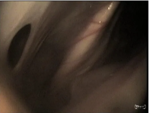
Twenty-four hours after hospitalization a second endoscopy was carried out. The dorsal wall of the pharynx was still occluding the larynx and pre-venting its visualization but no blood clots were present. The right guttural pouch was normal while the mucosal lining of the left guttural pouch was congest and edematous (indicating inflammation) and a viscous grey material covered the stylohyoid bone, part of the medial compartment and the me-dian septum (Figure 3). A yellowish-black, pointed, fungal plaque was located in the lateral compart-ment, on the wall of the maxillary artery (Figure 4). The plaque was about 1 cm in diameter and below it there was a focal aneurysmal dilatation of the ar-terial wall. A sample of guttural pouch lavage fluid was obtained by instillation and aspiration of 10 ml of sterile saline solution and used for cytological and bacteriological examinations. Endoscopy also showed the presence of food in the pharynx and trachea.
Cytological examination revealed the presence of some coccaceous bacteria that at microbiological examination were indentified as Streptococcus equi
subsp. zooepidemicus.
The same day the donkey showed a marked dysphagia, causing nasal food discharge but only occasional coughing. Nevertheless, crackles and wheezes were present at pulmonary
[image:2.595.64.292.86.256.2]ausculta-Figure 1. Bilateral epistaxis
[image:2.595.64.291.508.737.2] [image:2.595.310.526.510.718.2]tion (aspiration pneumonia). The owners did not consent to surgery for financial reasons and the decision was undertaken to implement topical an-tifungal therapy.
On the following day (three days from the first haemorrhage) a new endoscopy was performed and 15 ml of a 2% miconazole gel-emulsion was applied topically on the entire left guttural pouch mucosa through an indwelling catheter passed in the ac-cessory channel of the scope. On the next day the donkey had a new episode of haemorrhage and, in a few hours, aborted and developed a light laminitis treated with the appropriate hoof care, corrective shoes and antiinflammatory therapy. Despite the new haemorrhage, blood analysis did not show further worsening of the anaemia. Miconazole
application was repeated 48 hours later and for a further four times every 72 hours making a total of six applications. At the time of the fourth treat-ment the inflammation was absent and the plaque started to become smaller and lighter in colour. At the fifth treatment the erosion triggered by fungal infection caused a fistula in the median septum (Figure 5) but without fungal involvement in the controlateral pouch. At the last treatment no fun-gal lesions were visible but the aneurysm was still evident (Figure 6).
Dysphagia improved gradually and had complete-ly disappeared after 30 days, as did anisocoria and laminitis. A tracheotomy tube was kept inside until day six (2nd treatment), when the endoscopic
pha-ryngeal appearance did not show upper airway
ob-Figure 4. Detail of the mycotic plaque on the maxillary artery
[image:3.595.65.347.84.301.2] [image:3.595.64.351.539.755.2]struction. After thirty-two days of hospitalization the animal was discharged from the Hospital.
DISCUSSION AND CONCLUSIONS
Despite some personal communications regard-ing guttural pouch mycosis in donkeys, to our knowledge this is the first case report describing the progression of the diseases in this species.
Donkeys are normally treated like small horses but they have many peculiarities in incidence, presentation and treatment of diseases. Such dif-ferences have been highlighted also in our case; for example, when the patient presented only poor respiratory symptoms after developing aspiration pneumonia (little coughing and no fever).
Some peculiar clinical features of GPM in don-keys could be due to their important anatomical differences with horses: in the donkey the na-sopharynx is more constricted in its middle part and more flared dorso-ventrally than in the horse and, because of the shorter aryepiglottic folds, the epiglottis is normally closer to the arytenoids cartilages (Lindsay and Clayton, 1986; Fores et al., 2001). Furthermore, Delvaux et al. (2001) observed a partial collapse of the pharynx in 43% of normal donkeys endoscopically examined. These differ-ences could explain the severe dyspnea observed in the donkey at the first visit (normally not found in horses) when inflammation and bleeding in the guttural pouch could have caused the narrowing
of the pharynx as a result of both neurological and mechanical alterations, exacerbating the tendency to collapse. Since the history reported that epistaxis started a few hours after the dyspnea, the latter could be an early symptom of GPM in donkeys.
The donkey reported on in the present paper also presented neurological dysfunctions causing dysphagia and ipsilateral meiosis arising from dam-age to the nerves in contact with guttural pouches (cranial nerves IX, X, XI XII), similarly to what happens in horses. Also, the erosion of the median septum, rarely occurring in horses, has been found to be a possible complication in donkeys. Similar to what happens in the horse, also in our patient it was not possible to determine if the aneurism was a consequence of, or a predisposing factor for plaque formation.
Lindsay and Clayton (1986) found that to pass an endoscope through the pharyngeal opening of gut-tural pouches is more difficult in Spanish donkeys than in horses. In our case there were no differences in the technique between Martina Franca donkeys and horses.
[image:4.595.63.371.85.318.2]Topical treatment, normally considered inef-fective in horses (Ainsworth and Hackett, 2004), worked very well in our case. Topical treatment with enilconazole alone has been shown to be ef-fective in resolving dysphagia only in one single horse. The treatment schedule we used consisted in the endoscopical application of miconazole a total of six times, every 72 hours. Such a protocol is very short and less intensive that those suggested
Corresponding Author:
Dr. Fulvio Laus, University of Camerino, School of Veterinary Medical Sciences, Via Circonvallazione, 93/95 – 62024 Matelica (MC), Italy
Tel. +39 0737 403403, E-mail: fulvio.laus@unicam.it
for horses, where daily administration for longer periods (more than 30 days) is normally required. Although we cannot make any conclusions about the aetiology, the rapid and complete response of the donkey to the topical therapy could be due to the involvement of a more miconazole-sensitive fungus. Nevertheless, the gel-emulsion formulation of the drug may also have contributed to a better distribution of the drug on the mucosal surface of the pouch and, therefore, a more effective action.
More clinical cases will be necessary to better assess what are the differences in clinical manifesta-tion and therapeutic management of guttural pouch mycoses between horses and donkeys.
REFERENCES
Aisworth DM, Hackett RP (2004): Disorders of the res-piratory system. In: Reed SM, Bayly WM, Sellon DC (eds.): Equine Internal Medicine. 2nd ed. Saunders,
Missouri. 289–353.
Caron JP, Fretz PB, Bailey JV, Barber SM, Hurtig MB (1987): Balloon-tipped catheter arterial occlusion for prevention of hemorrhage caused by guttural pouch mycosis: 13 cases (1982–1985). Journal of American Veterinary Medical Association 191, 345–349.
Colles CM, Cook WR (1983): Carotid and cerebral angi-ography in the horse. Veterinary Record 113, 483–489. Cook WR (1968): The clinical features of guttural pouch
mycosis in the horse. Veterinary Record 83, 336–345. Delvaux V, Kirschvink N, Amory H, Busoni V, Art T,
Lekeux P (2001): Spécificités de la function cardiores-piratoire de l’âne (Equus asinus). Pratique Veterinaire Equine 33, 21–28. http://hdl.handle.net/2268/10351 Fores T, Lopes J, Rodriguez A, Harran R (2001):
Endos-copy of the upper airway and the proximal digestive tract in the donkey (Equus asinus). Journal of Equine Veterinary Sciences 21, 17–20.
Hardy J, Leveile R (2003): Diseases of the guttural pouches. Veterinary Clinic of North America-Equine Practice 19, 123–158.
Lane JG (1989): The management of guttural pouch mycosis in the horse. Equine Veterinary Journal 21, 321–324.
Lindsay F, Clayton H (1986): An anatomical and endo-scopic study of the nasopharynx and larynx of the don-key (Equus asinus). Journal of Anatomy 144, 123–132. Zinkl JG, Mae D, Guzman Merida P, Farver TB, Humble JA (1990): Reference ranges and the influence of age and sex on hematologic and serum biochemical values in donkeys (Equus asinus). American Journal of Vet-erinary Research 51, 408–413.