Effect of seven-day administration of carprofen
or meloxicam on renal function in clinically healthy
miniature pigs
P. Rauser, L. Stehlik, P. Proks, R. Srnec, A. Necas
Small Animal Clinic, University of Veterinary and Pharmaceutical Sciences, Brno, Czech Republic
ABSTRACT: Carprofen or meloxicam are nonsteroidal anti-inflammatory drugs (NSAIDs), which may elicit a variety of renal disturbances. Prior to this study, the effects of carprofen or meloxicam on renal function in pigs were unknown. A total of 21 clinically healthy Goettingen miniature pigs (36.9 ± 7.22 kg) were divided into three groups based on what they were administered – carprofen, meloxicam or saline. First, blood was collected from the jugular vein and urine by ultrasound-guided cystocentesis. Serum urea (U) and creatinine (CR), fractional clearance of sodium (FCNa), urine gamma-glutamyltransferase (GGT) and alkaline phosphatase (ALP) activities, urine protein/creatinine ratio (UP/UC), urine gamma-glutamyltransferase/creatinine ratio (GGT/CR) and urine alkaline phosphatase/creatinine ratio (ALP/CR) and urine analysis – urine specific gravity (USG) and sediment microscopy were assessed before and seven days after daily intramuscular administration (IM) of saline (1.5 ml per animal), carprofen (2 mg/kg) or meloxicam (0.1 mg/kg). All animals had identical housing, feeding and unlimited water intake and had not undergone surgery or been administered any medication for three months prior to this. All pigs served as control groups for an experimental study of fracture healing using transplantation of mesenchymal stem cells and scaffolds. The data were analyzed using a one way ANOVA and a Mann-Whitney test (P < 0.05). In pigs receiving carprofen, serum urea and creatinine were significantly decreased, compared to the control (P < 0.01) or meloxicam (P < 0.05) groups. In animals receiving meloxicam FCNa was significantly increased (P < 0.05) and urine specific gravity significantly decrease (P < 0.05) compared to the pretreatment values. Two carprofen-treated pigs had a slight increase in renal tubular epithelial cells upon urine sediment examination. Intramuscular administration of carprofen or meloxicam in healthy miniature pigs for seven days causes no clinically important changes in selected renal parameters (without azotemia). However these changes indicate mild damage of renal tubules. Despite these findings, meloxicam or carprofen are recommended for analgesia in healthy pigs.
Keywords: swine; non-steroidal anti-inflammatory drugs; renal safety
Supported by the Ministry of Education, Youth and Sports of the Czech Republic (NPV II, Grant No. 2B06130). Most surgeries require high-quality analgesia.
Due to damaged tissues, surgery often leads to inflammation (Lemke, 2004). In such cases anti-inflammatory analgesics are the best option. Non-steroidal antiinflammatory drugs (NSAIDs) are a good alternative to analgesia. Depending on the area of tissue damage and the duration of the pain-ful process, NSAIDs must often be administered for long periods of time. In small animals (dogs and cats) carprofen and meloxicam are used for
these purposes very often. There are many reports in the literature documenting both their adverse and beneficial effects (Curry et al., 2005; Luna et al., 2007). However, information regarding the ap-plication of meloxicam or carprofen in pigs is scant (Swindle, 2007).
generated by COX-2 play an important role in in-flammatory and painful reactions to tissue damage (Ferreira, 2002; McCrory and Lindahl, 2002). Apart from their positive effects, NSAIDs may also have numerous negative effects, impairment of kidney function being among the most severe. Hypotension and/or anesthesia during surgery leads to increased tonus of the sympathetic nervous system, resulting in prostaglandin-dependent changes in renal blood flow (Dunn and Zambraski, 1980). NSAID-induced inhibition of prostaglandins production may have a negative influence on renal hemodynamics and glomerular filtration rate with subsequent renal disorder (Oliver et al., 1981, 1983; Zambraski, 1995). Prostaglandins with a protective impact on the kidney are primarily based on the effectiveness of COX-1. If the kidney is in a prostaglandin-de-pendent condition, weaker adverse effects on renal hemodynamics are anticipated after application of meloxicam or carprofen compared to other NSAIDs. COX-2 also plays a major role in physiological kid-ney function (Harris, 2000). Carprofen and meloxi-cam are NSAIDs that preferentially inhibit COX-2, but partially also affect COX-1 (Kay-Mugford et al., 2000) with potential renal risks.
NSAIDs may cause numerous renal disorders, including acute renal failure (ARF), chronic renal failure (CRF), nephrotic syndrome, interstitial ne-phritis, water metabolism abnormalities, sodium retention and hypercalcemia (Clive and Stoff, 1984). However, studies on the effects of NSAIDs on renal function in anesthetized dogs after a sin-gle-dose administration of carprofen or meloxicam (Crandelli et al., 2004) did not show any clinically significant alteration in renal parameters. Similarly, a study by Lobetti and Joubert (2000) investigat-ing the influence of NSAIDs on renal function in anesthetized dogs did not detect adverse effects after administration of carprofen. Information on the effects of carprofen or meloxicam on the renal function of pigs is still missing. Junot et al. (2008) describe the influence of meloxicam on the renal function of piglets. While meloxicam was applied only once during the general anesthesia no devia-tions from monitored parameters were observed (urinary flow – UF, glomerular filtration rate – GFR and renal blood flow – RBF) after administration of meloxicam compared to the control group.
Patient renal function can be monitored by labora-tory tests of blood and urine. Biochemical tests include blood urea (U), creatinine (CR), fractional clearance of sodium (FCNa), urine
gamma-glutamytrans-ferase (GGT) and alkaline phosphatase (ALP) ac-tivities, urine protein/creatinine ratio (UP/UC), urine gamma-glutamyltransferase/creatinine ratio (GGT/CR) and urine alkaline phosphatase/creati-nine ratio (ALP/CR) which report on glomerular filtration and renal failure. Urine analysis includes urine specific gravity (USG) and sediment micros-copy with a focus on renal tubular epithelial (RTE) cells, which are informative with regard to renal tubular damage. Increases in the above-mentioned parameters (except USG) indicate possible renal failure (Osborne et al., 1995).
The purpose of this study was to compare the ef-fect of administration of carprofen and meloxicam in miniature pigs on selected serum and urine pa-rameters that might indicate impaired renal function. Both above-mentioned NSAIDs were, in contrast to previous studies, administered for a relatively long time (one week), which should outline the possibilities for prolonged NSAID therapy in miniature pigs. Thus, this analgesia could be used not only for orthopedic surgery in miniature pigs, but also for other surgical procedures carried out on these animals.
MATERIAL AND METHODS
Study group of animals
Twenty-one clinically healthy Goettingen mini-ature pigs – females weighing 36.9 ± 7.22 kg (mean ± SD) and aged 2.3 ± 0.62 years were used for the study. All animals were clinically normal, had identi-cal housing, feeding and unlimited water intake and were free from medication or surgical procedures for the three months prior to the commencement of experiments. All pigs served as control groups for an experimental study (NPV II Research Project 2B06130) on fracture healing using transplantation of mesenchymal stem cells (MSCs) and scaffolds, segmental bone defect stabilized by locking com-pression plate without MSC transplantation. All pro-cedures were carried out with consent of the Ethical Committee (No. 46613/2003-1020) of the University of Veterinary and Pharmaceutical Sciences.
Treatment groups and drug administration
The pigs were divided into three treatment groups:
group CAR – carprofen 2 mg/kg i.m. (Rimadyl;
i.m. (Metacam; Labiana Pharm.) and group CON – saline 0.9% 1.5 ml i.m. per animal (control). Each pig received drugs intramuscularly once a day for seven days in the above-mentioned doses. Blood and urine sample collection in all pigs were carried out before beginning with drug administration (T0) and seven days after daily drug administration. All drugs were applied daily at 2:00 p.m., samples were collected at 1:00 p.m.
Data collection
Blood was collected by venipucture from v. cava cranialis. Urine samples were collected aseptically by ultrasound-guided cystocenthesis. Urine and blood samples were collected before (T0) and seven days after medication (CAR, MEL, CON).
Serum blood variables evaluated included serum urea (U) and serum creatinine (CR). Urine variables evaluated included fractional clearance of sodium (FCNa), urine gamma-glutamyltransferase (GGT) and alkaline phosphatase (ALP) activities, urine protein/creatinine ratio (UP/UC), urine gamma-glutamyltransferase/creatinine ratio (GGT/CR) and urine alkaline phosphatase/creatinine ratio (ALP/ CR). Microscopic evaluation of the urine (performed by one investigator) was done on urine sediment stained with the Sternheimer-Malbin stain which enabled the differentiation of renal tubular
epithe-lial (RTE) cells from other urinary epitheepithe-lial cells (Osborne et al., 1995). The finding of RTE cells in the urine was subjectively scored on a scale of one to four. A score of one represented one RTE cell/two to three high power fields (HPF; 400× magnification), two represented 1−2 RTE cells/HPF, three represent-ed 2−4 RTE cells/HPF, and four representrepresent-ed more than five RTE cells/HPF (Vaden et al., 2009).
Statistical analyses
At first, variables of each group of animals seven days after medication were compared with vari-ables of the group before medication – at time T0 (CAR vs. T0, MEL vs. T0, CON vs. T0). Thereafter, variables evaluated seven days after medication were compared with each other (CAR vs. MEL, CAR vs. CON, MEL vs. CON). All variables were compared by means of one way ANOVA and the Mann-Whitney test. In all analyses, a value of P < 0.05 was considered significant.
RESULTS
[image:3.595.131.474.494.688.2]In pigs receiving carprofen, serum urea and se-rum creatinine were significantly decreased com-pared to the control (P < 0.01) or meloxicam (P < 0.05) groups (Figures 1 and 2).
Figure 1. Box plot showing median (line, value), 25th−75th percentile (box) and nonoutlier maximum and minimum
levels of serum urea in examined groups of pigs
T0 = before medication, CON = after administration of saline, CAR = after administration of carprofen, MEL = after administration of meloxicam
*significant decrease compared to the control (P < 0.01) or meloxicam (P < 0.05) groups
Serum urea
3.6
5.8
3.5*
4.9
0 1 2 3 4 5 6 7 8
T0 CON CAR MEL
Se
ru
m
le
ve
l (
m
m
ol
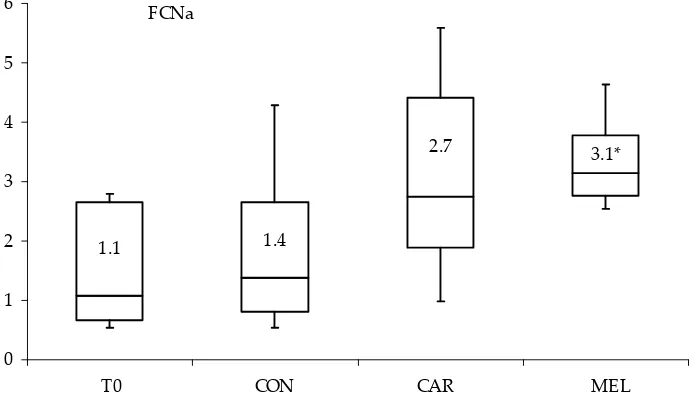
In pigs receiving meloxicam FCNa was signifi-cantly increased (P < 0.05) (Figure 3) and USG sig-nificantly decreased (P < 0.05) (Figure 4) compared to the pre-treatment values.
Two carprofen-treated pigs had a slight increase (scale 1) in RTE cells upon urine sediment
examina-tion compared to other groups including pretreat-ment values.
[image:4.595.130.475.86.280.2]There were no changes in urine enzyme (GGT, ALP) activity and urine GGT/CR and ALP/CR ratio as an indicator of renal dysfunction. All animals were without azotemia.
Figure 2. Box plot showing median (line, value), 25th−75th percentile (box) and nonoutlier maximum and minimum
levels of serum creatinine in examined groups of pigs
T0 = before medication, CON = after administration of saline, CAR = after administration of carprofen, MEL = after administration of meloxicam
*significant decrease compared to the control (P < 0.01) or meloxicam (P < 0.05) groups
FCNa
1.1 1.4
2.7 3.1*
0 1 2 3 4 5 6
[image:4.595.140.486.493.691.2]T0 CON CAR MEL
Figure 3. Box plot showing median (line, value), 25th−75th percentile (box) and nonoutlier maximum and minimum
levels of fractional clearance of sodium (FCNa) in examined groups of pigs
T0 = before medication, CON = after administration of saline, CAR = after administration of carprofen, MEL = after administration of meloxicam
*significant increase (P < 0.05) compared to the pretreatment values
Serumcreatinine
106.7 124.2
74.1*
99.7
0 20 40 60 80 100 120 140 160 180
T0 CON CAR MEL
Se
ru
m
le
ve
l (
um
ol
DISCUSSION
To assess the effects of long-term administra-tion of NSAID, it was first necessary to determine the suitable dosage for pigs, which had not been described previously in the literature.
For meloxicam, many authors mention only sin-gle-administration doses. Fosse et al. (2008) report a single dose of 0.4 mg/kg administered intrave-nously (i.v.) for the pharmacokinetic and pharma-codynamic study of meloxicam in piglets. The same single dose of meloxicam in adult pigs was used by Swindle (2007) and Friton et al. (2006) in endotox-emia therapy and by Hirsch et al. (2003) in masti-tis-metritis-agalactia syndrome therapy. A similar dosage (0.3 mg/kg) was administered by Girod et al. (2000, 2002) in their studies on the antiemetic effects of meloxicam. However, Reyes et al. (2002), used meloxicam in their neurological surgeries in piglets intravenously at the much higher dose of 1 mg/kg. Junot et al. (2008) applied meloxicam at a dose of 0.2 mg/kg when comparing its adverse effects to ketoprofen. Since the latter dose is close to the meloxicam dosage for dogs and cats (Mathews, 1997; Lascelles, 2000), we chose 0.1 mg/kg for week-long repeated administration of meloxicam dose.
Carprofen is often used in animals such as dogs, cats or horses (Mathews, 1997; Lascelles 2000).
However, only Swindle (2007) reports on its use with regard to analgesia in pigs. He reported the dose of 2−4 mg/kg subcutaneously (s.c.), but does not mention the possibility of repeated adminis-tration. For the week-long repeated administra-tion of carprofen in pigs we therefore chose the lower dose limit − 2 mg/kg. Crandelli et al. (2004) compared the effect of carprofen and meloxicam on renal function in dogs. They did not observe major deviations of selected parameters (GFR, U, CR) when comparing control and medicated groups. However, both substances were applied in single doses. We did not note any increase in pig serum U or CR in our study after a week-long medication with meloxicam or carprofen either. In pigs medicated with meloxicam we observed increased FCNa, which suggests possible altera-tions in renal tubular function (Osborne et al., 1995). However, the anticipated azotemia was not observed in animals with meloxicam or carprofen medication.
We believe that azotemia did not develop be-cause we administered only a low dose of meloxi-cam (0.1 mg/kg) even though it was administered repeatedly. After a single application of meloxi-cam in piglets during anesthesia, Junot et al. (2008) found only mild changes in UF, GFR and RBF. Blood and urine parameters, however, were not recorded.
Urine USG
1034 1034 1033
1023*
1000 1010 1020 1030 1040 1050
[image:5.595.135.477.84.288.2]T0 CON CAR MEL
Figure 4. Box plot showing median (line, value), 25th−75th percentile (box) and nonoutlier maximum and minimum
levels of urine specific gravity (USG) in examined groups of pigs
T0 = before medication, CON = after administration of saline, CAR = after administration of carprofen, MEL = after administration of meloxicam
The above-mentioned changes observed by Junot et al. (2008), though statistically insignificant, partial-ly correspond with our results. In pigs medicated with meloxicam we noted a significant increase in FCNa and decrease in urine USG. The lower urine USG values in pigs could be caused by sub-clinical renal damage or other non-kidney related diseases (Vaden et al., 2009). Lower urine USG could be also associated with limited secretion of antidiu-retic hormone (ADH; Greene and Grauer, 2007). The higher values for the urine GGT/CR ratio may also indicate functional renal disorder after mel-oxicam application. This increase, however, was not statistically significant. An increased GGT/CR ratio could be caused by decreased GFR or severe glomerular damage (Vaden et al., 2009).
The creatinine and urea blood levels were with-in limits with-in the group of animals medicated with meloxicam. Azotemia occurs only after 75% dam-age of renal parenchyma (Osborne et al., 1995). Physiological limits of urea and creatinine values are not specific to renal damage. Levels of blood urea and creatinine in animals were lower in carprofen-medicated animals. Together these two parameters can be regarded as markers of glomerular filtration (Osborne et al., 1995). We may therefore specu-late about increased glomerular renal filtration after carprofen medication, but we were not able to find or explain its cause. Ko et al. (2000) com-pared blood urea nitrogen (BUN, BUN × 2.14 = U) and creatinine values in dogs. They did not find any deviations from our values measured in pigs. This may be due to the different impact of carpro-fen on U and CR in dogs and pigs in relation to medication with carprofen. Unfortunately, a more comprehensive explanation or references are not available. The presence of RTE cells in the urine sediment indicates transitional renal damage. RTE cells are excreted into urine during normal exchange or as a result of renal damage (Lobetti and Joubert, 2000). In our groups of pigs we observed a statistically insignificant occurrence of RTE cells only in two animals medicated with carprofen, with no other abnormalities in the observed parameters. In animals medicated with meloxicam in which we observed increased FCNa and decreased urine USG indicating possible renal damage, RTE cells were not found in a single animal. For further as-sessment of renal function, some additional tests would be helpful. Nevertheless, as the animals were stabled in groups, we could not check individually water intake and urine production. Moreover, the
experiments were carried out with limited labora-tory equipment.
CONCLUSION
Intramuscular administration of carprofen or meloxicam in healthy miniature pigs for seven days causes no clinically important changes in selected renal parameters (without azotemia). However, these changes, especially in pigs after administra-tion of meloxicam, could indicate mild damage of renal tubules. Despite the findings, meloxicam or carprofen at the described dosages is recommended for analgesia in healthy miniature pigs.
This type of NSAID medication seems to be suit-able not only in miniature pigs used as fracture healing models in our research, but also on these animals in relation to other surgical procedures.
REFERENCES
Clive DM, Stoff JS (1984): Renal syndromes associated with nonsteroidal anti-inflammatory drugs. New Eng-land Journal of Medicine 310, 563–572.
Crandelli DE, Mathews KA, Dyson DH (2004): Effects of meloxicam and carprofen on renal function when administered to healthy dogs prior to anesthesia and painful stimulation. American Journal of Veterinary Research 65, 1384−1390.
Curry SL, Cogar SM, Cook JL (2005): Nonsteroidal an-tiinflammatory drugs: a review. Journal of American Animal Hospital Association 41, 298−309.
Dunn MJ, Zambraski EJ (1980): Renal effects of drugs that inhibit prostaglandin synthesis. Kidney Interna-tional 18, 609–622.
Ferreira SH (2002): Peripheral analgesic sites of action of antiinflammatory drugs. International Journal of Clinical Practice 7 (Suppl.), 2–10.
Fosse TK, Haga HA, Hormazabal V, Haugejorden G, Horsberg TE, Ranheim B (2008): Pharmacokinetics and pharmacodynamics of meloxicam in piglets. Jour-nal of Veterinary Pharmacology and Therapeutics 31, 246−252.
Friton GM, Schmidt H, Schroedl W (2006): Clinical and anti-inflammatory effects of treating endotoxin-chal-lenged pigs with meloxicam. Veterinary Record 159, 552−557.
Corresponding Author:
MVDr. Petr Rauser, Ph.D., University of Veterinary and Pharmaceutical Sciences, Faculty of Veterinary Medicine, Palackeho 1–3, 612 42 Brno, Czech Republic
Tel. +420 603 857 693, E-mail: rauserp@vfu.cz
inhibitors and a 5-HT(3) receptor antagonist.. Neu-ropharmacology 39, 2329−2335.
Girod V, Dapzol J, Bouvier M, Grelot L (2002): The COX inhibitors indomethacin and meloxicam exhibit anti-emetic activity against cisplatin-induced emesis in piglets. Neuropharmacology 42, 428−436.
Greene SA, Grauer GF (2007): Renal disease. In: Tran-quilli WJ, Thurman JC, Grimm KA (eds.): Lumb and Jones’ Veterinary Anaesthesia and Analgesia. 4th ed. Blackwel Publishing, Ames, Iowa. 915−919.
Harris RC (2000): Cyclooxygenase-2 in the kidney. Jour-nal of the American Society of Nephrology 11, 2387−2394.
Hirsch AC, Philipp H, Kleemann R (2003): Investigation on the efficacy of meloxicam in sows with mastitis-metritis-agalactia syndrome. Journal of Veterinary Pharmacology and Therapeutics 26, 355−360. Junot S, Troncy E, Keroack S, Gauvin D, del Castillo JR,
Boivin R, Bonnet JM (2008): Renal effect of meloxicam versus ketoprofen in anaesthetized pseudo-normovol-aemic piglets. Canadian Journal of Physiology and Pharmacology 86, 55−63.
Kay-Mugford P, Benn SJ, LaMarre J, Conton P (2000): In vitro effects of nonsteroidal anti-inflammatory drugs on cyclooxygenase activity in dogs. American Journal of Veterinary Research 61, 802–810.
Ko JC, Miyabiyashi T, Mandsager RE, Heaton-Jones TG, Mauragis DF (2000): Renal effects of carprofen admin-istered to healthy dogs anesthetized with propofol and isoflurane. Journal of American Veterinary Medical Association 217, 346–349.
Lascelles DX (2000): Clinical pharmacology of analgesic agents. In: Hellebrekers LJ (ed.): Animal Pain. Van der Wees, Utrecht. 85–116.
Lemke KA (2004): Understanding the patophysiology of perioperative pain. Canadian Veterinary Journal 45, 475−480.
Lobetti RG, Joubert KE (2000): Effect of administration of nonsteroidal anti-inflammatory drugs before sur-gery on renal function in clinically normal dogs. Amer-ican Journal of Veterinary Research 61, 1501−1507.
Luna SP, Basilio AC, Steagall PV, Machado LP, Moutinho FQ, Takahira RK, Brandao CV (2007): Evaluation of adverse effects of long-term oral administration of carprofen, etodolac, flunixin meglumine, ketoprofen, and meloxicam in dogs. American Journal of Veteri-nary Research 68, 258−264.
Mathews KA (1997): Non-steroidal antiinflammatory analgesics for acute pain management in dogs and cats. Veterinary and Comparative Orthopaedics and Trau-matology 10, 122−129.
McCrory CR, Lindahl SG (2002): Cyclooxygenase inhi-bition for postoperative analgesia. Anesthesia and Analgesia 95, 169–176.
Oliver JA, Sciacca RR, Pinto J, Cannon PJ (1981): Par-ticipation of the prostaglandins in the control of renal blood flow during acute reduction of cardiac output in the dog. Journal of Clinical Investigation 67, 229–237. Oliver JA, Sciacca RR, Cannon PJ (1983): Renal vasodi-lation by converting enzyme inhibition. Role of renal prostaglandins. Hypertension 5, 166–171.
Osborne CA, Stevens JB, Lulich JP, Ulrich LK, Bird KA, Koehler LA, Swanson LL (1995): A clinician’s analysis of urinalysis. In: Osborne CA, Finco DR (eds.): Canine and Feline Nephrology and Urology, Williams and Wilkins, Baltimore. 136–205.
Reyes L, Tinworth KD, Li KM, Yau DF, Waters KA (2002): Observer-blinded comparison of two nonopioid anal-gesics for postoperative pain in piglets. Pharmacology, Biochemistry and Behavior 73, 521–528.
Swindle MM (2007): Swine in the laboratory. Surgery, Anesthesia, Imaging, and Experimental Techniques. 2nd ed. CRC Press, Boca Raton. 35−79.
Vaden SL, Knoll JS, Smith FWK, Tilley J (eds.)(2009): Blackwell’s Five-Minutes Veterinary Consult: Labora-tory Tests and Diagnostic Procedures: Canine and Feline. Wiley-Blackwell, Ames, Iowa. 763 pp.
Zambraski EJ (1995): The effects of nonsteroidal anti-in-flammatory drugs on renal function: experimental stud-ies in animals. Seminars in Nephrology 15, 205–213.