POSTCENTRAL TOPECTOMY OPTIMIZATION BY SELECTIVE ABLATIONS OF THE PRIMARY SOMATOSENSORY CORTEX
Timothy David Challener
A dissertation submitted to the faculty at the University of North Carolina at Chapel Hill in partial fulfillment of the requirements for the degree of Doctor of Philosophy in the Joint
Department of Biomedical Engineering.
Chapel Hill 2018
Approved by:
Lianne Cartee
Robert Dennis
Oleg Favorov
Jeffrey Macdonald
Mark Tommerdahl
ii
© 2018
Timothy David Challener
ALL RIGHTS RESERVED
iii ABSTRACT
Timothy David Challener: Postcentral Topectomy Optimization by Selective Ablations of the Primary Somatosensory Cortex
(Under the direction of Oleg Favorov)
Postcentral topectomy is a neurosurgical procedure, practiced in the mid-20th century, in which surgical ablations of the primary somatosensory cortex (SI) were used as a therapeutic means of treating patients suffering from intractable chronic pain. While successful in treating many cases, the procedure was poorly understood and eventually became displaced by methods that more consistently stopped patient complaints of pain, such as opiates and frontal lobotomies.
SI contains nociresponsive neurons in two regions: one region in cytoarchitectonic area 1, with properties resembling sharp, discriminative, first pain; and the other in the anterior part of area 3a, with properties resembling burning, affective, second pain. To test the hypothesis that
permanent pain loss in postcentral topectomy was achieved when the nociresponsive part of area 3a was removed, we trained unrestrained squirrel monkeys to obtain a juice reward by pulling a noxiously heated metal rod. Pull duration shows high sensitivity to rod temperature and was used as a measure of each animal’s pain sensibility. After training, all monkeys gave consistent
baseline pull durations for each trial in a series of chosen neutral (control) and noxious temperatures.
Next, we made small electrolytic cortical lesions in 2 monkeys, targeting the hand region
of nociresponsive area 3a. These lesions significantly elevated pull durations for at least 4
iv
months (at which time the animals were euthanized), thus indicating permanently reduced pain sensibility. In contrast, ablation of motor cortex anterior to area 3a in the 3rd monkey
significantly reduced pull durations, indicating permanently elevated pain sensibility.
The current opioid crisis makes these results particularly relevant to clinical medicine.
Although the precise location of area 3a in the central sulcus varies extensively among humans, its nociresponsive region can be accurately localized in any given patient by using high-
resolution fMRI. Once localized, the area 3a nociresponsive region can be targeted for reversible or permanent inactivation. Such precisely targeted inactivation might greatly improve the success rate of the postcentral topectomy in amelioration of pathological pain, making it a highly
attractive means of treating otherwise intractable chronic pain.
v
TABLE OF CONTENTS
LIST OF FIGURES ... vi
CHAPTER 1: INTRODUCTION ...1
Pain and the Somatosensory Cortex ...1
CHAPTER 2: POSTCENTRAL TOPECTOMY ...4
Postcentral Topectomy: A Meta-Analysis ...4
Chronological Case Reports ...6
Previous Postcentral Topectomy Reviews ...18
Postcentral Topectomy Conclusions ...22
CHAPTER 3: BEHAVORIAL TEST OF PAIN SENSIBILITY ...24
Testing the 3c hypothesis ...24
Training Methodology ...24
Testing Procedure ...26
Initial Pull Training ...29
Lever Handedness Control ...30
Temperature Training ...32
Determining Individual Baselines ...32
Testing Regimen ...34
CHAPTER 4: EFFECT OF CORTICAL ABLATION OF PAIN SENSIBILITY ...37
Lesioning Preparations ...37
vi
Lesioning Procedure ...39
Lesioning Results ...41
CHAPTER 5: CONCLUSIONS ...45
REFERENCES ...48
vii
LIST OF FIGURES
Figure 1: Figure from The Cortical Localization of Cutaneous Sensations, Dana, 1888 ... 2
Figure 2: Histological slice at border between area 3 and 4 ... 3
Figure 3: Selected Case Reports ...5
Figure 4: View of machine from inside cage ... 31
Figure 5: Pull duration as a function of rod temperature for two subjects ... 33
Figure 6: Recording Chamber Schematic ... 38
Figure 7: Monkey with recording chamber attached. ... 38
Figure 8: Electrode sites, recordings, and histology. ... 40
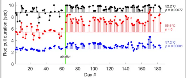
Figure 9: Results of 3c Ablation in Subject A... 41
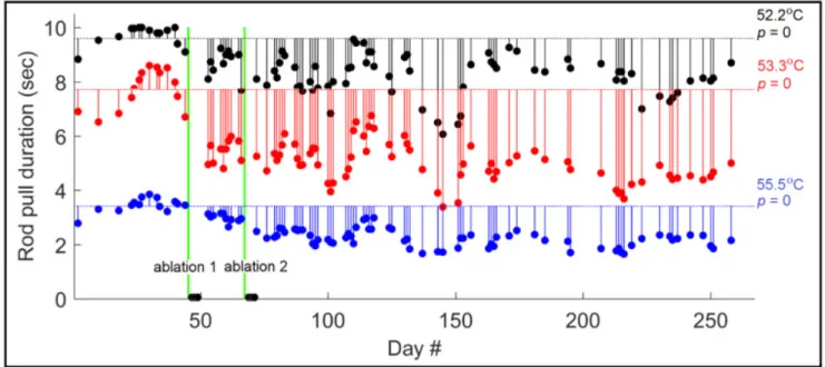
Figure 10: Results of 3c Ablation in Subject B... 42
Figure 11: Results of 3c Ablation in Subject C ... 43
Figure 12: Sagittal cross-sections of the human central sulcus ... 46
1
CHAPTER 1: INTRODUCTION
Pain and the Somatosensory Cortex
That the primary somatosensory cortex (SI) in the postcentral gyrus is involved in pain perception is uncontroversial. In 1888, Dana made one of the earliest maps of lesion locations which produced deficits in pain sensation (Dana et al, 1888). The clustering around the central sulcus is obvious (figure 1). Decades later, World War I provided Kleist with numerous cases of soldiers who had localized brain damage and suffered from a variety of symptoms. Having observed that superficial wounds to the somatosensory cortex spared temperature and pain sensibilities, but deeper wounds were far more likely to cause changes in those submodalities, Kleist concluded “pain and temperature sensations belong to the anterior, post-central area 3a and 3b” (Kleist, 1934). Marshall later conducted similar research on injured World War II soldiers, and came to the same conclusions (Marshall, 1951).
In the late 1990s and early 2000s, Barry Whitsel, Mark Tommerdahl, Oleg Favorov, and Chuck Vierck used optical intrinsic signal (OIS) imaging and microelectrode recordings to study area 3a in squirrel monkeys, directly demonstrating the involvement of anterior sector of area 3a in nociception (Tommerdahl et al, 1996, 1998; Whitsel et al, 2009). This function is distinct from posterior 3a’s well-known involvement in proprioception.
2
Figure 1: Figure from Dana (1888). All lesions marked were from patients who experienced analgesia while alive, and had the exact lesion locations recorded in postmortem autopsies.
Anterior 3a not only has a different function from posterior 3a, it is also a
cytoarchitecturally distinct region (figure 2). Traditionally, motor cortical area 4 is defined as a
region extending posteriorly as far as the large pyramidal neurons (Betz cells) in layer V, and
area 3a is defined as extending as far anterior as the thin band of small layer IV cells. However,
the somatosensory territory activated by noxious stimuli lies in the disputed region which meets
both these definitions. For sake of clarity, and because it has significant C-afferent input, I will
be referring to this specific transitional zone as “area 3c”.
3
Figure 2: Histological slice at border between area 3a and 4. The layer IV which defines the traditional extent of area 3a is highlighted in blue, the layer V defining area 4 in red. Arrow points to a lesion marking the location of a studied nociceptive neuron, which is located in area
“3c”. (Modified from Whitsel et al, 2009)
Not only is there sound theoretical support for 3c’s involvement in pain, there also is
practical experience modulating pain perception through surgical interventions in the SI cortex
(Vierck et al, 2013). These surgical interventions – the ‘postcentral topectomy’ – are worth
investigating in depth.
4
CHAPTER 2: POSTCENTRAL TOPECTOMY
Postcentral Topectomy: A Meta-Analysis
Postcentral topectomy is a surgical procedure which was used in the mid-20th century as a last-resort treatment for otherwise-incurable chronic pain. Surgeons opened the skull,
electrically stimulated the surface of the somatosensory cortex, and wherever the stimulation evoked pain, they ablated the cortex. While this treatment often provided long term pain relief, it was eventually displaced by treatment options that more consistently ended patient complaints of pain, such as narcotics and frontal lobotomies.
We identified 28 cases from 17 full-text reports in 5 languages (Gutierrez-Mahoney 1944;
Lhermitte and Puech 1946; Horrax 1946; Odom and Lyman 1946; Echols and Colclough 1947;
Gutierrez-Mahoney 1948; Wertheimer and Mansuy 1949; Stone 1950; Akhundov 1950; Sugar and Bucy 1951; Lewin and Phillips 1952; Pool and Bridges 1954; Török 1960; Carbonin 1961;
Deák and Tóth 1966; Lende and Druckman 1971; Woolsey et al 1979).
PubMed, JSTOR and Google Scholar searches were conducted using the terms
“postcentral topectomy”, “parietal topectomy” “parietal cortex pain surgery”, “parietal phantom limb surgery”, and “phantom pain surgical treatment”. No date boundaries or language
restrictions were set. Inclusion criteria for the studies were used. Those criteria were that the
study must be used to treat a chronic pain syndrome, give a firsthand description of the region
ablated, that the region ablated primarily consist of the postcentral gyrus, and that the outcome of
5
the surgery be known. Studies where the cortex was obviously damaged upon visual inspection of the surface were also excluded.
In 24 of the 28 patients the surgery was successful. Success was defined as the patient experiencing significant relief from the original pain at the end of the period recorded in the case report. Of these successes, 20 had a recording period that lasted at least six months after the surgery, and 14 were recorded for at least ten months. Of the failures, in 1 of the 28 cases, the surgery was entirely unsuccessful (with no pain relief whatsoever), and in 3 of 28 cases the original pain completely returned at some later point.
Figure 3: Selected Case Reports. Successes are cases with significant reduction of the original
pain at the last recorded point, “Lasting” successes indicating a follow-up of at least six months,
and “putative” successes a follow-up period of less than six months. Recurrence indicates the
complete return of the original pain.
6 Chronological Case Reports
The detailed postcentral topectomy case reports are scattered across the literature in five different languages. In order to do a proper analysis of the surgery, the case reports included in the statistical analysis have been summarized here.
In 1941, Gutierrez-Mahoney was the first to perform a postcentral topectomy (Gutierrez- Mahoney, 1944). His patient suffered from painful phantom fingers after a hand injury. Based on a previous report from Holmes, where a cerebral lesion abolished a phantom limb, Gutierrez- Mahoney decided to operate, assuming that the pain originated somewhere in the sensory cortex.
He explored the postcentral parietal cortex with electrostimulation to locate regions that evoked pain in the phantom limb. After those regions were identified and mapped, he excised them through subpial resection. The patient was cured, and remained so for at least two years after the operation.
Gutierrez-Mahoney went on to perform postcentral topectomies on three more patients (Gutierrez-Mahoney, 1948). The results were somewhat mixed. In the first of these cases there was considerable pain relief, with a small amount of remaining pain that could be controlled with aspirin. In the second, a patient with a mid-thigh amputation, the pain from the phantom limb was abolished, but the pain in his hip recurred within six months. In the final case, a variation upon the technique was performed; the precentral gyrus was removed along with the postcentral.
This surgery ultimately proved less effective, as the patient’s pain was abolished in the short term but recurred within six months.
Lhermitte and Puech (1945) were the second group to intentionally attempt a surgical
intervention. It was, unfortunately, unsuccessful; their parietal resection caused only a temporary
vanishing of the phantom limb, and also restored spontaneous movements of the stump, which
7
had previously been abolished by a myelotomy. Lhermitte suspected that the operation failed due to an insufficiently large resection, but didn’t provide exact details of the lesion’s location and extent beyond saying he ‘resected the left parietal lobe’.
The next year, Horrax (1946) reported on the four postcentral topectomies he performed.
In his first patient, he was unable to produce lasting pain relief, despite performing two separate surgeries and removing the pre- and post-central cortex both ipsilateral and contralateral to the phantom hand. The longest duration of pain relief lasted only twenty-two days. His later interventions, however, were more successful. The second patient experienced chronic pain in both arms after a spinal cord injury at the C6 vertebra, which had only been exacerbated by a cervical laminectomy. He received considerable relief from the pain in the arm contralateral to the topectomy (the more painful limb). Horrax’s third patient previously suffered from a glial tumor in the temporal lobe that damaged the basal ganglia and internal capsule. After its removal, he began experiencing pain in his right hand, arm, foot, and leg. Horrax mapped the motor cortex for the arm and leg and removed a large section of the postcentral cortex posterior to it. At five months, pain returned to the arm and hand, however as of fourteen months, there had been no pain in the leg and foot. The hand and arm were entirely anesthetic to external pain as well. The final patient had previously suffered from a fibrillary astrocytoma situated
parasagitally in the arm and leg centers, which had been removed surgically. Two years after its
removal, he suffered incapacitating pain in the right arm and hand, along with lesser pain in the
right face and leg and hypoesthesia in all four locations. The surgeons conducted electrical
stimulation of the postcentral cortex to evoke pain, followed by guided excisions. Relief of the
patient’s upper extremity pain was complete at ten months, when he died from a pontine
8
hemorrhage. Notably, until death the patient experienced a total loss of pain sensation in his right hand.
Odom and Lyman had an unusual case report later in 1946. Their patient, a 68-year-old woman, experienced left trigeminal nerve pain for four years. A complete left trigeminal
rhizotomy removed tic pains, but did not remove the burning pain on the left side of her face. A postcentral topectomy was performed in order to alleviate this pain. The motor and sensory areas for the face and left arm were mapped electrically, then removed. After the surgery, there was a loss of sensation on the left face and arm, except for the left eye, in which the patient complained of pain and photophobia. In addition, there was also a puzzling ataxic dysarthria observed, a symptom unique among postcentral topectomy patients. It is possible that the extensive removal of motor cortex is responsible. It is also possible that brain damage occurred outside the region of incision, as after the surgery the patient spent five days with a fever, and during that time was “confused and incoherent”. As she was only followed for a month post- surgery, it is impossible to know the long term results.
In 1946, Echols and Colclough (1947) performed a fully successful postcentral topectomy
on a 53 year old male patient suffering from phantom limb pain in his leg, after an attempt at
spinal anesthetic proved effective for only five minutes. Stimulation of the leg’s motor cortex
made the stump jerk, while stimulation of the corresponding sensory cortex made the phantom
foot feel hot. Both the gray matter and some white matter of the leg and foot sensory cortex
were removed, with the patient spontaneously announcing partway through the ablation that the
phantom foot and pain had vanished. Although the patient experienced some Jacksonian seizures
and aphasia due to an extradural blood clot, after it was removed in a second surgery he made a
9
full recovery. Eleven months later he remained completely free of both the phantom and the pain.
Wertheimer and Mansuy (1949) operated on a 58-year old male patient whose leg was amputated in World War One. He had experienced phantom limb pain in his foot since 1916, and had undergone multiple surgical treatments in the intervening 31 years, all unsuccessful. During the surgery, the motor cortex was explored with electrical stimulation, and the corresponding region of the postcentral cortex was ablated, resecting two square centimeters with deep
coagulation at the top and inside. The patient felt nonpainful jerks of the stump when stimulated, and the pain vanished at the end of the procedure. However, shortly after the surgery, the patient soon grew increasingly agitated, confused, and restless. The surgical staff discovered that he had—foiling all attempts at surveillance—secretly been treating his pain with daily doses of up to 24 cg of morphine, and was now suffering the withdrawal symptoms. He was released after ten days of detoxification, and six months later was in excellent condition.
Stone (1950) and John Martin operated on three phantom limb patients: two males, 27 and 28, and one female, 58. The first had his left forearm amputated after a shell fragment wound in 1944. After several failed procedures, including three neuroma operations, he was eventually taken in for a postcentral topectomy. When Martin attempted to identify the correct brain region through electrical stimulation, the patient had a generalized convulsion and lost consciousness. Nevertheless, Martin excised the region which seemed to be the sensory cortex for the hand, approximately 2 cm by 1 cm, to 1 cm of depth. The patient had no pain or
awareness of his phantom limb post-surgery, and while the phantom itself reappeared four weeks after discharge, the pain was still absent ten months later. The second patient had his leg
amputated at the thigh after a motorcycle accident. His phantom limb pain was concentrated in
10
the heel, and any movement of the stump, including wearing his artificial leg, exacerbated it. A postcentral gyrectomy was decided upon. Electrical stimulation of the postcentral gyrus exactly at the midline produced “exquisite pain and tingling” in the phantom limb. When the excision was completed, the patient said he no longer felt the phantom. He remained free of the pain for at least eleven months post-surgery. The final patient, a 58 year old woman, suffered from both central pain and phantom pain, and because of this pain had attempted suicide on three
occasions. The surgery excised the entire sensory gyrus, from the posterior border to the next gyrus. Although the patient had a “truly stormy convalescence”, and temporarily suffered motor aphasia, her phantom limb pain was immediately cured, and remained so at the fourteen month mark.
Also in 1950, Akhundov treated a patient (male, 41) who was run over by a car and subsequently suffered from a form of supernumerary phantom limb pain (Akhundov, 1950).
While his arm was physically intact, the nerves had been severed, and he felt a phantom with a harshly contracted elbow and hand, with both a continuous burning pain and intermittent increases in the ulnar or radial areas. Surgery was conducted four and a half months after the accident. Stimulation in the postcentral gyrus for the hand caused ‘pricking’ pains that
immediately grew into the full phantom pain. They then electrocoagulated all the blood vessels in the region, and removed cortical matter 1 cm deep, 1 cm medial, and 2.5 cm posterior, all the way to the lower temporal lobe (removing not only S1, but also areas 5, 7, and 40). Recovery followed an unusual course. After one hour, the phantom was still present, with pain only
present in digit 5, much diminished and periodic instead of continuous. At day four, the pain and sensation in the fingers was totally gone. However, on day 9 the phantom hand and pain
abruptly reappeared, somewhat diminished, and then started vanishing again on day 12. As of
11
the last recorded time - seven months after surgery - the phantom arm and pain were both completely abolished.
Sugar and Bucy (1951) attempted to treat postherpetic trigeminal neuralgia with a
postcentral topectomy in 1946. The patient (male, 67) had a painful vesicular eruption on the left side of his face, with constant burning pain that persisted even after the rash vanished. Over the course of the next three years, several operations on the left trigeminal nerve proved ineffective, eventually leaving the area numb, but with continuing pain. Severing all the root fibers of the gasserian ganglion resulted in pain relief for only two days. All other courses having failed, they decided to perform a postcentral topectomy. Despite extensive attempts to stimulate both the precentral and postcentral gyri, they could not evoke the same kind of pain from the patient (one location caused a feeling of ‘strangulation’ accompanied by involuntary clearing of the throat).
All of the postcentral gyrus was removed between the representation of the throat, and the representation of the thumb and index finger. Repeated stimulation still could not evoke pain, and the patient spontaneously said that the pain in the face had been completely relieved.
However, on the fifth day a slight stinging pain recurred, spreading to including all the initial area on the twelfth day, and eventually returning to its original strength. Speculating that the failure might have been caused by the bilateral representation of the face, they attempted another topectomy five months later, on the ipsilateral side. However their region of excision was too posterior, and missed the postcentral gyrus entirely, resulting in an unsuccessful surgery.
Lewin and Phillips (1952) operated on two patients injured in World War I. The first
patient (male, 66) had a left thigh amputation after a torpedo explosion. While his phantom limb
was initially painless, by 1941 the phantom started hurting, and by the time of admission in 1951
he had undergone numerous local operations to treat the pain, all unsuccessful. Electrical
12
stimulation of the postcentral gyrus immediately and repeatedly reproduced his pain. Excision of an area 1 cm in diameter had no effect on the pain, and stimulation deep in the anterior cut edge (on the posterior bank of the sulcus) still produced pain. The excision was increased in size until a 3 cm x 1 cm x 1 cm piece of cortex was removed. At this point the patient’s pain had been considerably reduced (though not fully eliminated), and the operation was ended. This minor pain faded over the course of the next few weeks, but returned to a small extent six months later, controllable with aspirin. Their second patient (male, 58) had a mid-thigh amputation due to a gunshot wound. For 27 years he had no phantom limb sensation, however in 1946 the stump abruptly became extremely painful, and by the time of admission in 1951 he had numerous local operations, a cordotomy, and a spinal analgesic. Only the cordotomy offered any relief, which lasted for three weeks. Stimulation of the postcentral gyrus immediately and repeatedly
reproduced the ‘gripping’ pain in his stump. Once again, the initial excision of 1 cm in diameter was insufficient to alter the pain. Unusually, using forceps to pinch a small blood vessel at the bottom of this excision reproduced the pain. As deeper gray matter was removed, the pain disappeared, leaving a final excision 2.0 cm × 1.5 cm × 1.5 cm deep. Following the operation the patient had some superficial tingling, with an attack of burning pain five days later, “quite different from any pain he had had previously”, which vanished after 17 hours. Five weeks post- operation, he had been completely relieved of the pain existing before the operation, although he now had a few new and minor aches over a small area of the stump 1 .
Pool and Bridges (1954) took a similar approach to Akhundov, removing both the postcentral gyrus and a large portion of the cortex posterior to it. The patient (female, 66) was
1
: The same paper includes a third successful (4 years) case report not included in the overall analysis. The surgery
removed abnormal postcentral cortex to prevent painful epileptic fits - the epilepsy is a complication which makes
this case insufficiently unambiguous to include as an example.
13
admitted in 1952 for constant burning and cramping phantom arm pain. Pool first electrically stimulated the motor cortex to identify it, then drove a ‘fence’ of needles 2.5 cm down into the sensory cortex along the border with the motor cortex. Pool then started undercutting from a point 7.5 cm posterior to the central fissure, continuing the cut to the anterior until the pins blocked his blade. Recovery was difficult for the first six weeks, due to withdrawal symptoms from taking the patient off narcotics too quickly. By the sixth week, there were occasional twinges of phantom pain, but the original constant pain did not recur, and the occasional pains gradually decreased over the next four months. The patient continued to experience relief from her phantom limb syndrome at least seventeen months after surgery.
Török (1960) treated a patient (male, 59) who had suffered numerous accidents, leaving him blind and resulting in an arm amputation. Phantom pain appeared in the arm immediately after surgery, could not be controlled by drugs and prevented him from sleeping. After eight days of conservative treatments proved unsuccessful, a postcentral gyrectomy was performed.
Electrical stimulation outlined the area of interest, but did not provoke any pain. The topectomy was performed on both pre and post central gyri for both the hand and arm areas. Immediately after, the patient was a little stunned, and still complained about pain but without any emotion - by the next day, however, he was relieved that he had no more pain and his stump no longer convulsed. After a month, the patient started occasionally feeling a different kind of dull, drawn- out pain in his hand at irregularly spaced intervals. He reported going days without any need for analgesics, and during his weeklong follow-up clinic visit six months later, there were no
complaints of pain. As the original phantom pain was gone, and the patient could now sleep at
night, Török considered the surgery a success.
14
Carbonin (1961) treated a patient (male, 64) who had suffered an amputation of the lower two thirds of his arm in 1953. Three weeks after the amputation, the patient began experiencing a painful burning electric current sensation from the shoulder stump to the tip of his fingers.
Novocainization of the stump, infiltration of the stellate ganglion, and resection of the median nerve all proved ineffective, granting only short-term temporary relief. Over the next few years the patient underwent ten different operations to treat his intolerable pain, all unsuccessful. In August 1959 a postcentral topectomy was performed. The sensory cortex corresponding to the arm was removed, with a total excision of 3 cm along the length of the convolution, 1 cm wide and 1 cm deep. Immediately upon waking, the patient was happy to find the pain had
disappeared, except for a small point on his wrist and little finger. Two days later, he had a Bravais-Jacksonniene seizure. The surgeons reopened the site, removed coagulated blood from the cortical resection, and while they were there, removed a couple more millimeters of gray and white matter from the upper and lower limits of the incision. This succeeded at removing the wrist pain and all of the finger’s pain but a small dot under the fingernail. Notably, a period of time after the surgery, the patient returned to the surgeons, claiming to be suffering from horrible pain. However, further investigation revealed the true source of this complaint lay in his severe opioid addiction, which had reached 219 codeine pills in 15 days. After a round of detox, the addiction and claims of pain were both gone, and as of a year later the patient remained cured of his pain.
Deák and Tóth (1966) attempted to treat two patients with postcentral topectomy (female,
49 and male, 48). The first had her arm amputated due to malignant tumor in the shoulder joint,
which became painful over the course of the next four months. Injections of procaine locally and
to the plexus brachialis were ineffective, giving only a few hours of relief. The surgeons
15
performed a postcentral topectomy, stimulating the brain for mapping, excising the sensory representations of the face and arm, and getting no reaction from the resected areas. Recovery was uneventful, and as of five years later, the patient was entirely free of symptoms, aside from occasional headaches. The second patient had his foot amputated in world war two, phantom pain resulting in three additional reamputations and removal of a neurinoma, all unsuccessful.
Removal of two additional neuromas halted the pain for two months, after which severe pain resurfaced and the patient attempted suicide. A right posterior radicotomy provided pain relief for a few more months, then failed. Severing and reconnecting the sciatic nerve also proved ineffective. As all peripheral options had failed, a postcentral topectomy was attempted. The sensory area corresponding to the foot was stimulated, paraesthesia was evoked, and the
corresponding cortical areas was excised. While the pain stopped immediately, two months later osteomyelitis developed in the stump, and the pain returned. After this failure, he refused further operations.
Lende and Druckman (1971) decided to treat two patients with intractable facial pain (male, 61 and female, 41) by ablating both the postcentral and precentral cortex. The first patient had what was believed to be a pontine lesion, resulting in continuous burning left facial pain.
Sectioning the trigeminal nerve had no effect. The mapping stimulation provoked a burning
feeling in the hand, but not the face. The facial region’s representation in both the precentral and
postcentral gyri was removed, extending from the border of hand representation inferiorly to the
Sylvian fissure, and exposing the insular gyri. The patient’s pain was fully relieved after the
operation, although he suffered from some slight weakness of the left hand he remained free of
pain until his death from a coronary thrombosis 20 months later. The second patient suffered
from constant facial pain caused by trigeminal neuralgia. She underwent multiple alcohol
16
injections, resections of the trigeminal sensory root, numerous medications, and a frontal lobotomy, all unsuccessful. A postcentral gyrectomy was performed, and as with the other patient, the excision extended from the area of arm representation to the Sylvian fissure,
exposing the insular gyri. After the procedure, the patient was completely free of pain, suffered from some weakness of the left hand, and pain could no longer be elicited from the left side of the face. As of the report two and half years later, the patient remained completely free of facial pain.
Woolsey published the most recent report on postcentral gyrectomies in 1979 (Woolsey et
al, 1979). Two patients (female and male, age unknown) were treated for intractable phantom
pain. The first had her leg amputated 13 years earlier, and now suffered from pain in the
phantom leg. Stimulation of the postcentral gyrus was capable of evoking the pain at multiple
locations. The region of interest was then excised, which immediately caused the patient to lose
awareness of the phantom limb entirely. Six month later, the phantom was still present, but
greatly diminished in size, and while the phantom pain had not entirely disappeared, it was less
intense. The second patient had his arm amputated after getting it stuck in a hay-baler. Since the
accident, he had severe chronic pain in his hand, sometimes intensifying to the point where it
caused nausea. Every point in the hand stimulated in the postcentral cortex gave rise to a
burning sensation, which the patient described as like his normal pain. The whole postcentral
arm area was removed surgically. While no recurrence was noted, long term follow-up was
impossible, as the patient died within a year.
17 Additional Case Reports
Bornstein provides the most dramatic postcentral topectomy case report (Bornstein, 1949). Serving as a medic in World War II, he treated a Russian officer whose leg had been shot and amputated. This officer now suffered from crippling phantom limb pain which grew even more intense at night. One day, Bornstein walked in to find the officer collapsed on the floor, unconscious, in a pool of his own blood. He had tried to take his own life. However, the suicide attempt was unsuccessful, and when the officer stabbed himself in the brain, he successfully wounded the contralateral parietal lobe, apparently precisely in the sensory cortex associated with the missing leg. When he regained consciousness a few days later, the phantom limb – and the pain – were both gone, and remained so for the rest of his life. As this occurred in 1942, by sheer dumb luck, this Russian officer was the second person to successfully perform a
postcentral topectomy. Unfortunately, the exact details of this impromptu operation weren’t particularly detailed, so this case is entirely omitted from the overall analysis of the surgery’s success or failure.
Several other case reports were also omitted from overall analysis for a variety of
reasons. Some were insufficiently detailed. Echols reports “5 consecutive successful cases”, but only went into detail for one of them. John Martin reports three of his five patients had
“immediate and lasting relief”, but did not go into enough detail to clarify how this overlaps with
the procedures he performed with Stone (Martin, 1952). Sorgo gives a one sentence summary
detailing a relapse at three months (Sorgo, 1951). Penfield and Welch mention a relapse at 18
months, but were primarily concerned with the supplementary motor cortex (Penfield and Welch,
1951). H.C. Trumble produced temporary relief in a paraplegic patient suffering from chronic
pain with some variety of cortical excision, but never published (Sunderland and Kelly, 1948).
18
Sweet and White made several secondhand references to cases from their personal communications as well (Sweet and White, 1969).
Cases where noticeably abnormal cortex was excised, or where seizures were the primary cause of pain, were also omitted. Lewin and Phillips included a third patient suffering from painful seizures in their case reports, who was successfully treated by a postcentral gyrectomy.
Hamby treated a patient who suffered from central pain after a car accident with a post-central topectomy, resulting in a total release from pain which lasted at least ten years after surgery (Hamby, 1961). However, the surface of the postcentral cortex removed appeared “leathery and atrophic”, so the reason for the success cannot be unequivocally determined. Hécaen and
Penfield also had a case that was successful at a four and half year follow-up, but the postcentral cortex they removed was heavily scarred (Hécaen et al, 1956).
In addition, a few frequently-cited case reports were of historical interest, but were not entirely comparable to a traditional postcentral topectomy. Leriche (1949) was the first to attempt a postcentral treatment for phantom pain, however he injected procaine instead of ablating the area, and the treatment only lasted for two months. Lenshoek (1959) treated three patients, one of which was still cured ten years post-surgery; however, he ablated multiple regions of the brain in addition to the postcentral cortex, and the secondary somatosensory area appears to have been more important for his cases. Similarly, Sano (1977) operated in the cm/pf complex, outside the region of interest.
Previous Postcentral Topectomy Reviews
There have previously been three major reviews conducted assessing the postcentral
topectomy’s effectiveness. Gutierrez-Mahoney, as the first person to perform the surgery, stayed
19
in contact with the surgeons who attempted to replicate his early successes. His initial review was delivered at a combined meeting of the New York Academy of Medicine and the New York Neurological Society in 1949. Of the 28 patients treated by resection of the somatosensory cortex, the results were ‘good’ in 19, ‘fair’ in 4, and ‘poor’ in 5 (Gutierrez-Mahoney, 1950).
Unfortunately, despite continuing work on the review for over six years (Sweet and White, 1955), he never published his completed results. Attempts to locate it proved unsuccessful (including the archives of the New York Neurology Society, the New York Academy of Medicine, the former Harvey Cushing Society, the National Library of Medicine, and the Gutierrez-Mahoney papers at the Georg-August-Universität Göttingen).
Talairach (inventor of the eponymous coordinate scheme) reviewed the postcentral topectomy as part of a more comprehensive analysis of parietal involvement in pain sensation (Talairach, 1959). He viewed the postcentral topectomy as a surgical technique based more upon experimental and empirical results than logical reasoning from a theory. This was partially because the literature has very few detailed surgical examples, patients are not followed up on for long periods, and evaluation of the long-term results of a surgery are thus often left to chance.
He also noted that actually performing the surgery properly is quite difficult, as minimizing the damage to the pia mater limits how effectively the involved cortex can be excised. He was more complimentary when assessing the surgery’s results, judging based on 40 cases that the surgery fully succeeded in roughly half, with the caveat that many of the ‘failures’ in the other half still provided extended temporary relief, which should still be regarded as valuable to the patient.
The final review, Sweet and White’s 1969 section in Pain and the Neurosurgeon,
diverged significantly from the others, and contributed significantly to the decline of the
surgery’s use. Sweet and White assessed the procedure as having been successful in only 4 out
20
of 23 cases of phantom pain, and deemed its side effects too risky, due to the chance of seizures produced by scarring of the excision site, and a single patient of the 38 they investigated dying four days after surgery. They recommended treating phantom pain patients by performing a tractotomy, and cutting into the medulla instead. For thalamic pain and postcordotomy
dysesthesia, on the other hand, Sweet and White recommended a conservative frontal lobotomy instead, claiming the procedure didn’t risk “significant mental deterioration”.
While influential, this review had several problematic elements. The methodology used to get the statistic of ‘4 successes out of 23 cases’ for a phantom limb treatment was extremely misleading. They only counted successes from case reports that had a follow-up at least one year later, but they include all case reports in the denominator. If, for example, every case was last followed up on exactly 11 months later, and the patient was entirely free of pain at that time, this methodology would yield a rate of 0 successes out of 23 cases. Sweet and White also seem to have applied a binary success/failure criterion, where recurrence of any amount of pain is considered a failure; this included patients of Gutierrez-Mahoney and Lewin and Phillips who had mild pain which could be controlled with aspirin. In addition, they classified surgeries performed in multiple different locations in the cortex outside of the postcentral gyrus as
‘postcentral topectomies’.
Lenshoek is one of the authors included in their phantom pain assessment. In an earlier 1955 paper, Sweet and White requested that pain surgeries be followed up on for longer
durations. In response, Lenshoek published a ten-year follow-up to one of his pain surgeries in
1959, specifically mentioning Sweet and White’s request in his introduction. Ironically enough,
Sweet and White appear to have missed this paper, as their 1969 review listed Lenshoek as
having made ‘no late observations’, despite one of his patients remaining pain free ten years after
21
the surgery. Lenshoek is therefore inaccurately listed as having performed three surgeries with zero long-term successes. This number is especially misleading because, of the three, only the successful surgery had any excisions in the postcentral cortex, and even in that case Lenshoek ascribes the pain relief to one of the excisions he made in the secondary somatosensory cortex.
Another particularly odd element is the Robert S. case report. Robert S. was a patient suffering from intractable phantom pain in the thumb, index, and middle fingers of his right hand. He was treated by Sweet and Carmody on January 9th, 1946, through a postcentral topectomy. The success of this procedure was reported inconsistently. In Sweet’s 1947 article Relief of Pain by Operations on Nervous System, he described the outcome of the surgery, reporting that it lasted more than a year, a very promising result: “The patient has remained practically free of his pain for the fourteen months which have elapsed since operation”. In the 1955 Pain: Its Mechanisms and Surgical Control, Sweet stated “the disagreeable phantom disappeared, but only for a period of several months” (Sweet, 1955). Finally, in 1969, Pain and The Neurosurgeon listed Robert S. in Table CIII as having “recurred after 2 months” (Sweet and White 1969). This is a patient personally treated by Sweet, and should ideally be the most accurate data he included.
There are a few other differences between the published papers and what Sweet and White report. Some of these are intentional; Sweet and White reference personal
communications with other scientists as follow-ups, although they do not include any direct quotations and did not create a consolidated list of what additional information came from these secondhand sources. Their apparent criteria that any degree of pain returning in the
postoperative period rendered the surgery a failure explains some of the other differences.
However, even taking those into account, the reason why a few cases diverge is still unclear.
22
One of Lewin and Phillip’s patients being listed as having recurred despite only needing aspirin to control his pain has been previously discussed; the other one, however, was reported by Lewin and Phillips as “completely relieved of his pre-operative pain”, but is listed as having recurred.
One of Erickson’s patients is listed as being free of pain for three months, instead of until the time of death at five months; they also omit the ‘until time of death’ for one of Erickson’s other patients, despite recording that elsewhere in their chart. One of Horrax’s patients had both his arm and leg pain cured; the arm pain recurred at five months, but as of the last record he was free of leg pain at 14 months; Sweet and White record this as a recurrence at 14 months.
One of the cases that made Sweet and White worry about the procedure’s safety, an attempted chronic pain surgery by Dimitri and Balado which led to the death of the patient four days later, is also odd. This surgery actually ablated the inferior parietal lobule; despite the operation taking place outside the postcentral gyrus this case is still included in the analysis (David et al, 1947).
As Sweet and White’s review makes it difficult to assess the outcomes of the surgeries it references, the conclusions it draws are hard to justify. In several cases it appears to not just misrepresent the results of surgical procedures, but also what procedures were being performed.
In light of this, Sweet and White’s pessimistic assessment of the Postcentral Topectomy does not appear to be justified.
Postcentral Topectomy Conclusions
Our analysis of the postcentral topectomy’s effectiveness is in line with Gutierrez- Mahoney’s original review of the surgery. Out of Mahoney’s 28 patients, he declared ‘good’
results in 19, ‘fair’ in 4, and ‘poor’ in 5. In our analysis of 28 patients, we found success in 20,
23
putative success (less than six months of tracking) in 4, and total failure or relapse in 4. While Gutierrez-Mahoney’s exact category definitions are unknown, these numbers are very close, particularly since 16 of our 28 cases were published after his 1949 review. Our analysis is least consistent with Sweet and White’s review. The discrepancy between these reviews appears to arise from differences in our methodologies.
The results of the postcentral topectomy are consistent with the hypothesis that its effects were due to area 3c being ablated by surgeons. In the cases where the ablation was successful, the somatotopically related region of 3c was excised; in cases where the ablation was
unsuccessful, 3c was only temporarily suppressed, or missed entirely. As 3c is extremely
difficult to find in the convolutions of the human central sulcus, it was not possible to precisely
identify the region to be cut with the technology available. This hypothesis, however, is much
easier to test in squirrel monkeys, as in squirrel monkeys area 3c is on the surface of the cortex
and easily accessible.
24
CHAPTER 3: BEHAVORIAL TEST OF PAIN SENSIBILITY
Testing the area 3c hypothesis
Testing area 3c’s involvement in pain perception is straightforward on a surface level. It is as simple as ablating 3c, then testing to see if pain perception changes. However, measuring subjective perceptions in non-human subjects can be quite difficult. Thus, before determining whether or not area 3c ablation reduces pain sensibility in squirrel monkeys, first we need to determine how to measure pain sensibility in squirrel monkeys.
Training Methodology
Pain assessment – and, in particular, measurements in animal models of pain – has been a topic of considerable discussion in nociception research. There are many challenges with current methods, ranging from the obvious difficulties in interpreting animal behavior to animals
deliberately attempting to avoid noxious stimuli (Anil et al, 2002).
Currently, hot plate tests are commonly used to assess C-afferent pain response (Vogel,
1997). In a hot plate test, the animal is put onto a metal floor, which is gradually heated up
(Hunskaar, 1986). The researcher observes with a stopwatch and times when the animal shows
signs of discomfort (such as jumping off the hot surface). This form of test has some elements
which make it difficult to apply and interpret. First, the animal can easily become accustomed to
removing its limb or tail from the plate upon sensing an increase in temperature, avoiding the
painful stimulus before it occurs. Second, the basic form of the hot plate test is subjective; what
25
a researcher interprets as signs of discomfort can be more a judgment call than an objective measurement. Variations using an automated system are less subjective, but it remains unclear how closely paw licking or jumping behaviors reflect pain perception, even when accurately timed (Yezierski and Vierck, 2011). Counterintuitively, behaviors that are more conscious appear to have more consistent results than reflexive behaviors; rats will exhibit escape responding to even mild heat (44 C) consistently, but at the same temperature do not
consistently alter their licking, guarding, or reflex responses (Yezierski and Vierck, 2015). The classical hot plate test also cannot test individual limbs, and risks stress and anxiety confounding results. Tests using a Hargreaves method, which uses intense light to heat a smaller area of the body, solve some of the issues of a hot plate test but are largely similar (Ma et al, 2015).
With the input of Dr. Vierck, we developed our own C-afferent nociception test to address these problems: the heated lever test. In this pain tolerance test, the animal pulls on a metal lever heated to a particular temperature, and while the lever is actively pulled a juice reward is
delivered. The animal stops pulling (and thus stops receiving its reward) when the lever becomes too uncomfortable to hold. This addresses many of the issues of the hot plate test.
First, the use of a positive reinforcer incentivizes the animal to hold onto the lever even after the first painful sensation - the animals get more juice if they ‘tough it out’. As actively pulling the lever is necessary for the reward to be dispensed, the animal must maintain firm contact with the lever’s heated surface. Second, the lever pull durations can be accurately and easily timed.
Third, this test relies upon the animal’s conscious behavior, rather than its reflexive actions.
Finally, as this is a volitional test that the animal initiates itself, and requires no restraint, the test
is less stressful for both the animal and researcher. In fact, the animals are both willing and eager
to participate, and usually run into the training cage the moment it is opened.
26
The heated lever test is relatively quick to train (2-4 weeks), and gives results that are remarkably consistent over time. It also improves upon the hot plate task by allowing a wider variety of experiments; analgesic effects upon specific limbs can easily be tested. More
elaborate versions of the task – such as testing left and right hands on alternating days – are not only feasible, but easy to perform.
Because squirrel monkeys have a cortex that closely resembles that of humans, they can be used as an effective model for human C-afferent nociception. We trained four squirrel monkeys to perform this test, although we only gathered neural data from three of them.
All experiments were conducted at the University of North Carolina. The experimental protocol was in compliance with NIH guidelines for the care and use of animals in research, and was approved by the University of North Carolina Animal Care and Use Committee.
Testing Procedure
The heated lever test relies upon a food reward for motivation. Therefore, during the four hours preceding, as well as during the training/testing period, the subjects were deprived of food.
During a typical training session, each subject could earn up to 10 mL of apricot juice 2 . The juice dispenser was gravity fed, controlled by a solenoid valve, with a flow rate of approximately 0.1 mL/s. A plastic tube from the dispenser extended into the cage, positioned within easy reach of the monkey's mouth. Testing was typically conducted on each weekday (with rest days on weekends), five days a week. After testing of all animals was complete, the monkeys were given treats and their solid food dispensers were returned to the cages.
2
Finding a juice reward the monkeys all liked was somewhat difficult. After testing several varieties, Looza brand
apricot juice was the one that made them most enthusiastic.
27
Before the test, the subject was separated from the other animals by opening the door connecting the main cage to the testing chamber. The subject then entered the testing chamber, and the door was closed behind it. This was necessary because the animals were highly
motivated to receive their juice reward, and given the opportunity they would shove each other away from the apparatus and try to steal each other’s juice. During the first few weeks, it was sometimes necessary to give the monkeys 3-5 minutes to settle themselves in the testing chamber before testing began. However, the monkeys soon became accustomed to the task, and upon entering the chamber would immediately attempt to pull the lever before testing began (or attempted to remove the panel separating them from the lever). In this case, to prevent frustration, the tests were started immediately.
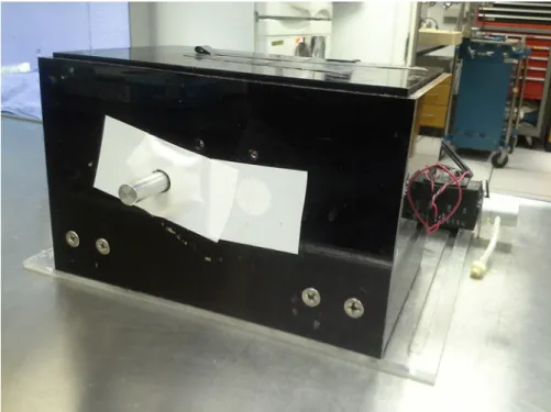
Figure 3: Testing Device. Heated lever is visible in center, valve controlling juice flow visible at right.
When in use, a longer tube is screwed onto the valve and attached to the cage.
28
The lever was a hollow aluminum rod sized to make it easy for the squirrel monkey to grip with its whole hand (a “power grip”). Inside the rod was a heating element attached to a thermocouple and a PID controller. Between the lever and the cage was an opaque plastic panel, physically separating the monkey from the lever.
At the beginning of each trial in the testing battery
*An electronic tone played
*The panel was removed, revealing the lever and juice dispenser.
When the monkey first pulled the lever
*The timer started
So long as the monkey actively continued pulling
*A different tone played
*The juice dispenser dispensed juice
The trial ended after any of the following happened:
*Ten seconds elapsed since the start, or
*The monkey physically let go of the lever, or
*Two seconds passed since the monkey last actively pulled the lever When the trial was concluded
*The time elapsed between the first and last pull was recorded to the nearest 0.1 seconds.
*A buzzer sounded
*The tone and juice dispensing stopped (even if the monkey was still pulling the lever)
*The panel shielding the lever was restored
*The lever temperature was set to the value needed for the next trial.
*The 90 second countdown until the beginning of the next trial started
29 Initial Pull Training
The machine’s design was relatively simple, making it fairly straightforward to train the monkeys to pull the lever and engage in testing. Squirrel monkeys are very “grabby”, and will attempt to stick their hands out of the cage and handle any foreign object that is within sight.
This behavior could easily be reinforced on a specific object, then transferred.
The first phase of training was conditioning the monkey to pull on a pipette to get juice.
Training began by holding a soft disposable plastic pipette full of apricot juice just outside the animal's cage, with the tip pointed inwards. Soon, a monkey would grab onto the tip and attempt to pull it into the cage. Experimenter allowed this, while maintaining a firm grip on the other half of the pipette. Once the pipette's tip was inside the cage, the experimenters slightly squeezed the bulb, dispensing juice for the monkey to drink. The pipette was pulled out of the cage either after one mL of juice is dispensed, or if the monkey stopped pulling or released the pipette. Training for the day continued until each monkey had received 10 mL of juice. Within three days, the monkeys became very proficient in this behavior, and eagerly engaged in it.
Once the pipette pulling skill had been acquired, the monkey could be separated from the others for testing. The monkey was transferred into the smaller testing cage attached to the main one, and the same procedure was applied once again, until the monkey was familiarized with the change in setting and presence of experimental equipment. Because the new cage was now where they received juice, the monkeys formed positive associations with going into it.
Transference of the pipette-pulling skill to the heated lever was fairly quick. Once a
monkey was in the testing cage, the juice dispenser nozzle could be triggered to give small
amounts of juice, so the monkey recognized the nozzle as a juice source. The pipette was then
30
physically attached to the unheated metal lever, such that it could be grabbed onto from inside the cage, but the pipette tip was not close enough to drink from. The nozzle for the juice dispenser was placed for easy ergonomic access to the monkey’s mouth when its arm was extended to pull on the pipette.
When the monkey pulled on the pipette, it was also pulling on the lever, causing juice to flow out of the nozzle in front of the monkey’s mouth. The monkeys soon learned to transfer the location they drank from to the nozzle. Only 3 days of training were necessary until the
monkeys consistently pulled the lever for a full 10 second trial. Over the course of the next two weeks, the plastic pipette could then be shortened by a quarter of an inch in each test, until each monkey was pulling on the lever directly. At this point, the monkey was successfully completing a lever-pulling task.
Lever Handedness Control
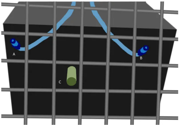
In order to control the hand used by the monkey for the task, the nozzle was placed off
center from the lever (figure 4). As the monkey will put its mouth on the juice nozzle, if the
lever is to the left of the nozzle, the monkey must use the left hand to operate it. If the nozzle is
moved to the other side of the lever, it becomes far easier to use the right hand instead.
31
Figure 4: View of machine from inside cage. Nozzle position can be moved to select which hand is used. For the right hand the nozzle is in position A, for the left hand the nozzle is in position B. The heated lever always remains at location C.
In training, subjects initially favored one hand over the other, and attempted to twist their bodies to use the “wrong” hand. However, the poor ergonomics of using the wrong hand made even a 'successful' pull uncomfortable, and the increased difficulty meant they also received less juice reward. As such, they quickly used the proper hand exclusively. After six days of 10-trial batteries (alternating between left-hand-only and right-hand-only days 3 ), the monkeys
successfully pulled for a full 10 seconds using only the proper arm for each of the 10 trials.
3
While we trained them to be able to use both hands, we only performed tests on the right hand of each monkey.
32 Temperature Training
Once a monkey was trained to pull the lever with the proper arm for full 10-second trial periods, the next stage of training was to introduce varying temperatures. To avoid the monkey associating every warm sensation on the lever with a painful experience, non-noxious
temperatures were introduced first.
The lever started at an initial non-noxious temperature of 114 F. If the monkey pulled the lever for a full trial five times within the first seven trials (allowing for two mistakes), the temperature was increased to 120 F for the remaining trials.
This continued until the monkey succeeded at full 10 second trials for all ten tests.
After the monkey was acclimated to warmed levers, the next step was introducing changes in temperature between tests. The monkeys were given two test days of batteries with temperature patterns alternating between 114 and 120 F tests – a difference sufficient to be noticeable, but still outside the noxious range. They did not have any difficulties completing these tests.
Determining Individual Baselines
While individual monkeys had consistent pull durations at a given temperature, these
durations varied from monkey to monkey. While one monkey would pull on the lever for 8
seconds at 128 F, another would pull for only 5 seconds. As this test was being used to assess
changes in perceptual pain response, individualized baseline temperatures needed to be found
that produced the same pull durations across different monkeys. In this way, changes in
individual pain sensibility could be controlled for.
33
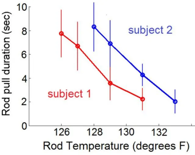
An exploratory battery was used in order to discover the individual set of temperatures which result in desired pull durations for a given monkey. This was done by locating the highest temperatures at which the monkey would consistently hold onto the rod for 10 seconds, 8
seconds, 5 seconds, and 2 seconds. These are defined as the ‘threshold’, 'low', 'medium', and 'high' temperatures, respectively. The 'neutral' temperature is defined as two degrees below the threshold temperature, to ensure that the neutral temperature is non-noxious.
Figure 5: Pull duration as a function of rod temperature for two subjects. Notably, the decline in
duration as a function of temperature is similar across both subjects, but is horizontally offset.
34
These temperature responses were found through staggered ramp-up trials. A series of increasing test temperatures alternated with neutral temperatures, allowing the animal to get an easy reward half the time, and preventing the rod pulling being associated only with pain. In addition, all test sessions began and ended with a much lower temperature ‘reward’ test, to ensure the first and last impressions were of the test being easy, and to prevent the animal from seeing the test as constantly increasing in difficulty.
For instance, a typical exploratory battery was 114F, 120F, 124F, 120F, 126F, 120F, 128F, 120F, 130F, 114F.
Once temperatures with the appropriate pull duration were identified, the final testing regimen was established.
Testing Regimen
The final testing regimen was structured similarly to the exploratory battery, as the
considerations were largely the same. However, as the testing regimen also needed to control for different levels of animal motivation, a warm-up period was added.
The monkeys were often unfocused at the beginning of a series of trials. They spent the first one or two tests unmotivated and easily distracted, but after that point became attentive and well-behaved. In some rare cases, however, after one or two tests the monkey remained
completely unmotivated and unwilling to participate. Sometimes this was due to an obvious environmental factor influencing its mood, such as having a new member of the staff assigned to the room. Other times they just seemed “cranky” for no understandable reason.
The three test warmup existed to prevent this initial lack of motivation from skewing the
early test results. The first two tests were entirely non-noxious, and the third was a low
35
temperature test which is only borderline noxious. If the monkey had low performance on the warmup: either pulling less than nine seconds at the reward or neutral temperature trial, or less than five seconds at the low temperature trial, then that test was repeated, up to a maximum of three total repetitions.
Most of the time the warm-ups were successfully completed, and the monkeys had no trouble completing the following tests. In the rare cases the monkey was too unmotivated to drink ‘free’ juice, and failed the warm-up, the door of the test cage was opened and they were let back into their home cage.
Thus, the final testing regime was Warmup:
Reward Neutral Low Main Test:
Neutral Medium
Neutral High Neutral Medium
Neutral High Neutral
Low
Neutral
36
This test pattern collected two data points at each noxious temperature. As with the exploratory tests, the temperatures were not a continuous ramp-up, and the most noxious temperatures were separated. 4 The neutral tests served multiple purposes – they gave an
additional incentive for participating in testing, allowed for temporary thermal sensitization from high temperature exposure to wear off, and also helped to assess whether or not some
environmental factor has disturbed the monkey partway through the test set. If the monkey pulled for only a short duration on a non-noxious test, that indicated either lack of motivation or distraction. In either case, repeated unwillingness to pull for a full neutral test indicated that the test results for the day were unreliable, and should be discarded. Discarding a trial set was rare, but enabled controlling for mid-test changes in mood.
4