Original Article
Methylation and mRNA expression of WWOX and
smad4 gene in gastric cardia adenocarcinoma
Hong Wei, Hong-Yan She, Wen-Li Liu, Li-Hua Zhao, Li-Dong Li, Liang Xu, Zhi-Jun Liu
The Affiliated Hospital of Hebei University of Engineering, Congtai Congtai Road No. 80, Handan 056002, Hebei, China
Received October 8, 2015; Accepted December 29, 2015; Epub February 15, 2016; Published February 29, 2016
Abstract: Methylation status at the promoter regions of WWOX and Smad4 gene and their expression in gastric car-dia adenocarcinoma (GCA) were investigated. And their relationship with the development of GCA and the possible mechanism were also discussed. The methylation status of WWOX and Smad4 gene and their mRNA expression
levels in the GCA and the corresponding normal mucosal tissues were investigated by nested methylation specific
PCR (MSP) and RT-PCR, respectively. Methylation rate of WWOX gene in the GCA and para-carcinoma tissues was
26.1% and 4.2%, respectively, with significant difference (P < 0.05); methylation rate of WWOX gene in the GCA pa
-tients of stage III/IV (43.3%) was higher than that in the pa-tients of stage I/II (12.8%), with significant difference (P
= 0.004). The relative expression level of WWOX mRNA in the GCA and para-carcinoma tissues was 0.77±0.16 and
0.89±0.12, respectively, with significant difference (P < 0.05); the relative expression level of Smad4 mRNA in the GCA and the para-carcinoma tissues was 0.75±0.17 and 0.88±0.14, respectively, showing significant difference (P < 0.05). WWOX gene methylation probably contributes to pathogenesis of GCA and promotes progression of GCA.
Keywords: GCA, methylation, WWOX, Smad4, gene, RT-PCR
Introduction
Epidemiological surveys show that incidence and mortality of gastric cancer are on the decline, but the incidence of gastric cardia ade-nocarcinoma (GCA) is on the rise [1]. Therefore, the etiological study on GCA cannot be ignored. WWOX is a newfound tumor suppressor gene, and its downregulated expression increases the susceptibility of cells to cancer cells. Its abnormal expression is common in breast can-cer, ovarian cancan-cer, lung cancan-cer, multiple myeloma and esophageal squamous cell carci-noma but rarely reported in GCA. Smads are important signaling intermediates and regula-tors in TGF-β signal transduction system, which can directly transmit the TGF-β signal from the cell membrane into the nucleus. Disorder of TGF-β signal transduction pathway is inevitably caused by the inactivation of Smad4, and the inhibitory effect on cell proliferation is lost, leading to cancer. In this research, we investi-gated the methylation status in the promoter regions of WWOX and Smad4 gene, and their expression in GCA to reveal the relationship
between the methylation of WWOX and Smad4 gene and the development of GCA as well as the possible mechanism, providing a significant candidate indicator for the early detection and treatment of GCA.
Materials and methods
Materials
Main reagents: Proteinase K (Merck), hydroqui-none, sodium bisulfite (Sigma), Wizard DNA purification kit (Promega), Trizol (Invitrogen, USA). All primers were synthesized by Beijing SBS Genetech.
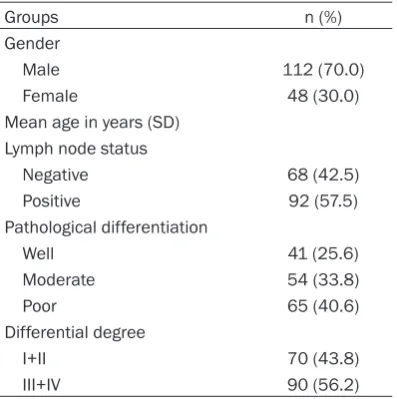
60 years. All patients had not been treated by chemotherapy or radiotherapy before surgery. HE staining verified that the tumor samples were mostly tumor tissues and the para-carci-noma tissues were free from tumor cell infiltra -tion. The primary tumor tissues were sampled from each GCA patient and some of the sam-ples were collected from the para-carcinoma mucosal tissues (5 cm from tumor). All collect-ed samples were immcollect-ediately storcollect-ed in liquid nitrogen. The samples were transferred from liquid nitrogen to a -80°C refrigerator for tissue DNA and RNA extraction. There were 90 cases of stage I/II and 70 cases of stage III/IV accord-ing to UICC TNM stagaccord-ing system; they were divided into three groups according to patho-logical stage, well differentiated group (41 cases), moderately differentiated group (54 cases) and poor differentiated group (65 cases); there were 92 cases with regional lymph node metastasis and 68 without region-al lymph node metastasis. The protocol was approved by the Ethics Committee of the Affiliated Hospital of Hebei University of Engineering, and informed consent was obtained from all patients.
Methods
Nested methylation specific PCR (MSP): Proper amount of sample DNA was purified by Wizard DNA purification kit according to instructions. Treated by sodium bisulfite, C of DNA was trans -formed into U unless the CpG island was meth-ylated. The corresponding primers designed
based on this principle (Table 2) were used to determine whether the genes were methylated or not. Reaction conditions: predenaturation at 95°C for 10 min, denaturation at 94°C for 45 s, annealing at 60.5°C (methylated primer) or 55.2°C (unmethylated primer) for 50 s, exten-sion at 72°C for 45 s, 40 circles; final extenexten-sion at 72°C for 10 min. After 2% agarose gel elec-trophoresis was performed on the amplified products, the result was subjected to image analysis by the UV gel imaging and analysis sys-tem. The genomic DNA treated by methylase (Sss I, New England BioLabs) was used as the positive control of MSP and subjected to PCR. Sterilized double-distilled water was used as the negative control in place of the DNA tem-plate and subjected to PCR. 10% of the sam-ples were selected for repeated trials as the quality control of the MSP test.
RT-PCR test: The total RNA was extracted according to the instructions of Trizol (Invitrogen), and transformed into cDNA by reverse transcription according to the instruc-tions of the RT-PCR kit (Reverse Transcription System A3500, Promega). Synthesized primers of WWOX and Smad4 are show in Table 3. Reaction conditions: preheating of RNA at 70°C for 10 min, 25°C for 10 s, 42°C for 60 min, 99°C for 5 min, 4°C for 5 min, 40 circles. 2% agarose gel electrophoresis was performed on the PCR products, with GAPDH as the internal reference.
Results determination and statisticals
[image:2.629.99.298.104.304.2]The methylated amplified fragments of expect -ed lengths were generat-ed by the amplification of the DNA which was treated by Sss I methyl-ase using the methylation specific primer (M), but nothing was generated in the blank control trial, indicating that the primers and reagents used were appropriate and the results were plausible. Three different results of the methyl-ation state of WWOX and Smad4 gene in the GCA tissues may appear: firstly, the objective band was generated by the methylation specific primer (M), while no band was generated by the unmethylated specific primer (U), as was speci -fied as methylation; secondly, the objective band was generated by the unmethylation spe-cific primer (U), while no band was generated by the methylated specific primer (M), as was specified as unmethylation; thirdly, the objec -tive bands were generated by both primers, as Table 1. Clinical and pathological
characteris-tics of GCA patients
Groups n (%)
Gender
Male 112 (70.0)
Female 48 (30.0) Mean age in years (SD)
Lymph node status
Negative 68 (42.5) Positive 92 (57.5) Pathological differentiation
Well 41 (25.6)
Moderate 54 (33.8)
Poor 65 (40.6)
Differential degree
I+II 70 (43.8)
was specified as hemimethylation, recorded in methylation.
Determination of the relative content of the PCR amplified products: After 1.5% agarose gel electrophoresis was performed on the PCR amplified products (6 μL), the result was photo -graphed by the gel imaging and analysis sys-tem; the grey value was measured by Gel Pro Analysizer 3.1; the 1OD value of WWOX and Smad4 gene was standardized using the 1OD value of the internal reference GAPDH, and the relative content was obtained and analyzed. The formula is given below:
Relative expression of mRNA = (1OD value of WWOX/Smad4)/(1OD value of GAPDH)
Statistics
The data were processed with SPSS 13.0
soft-carcinoma tissues (Table 4)
WWOX gene methylation rate of the tumor tis-sues and the corresponding para-carcinoma tissues among the 160 GCA patients was 26.1% and 4.2%, respectively, showing signifi -cant difference (P < 0.05) (Figure 1).
Smad4 gene methylation rate of the tumor tis-sues and the corresponding para-carcinoma tissues among the 160 GCA patients was 2.9% and 8.3%, respectively, showing no significant difference (Figure 2).
Correlation between methylation of the two genes and clinical data
Relations between WWOX gene and clinical pathological data: No significant difference was observed between WWOX gene methylation in the GCA tissues and clinical data such as gen-der, age, pathological grading, presence of lymph node metastasis, etc. (P > 0.05). The statistics based on TNM staging showed that WWOX gene methylation rate among the GCA patients of stage III/IV (43.3%) was higher than that among the patients of stage I/II (12.8%), with significant difference (P = 0.004).
[image:3.629.100.530.93.213.2]Relations between Smad4 gene and clinical pathological data: No significant difference was Table 2. WWOX, Smad4 gene primer in MSP
Gene CpGStatus Primer sequence (5’-3’) Annealing Temperatur (°C) Product Size (bp)
WWOX Unmethylated TATGGGTGTTGTTTTTTA 56 347
CAATCTCCACAATATCACAAC
Methylated TATGGGCGTCGTTTTTTAGTC 51 347
CAATCTCCGCAATATCGCGAC
Smad4 Unmethylated TTTGTAATGAGATGTTAATTTTTTTGGT 55.7 274 CAACTTATCAAAAAACCACTAAACATACA
[image:3.629.100.371.248.351.2]Methylated GTAACGAGATGTTAATTTTTTCGGC 49 269 ACTTATCGAAAAACCACTAAACATACG
Table 3. WWOX, Smad4 gene Primers used in RT-PCR
Gene Primer sequence (5’-3’) size (bp)Product temperature (°C)Annealing GAPDH 5‘CGGGAAGCTTGTGATCAATGG 3’ 342 60
5‘GGCAGTGATGGCATGGACTG 3’
WWOX 5‘GAGCAAACACATCTGACCTAC 3’ 258 58 5‘CAAAATGAGCATCCCCTCC 3’
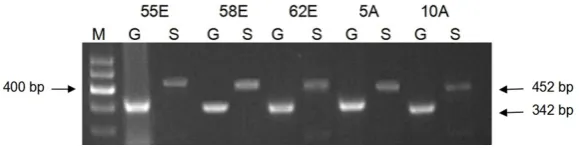
Smad4 5‘ATCTGAGTCTAATGCTACC3’ 452 58 5‘CGTATCCATCAACAGTAAC3’
Table 4. Frequency of WWOX, Smad4 meth-ylation in GCA and GCA
Gene
Methylation ratio
χ2 P
GCA Corresponding nonmalignant
WWOX 26.1% 4.2% 9.598 0.002* Smad4 2.9% 8.3% 0.783 0.376*
*: P value of GCA against corresponding nonmalignant.
formed by Chi-square test or cor-rected Chi-square test. The inter-group compare of the relative ex- pression was conducted by t test or variance analysis. P < 0.05 indicated significant difference.
Results
[image:3.629.99.297.396.462.2]para-the GCA tissues and clinical data such as gen-der, age, pathological grading, TNM staging, presence of lymph node metastasis, etc. (P > 0.05).
MRNA expression of WWOX and Smad4 gene in the GCA and the para-carcinoma tissues (Table 5)
The relative expression level of WWOX mRNA in the GCA and the para-carcinoma tissues was 0.77±0.16 and 0.89±0.12, respectively, with significant difference (P < 0.05) (Figure 3); the relative expression level of Smad4 mRNA in the GCA and the para-carcinoma tissues was 0.75±0.17 and 0.88±0.14, respectively, showing significant difference (P < 0.05) (Figure 4).
Relationship between WWOX gene methylation and mRNA expression
The relative mRNA expression level of WWOX gene in the methylation positive GCA tissues (0.73±0.15) was lower than that in the methyla-tion negative GCA tissues (0.76±0.17), but the
genetic alterations including DNA methylation, histone acetylation, etc. In recent years, DNA methylation has become a hot topic in the research area of tumor pathogenesis and treatment.
WW domain containing oxidoreductase (WWOX) gene is a newfound candidate tumor suppres-sor gene, situated at the chromosomal fragile site FRA16D and encoding a 414-amino acid protein of 46 kDa. The WWOX protein contains two N-terminal WW domains (residues 18-47, 59-88) and a short-chain dehydrogenation reductase site at the C-terminal, and its essen-tial function is signal transmission and informa-tion transducinforma-tion.
[image:4.629.99.387.80.146.2]WWOX protein is a pro-apoptotic protein engag-ing in multiple signal transduction pathways to inhibit oncogenesis. Chang et al. [2, 3] found that mitochondrial WWOX probably engaged in the molecular pathway of apoptosis triggered by p53 in the mitochondrion, increasing the susceptibility of the tumor cell to tumor necro-sis factor (TNF) cytotoxin; meanwhile, Aqeilan et al. [4, 5] found that WWOX could enhance the pro-apoptotic effect of P73. However, the gene silencing and down-regulated expression caused by genetic and epigenetic alterations of the WWOX gene lessen its pro-apoptotic effect, promoting tumor formation and progression. During the process, epigenetic alterations, especially the methylation of the promoter region of the gene, may play a significant role. Figure 1. Methylation analysis of WWOX gene in GCA tissues. Case1:
Hemi-Methylated; Case2: Hemi-Methylated; Case3: Unmethylated; PC: Positive control;
[image:4.629.98.388.207.272.2]BC: Blank control; M: Methylated; U: Unmethylated; MA: Marker.
Figure 2. Methylation analysis of Smad4 gene in GCA tissues. Case1: Hemi-Methylated; Case2: Hemi-Methylated; Case3: Unmethylated; PC: Positive control;
BC: Blank control; M: Methylated; U: Unmethylated; MA: Marker.
Table 5. The expression of WWOX, Smad4 mRNA in adjacent carcinoma andGCA
Group N WWOX Smad4
Adjacent cancer 44 0.89±0.12 0.88±0.14 Cancer 15 0.77±0.16 0.75±0.17
difference was not significant (P = 0.13).
No correlation was found between the relative expres-sion levels of WWOX and Smad4 gene.
Discussion
[image:4.629.99.298.360.400.2]epi-DonatiV et al. [6] detected methylation at the WWOX promoter in the study on non-small cell lung cancer, and found a close relationship between WWOX gene methylation and its nega-tive expression in lung cancer tissues, with WWOX protein lost in 62% of the lung cancer cells due to the gene methylation. Qin HR et al. [7] found a significantly lower WWOX mRNA and protein expression in the prostate cancer cell lines (LNCaP, DU145, PC-3) due to the DNA methylation in the WWOX regulatory domain than that in the non-tumorous prostate cell (PWR-1E), and found a significant increase in WWOX mRNA and protein expression in the prostate cancer cell lines treated with the DNA methylation inhibitor AZA, especially in DU145. In a study on WWOX gene methylation and breast cancer, Wang X et al. [8] found that methylation rate of the promoter region of WWOX gene was 55% in breast cancer. But no methylation was found in the promoter region of WWOX gene in the normal para-carcinoma tissues. Moreover, WWOX gene methylation was closely correlated with the down-regulated mRNA expression in tumor tissues.
In this research, we found a significantly higher methylation rate of WWOX gene in the GCA tis-sues of the 160 cases (26.1%) compared to the normal para-carcinoma tissues (4.2%), indicat-ing a probable relationship between WWOX gene methylation and GCA development possi-bly with the hypermethylation of the WWOX
results of the study also showed that the rela-tive expression of the mRNA in the tumor tis-sues was significantly lower than that in the para-carcinoma tissues, but the relative expression level of the mRNA in the methyla-tion positive GCA tissues was not significantly lower than that in the methylation negative GCA tissues. The possible reasons are as follows: Hemimethylation might contribute little to the down-regulated expression of the gene; other genetic alterations mediating down-regulated expression of the WWOX gene might exist in GCA besides methylation. Except methylated tumor cells in the investigated tumor tissues, unmethylated tumor cells and abundant inter-stitial cells with normal expression of WWOX gene coexisted, which obscured the impacts of methylation on expression. No correlation was found in this study between the relative expres-sion of the mRNA and clinical data, indicating that the WWOX deletion might occur at the early stages of the GCA progression. The con-crete mechanism of the abnormal methylation in the promoter region of the WWOX gene caus-ing malfunction to WWOX mRNA transcription remains to be further studied.
[image:5.629.100.387.80.166.2]The TGFβ-Smads signal transduction pathway displays various biological activities, playing an important role in regulating cellular growth, dif-ferentiation, apoptosis, adhesion, the synthe-sis and deposition of extracellular matrix, embryogenesis and tissue repair, inflammatory responses and fibrosis. It plays a significant Figure 3. mRNA analysis of WWOX gene and GAPDH. M: Marker; A: Adjacent
non-cancerous tissue; E: GCA tissue; G: GAPDH; W: WWOX.
Figure 4. mRNA analysis of Smad4 gene and GAPDH. M: Marker; A: Adjacent non-cancerous tissue; E: GCA tissue; G: GAPDH; S: Smad4.
[image:5.629.98.388.217.292.2]role in inhibiting oncogenesis predominantly through regulating intranuclear CKI (cyclin dependent kinase inhibitor) expression, pre-venting cell cycle running, inhibiting cellular malignant transformation, which requires the Smad family in the cytoplasm to serve as a link-ing bridge. The Smad4 gene and the encoded protein play a central role in the TGFβ-Smads pathway as a tumor suppressor gene and the only Co-Smad in the pathway, respectively. Once Smad4 expression is negative or down-regulated, the whole TGF-β signal transduction pathway will be destroyed, thereby unable to inhibit cell proliferation and losing the potential ability to suppress oncogenesis.
The abnormal expression of Smad4 causes malfunction in TGF-β signal transduction path -way and eventually leads to oncogenesis, which is most commonly found in pancreatic cancer [9], with a mutation rate of 50%. It is secondly common in digestive system cancers such as colon cancer, gastric cancer, bile duct cancer, liver cancer, etc. And it is also found in ovarian cancer, acute myeloid leukemia, lung cancer, head and neck squamous cell carcinoma, etc. [10]. Xu et al. [11] verified that the haploid dele -tion on the mouse Smad4 gene could promote the development of gastric polyps and gastric cancer. Takagi et al. [12] found missense muta-tion of the Smad4 gene and its abnormal expression were caused by gene deletion in the study on primary colorectal cancer. Koyama et al. [13] found the loss of heterozygosity of the Smad4 gene in 78% of the patients in the study on primary colorectal cancer, with mutation and insertion in several cases. Furthermore, researches abroad showed that mutation was not the primary cause of Smad4 gene inactiva-tion. Therefore, it is estimated that hypermeth-ylation in the promoter region of the gene func-tions as the second blow leading to the abnor-mal expression of Smad4. Onwuebusi et al. [14] conducted methylation analysis on the promot-er sequence of Smad4 in the study about esophageal adenocarcinoma, and found a sig-nificant correlation between the abnormal methylation of the promoter sequence and the negative expression of the Smad4 protein. It was estimated that epigenetic modifications silenced the gene, inhibited the gene expres-sion and hindered TGF-β1/Smad4 signal trans -duction. However, there are few reports about methylation of Smad4 gene both at home and abroad. Methylation of Smad4 gene remains to be further studied.
This trial was designed and carried out in an effort to investigate the role of methylation of Smad4 gene in the pathogenesis and develop-ment of GCA. The results showed that the Smad4 gene methylation rates in the tumor tis-sues and the corresponding para-carcinoma tissues among the 160 GCA patients were 2.9% and 8.3%, respectively, without signifi -cant difference (P > 0.05), probably indicating no correlation between methylation of Smad4 gene and the pathogenesis of GCA. In this study, the relative expression of Smad4 mRNA in the GCA tissues was significantly lower than that in the para-carcinoma tissues; however, no correlation was found between the relative mRNA expression and clinical data as well as between methylation of Smad4 and the down-regulated mRNA expression, probably indicat-ing a certain relationship between the down-regulated expression of the Smad4 mRNA and genetic alterations such as the gene deletion. KiKD et al. [15] detected the down-regulated expression of Smad4 mRNA in tumor tissues in the study on the expression and mutation of TGFβ-Smads in human cervical carcinoma; Leng A et al. [16] found a significantly lower expression of Smad4 in tumor tissues than that in para-carcinoma tissues, a significantly lower expression of Smad4 in the poor differentiated group than that in the well and the moderately differentiated groups, and a lower expression in the group with lymph node metastasis than that in the group without lymph node metasta-sis. All of the findings were consistent with the results of this study.
Tumor pathogenesis and development are an extremely complicated process involving multi-ple genes, predominantly concerning onco-genes activation and anti-oncoonco-genes inactiva-tion. The results of this study indicated that the hypermethylation of WWOX gene in tumor tis-sues was correlated with the pathogenesis and development of GCA. However, the correlation between the methylation of Smad4 gene and the development of GCA remains uncertain. Acknowledgements
This work was supported by the Hebei province science and technology research program, project number: 14277765D.
Disclosure of conflict of interest
None.
Address correspondence to: Zhi-Jun Liu, The
Affi-liated Hospital of Hebei University of Engineering, Congtai Congtai Road No. 80, Handan 056002, Hebei, China. Tel: +86- 0310-8572360; Fax: +86-
0310-8572360; E-mail: liuzhijun_zj@163.com
References
[1] Chen ZF, Hou J, He YT and Wang SJ. Gastroesophageal junction adenocarcinoma-new issues faced by cancer registries. Zhong Guo Zhong Liu Lin Chuang 2007; 34: 1381-1382.
[2] Chang NS, Pratt N, Heath J, Schultz L, Sleve D, Carey GB and Zevotek N. Hyaluronidase induc -tion of a WW domain-containing oxidoreduc-tase that enhances tumor necrosis factor
cyto-toxicity. J Biol Chem 2001; 276: 3361-3370.
[3] Chang NS, Doherty J, Ensign A, Schultz L, Hsu
LJ and Hong Q. WOX1 is essential for tumor ne-crosis factor-, UV light-, staurosporine-, and p53-mediated cell death, and its tyrosine
33-phosphorylated form binds and stabilizes serine 46-phosphorylated p53. J Biol Chem
2005; 280: 43100-43108.
[4] Aqeilan RI, Palamarchuk A, Weigel RJ, Herrero JJ, Pekarsky Y and Croce CM. Physical and functional interactions between theWWOX tu-mor suppressor pro- tein and the AP-2gamma transcription factor. Cancer Res 2004; 64: 8256-8261
[5] Aqeilan RI, Pekarsky Y, Herrero JJ, Palamarchuk A, Letofsky J, Druck T, Trapasso F, Han SY, Melino G, Huebner K and Croce CM. Functional association between Wwox tumor suppressor protein and p73, a p53 homolog. Proc Natl Acad Sci U S A 2004; 101: 4401-4406.
[6] Donati V, Fontanini G, Dell’Omodarme M, Prati MC, Nuti S, Lucchi M, Mussi A, Fabbri M,
Basolo F, Croce CM and Aqeilan RI. WWOX ex -pression in different histologic types and sub-types of non-small cell lung cancer. Clin Cancer Res 2007; 13: 884-891.
[7] Qin HR, Iliopoulos D, Semba S, Fabbri M, Druck T, Volinia S, Croce CM, Morrison CD, Klein RD and Huebner K. A role for the WWOX gene in prostate cancer. Cancer Res 2006; 66: 6477-6481.
[8] Wang X, Chao L, Jin G, Ma G, Zang Y and Sun J. Association between CpG island methylation of the WWOX gene and its expression in breast
cancers. Tumour Biol 2009; 30: 8-14.
[9] Wang W, Owen SM, Rudolph DL, Cole AM,
Hong T, Waring AJ, Lal RB, Lehrer RI. Activity of
alpha- and theta-defensins against primary isolates of HIV-1. J Immunol 2004; 173: 515-520.
[10] Cunliffe RN, Kamal M, Rose FR, James PD and Mahida YR. Expression of antimicrobial neutro-phil defensins in epithelial cells of active
in-flammatory bowel disease mucosa. J Clin
Pathol 2002; 55: 298-304.
[11] Xu X, Brodie SG, Yang X, Im YH, Parks WT, Chen
L, Zhou YX, Weinstein M, Kim SJ and Deng CX. Oncogene 2000; 19: 1868-1874.
[12] Takagi Y, Kohmura H, Futamura M, Kida H, Tanemura H, Shimokawa K and Saji S. Somatic alterations of the DPC4 gene in human colorec-tal cancers in vivo. Gastroenterology 1996; 111: 1369-1372.
[13] Koyama M, Ito M, Nagai H, Emi M and Moriyama Y. Inactivation of both alleles of the DPC4/SMAD4 gene in advanced colorectal
cancers: identification of seven novel somatic
mutations in tumors from Japanese patients. Mutat Res 1999; 406: 71-77.
[14] Onwuegbusi BA, Aitchison A, Chin SF, Kranjac
T, Mills I, Huang Y, Lao-Sirieix P, Caldas C and
Fitzgerald RC. Impaired transforming growth factor beta signalling in Barrett’s carcinogene -sis due to frequent SMAD4 inactivation. Gut 2006; 55: 764-774.
[15] Ki KD, Tong SY, Huh CY, Lee JM, Lee SK and Chi SG. Expression and mutational analysis of TGF-beta/Smads signaling in human cervical cancers. J Gynecol Oncol 2009; 20: 117-121. [16] Leng A, Liu T, He Y, Li Q and Zhang G. Smad4/
Smad7 balance: a role of tumorigenesis in gastric cancer. Exp Mol Pathol 2009; 87: 48-53.
[17] Hsu LJ, Schultz L, Hong Q, Van Moer K, Heath
J, Li MY, Lai FJ, Lin SR, Lee MH, Lo CP, Lin YS, Chen ST and Chang NS. Transforming growth factor beta1 signaling via interaction with cell surface Hyal-2 and recruitment of WWOX/