Sitsapesan
John Parrington, Antony Galione and Rebecca
Gosain, Grant C. Churchill, Michael X. Zhu,
Margarida Ruas, A. Ganesan, Rajendra
Sitsapesan, Elisa Venturi, Katja Rietdorf,
Samantha J. Pitt, Tim M. Funnell, Mano
2+
Sensor of Luminal pH and Ca
Release Channel, Operating as a Dual
2+
TPC2 Is a Novel NAADP-sensitive Ca
Molecular Biophysics:
doi: 10.1074/jbc.M110.156927 originally published online August 18, 2010 2010, 285:35039-35046.
J. Biol. Chem.
10.1074/jbc.M110.156927 Access the most updated version of this article at doi:
. JBC Affinity Sites Find articles, minireviews, Reflections and Classics on similar topics on the
Alerts:
When a correction for this article is posted •
When this article is cited •
to choose from all of JBC's e-mail alerts Click here
Supplemental material:
http://www.jbc.org/content/suppl/2010/09/03/M110.156927.DC1.html
http://www.jbc.org/content/285/45/35039.full.html#ref-list-1
This article cites 46 references, 21 of which can be accessed free at
at Univ of St Andrews on December 16, 2013
http://www.jbc.org/
Downloaded from
at Univ of St Andrews on December 16, 2013
http://www.jbc.org/
TPC2 Is a Novel NAADP-sensitive Ca
2
ⴙ
Release Channel,
Operating as a Dual Sensor of Luminal pH and Ca
2
ⴙ
*
□SReceived for publication, June 22, 2010, and in revised form, August 17, 2010Published, JBC Papers in Press, August 18, 2010, DOI 10.1074/jbc.M110.156927
Samantha J. Pitt‡1, Tim M. Funnell§1, Mano Sitsapesan‡, Elisa Venturi‡, Katja Rietdorf§, Margarida Ruas§, A. Ganesan¶, Rajendra Gosain¶, Grant C. Churchill§, Michael X. Zhu储, John Parrington§, Antony Galione§2, and Rebecca Sitsapesan‡3
From the‡School of Physiology and Pharmacology, Medical Sciences Building, and Center for Nanoscience and Quantum Information, University of Bristol, Bristol BS8 1TD, United Kingdom, the§Department of Pharmacology, University of Oxford,
Mansfield Road, Oxford OX1 3QT, United Kingdom, the¶School of Chemistry, University of Southampton, Southampton SO17 1BJ, United Kingdom, and the储Department of Integrative Biology and Pharmacology, The University of Texas Health Science Center, Houston, Texas 77030
Nicotinic acid adenine dinucleotide phosphate (NAADP) is a molecule capable of initiating the release of intracellular Ca2ⴙrequired for many essential cellular processes. Recent evidence links two-pore channels (TPCs) with NAADP-in-duced release of Ca2ⴙfrom lysosome-like acidic organelles; however, there has been no direct demonstration that TPCs can act as NAADP-sensitive Ca2ⴙrelease channels. Contro-versial evidence also proposes ryanodine receptors as the pri-mary target of NAADP. We show that TPC2, the major lyso-somal targeted isoform, is a cation channel with selectivity for Ca2ⴙthat will enable it to act as a Ca2ⴙrelease channel in the cellular environment. NAADP opens TPC2 channels in a con-centration-dependent manner, binding to high affinity acti-vation and low affinity inhibition sites. At the core of this process is the luminal environment of the channel. The sen-sitivity of TPC2 to NAADP is steeply dependent on the lumi-nal [Ca2ⴙ] allowing extremely low levels of NAADP to open the channel. In parallel, luminal pH controls NAADP affinity for TPC2 by switching from reversible activation of TPC2 at low pH to irreversible activation at neutral pH. Further evi-dence earmarking TPCs as the likely pathway for NAADP-induced intracellular Ca2ⴙrelease is obtained from the use of Ned-19, the selective blocker of cellular NAADP-induced Ca2ⴙ release. Ned-19 antagonizes NAADP-activation of TPC2 in a non-competitive manner at 1Mbut potentiates NAADP activation at nanomolar concentrations. This single-channel study provides a long awaited molecular basis for the peculiar mechanistic features of NAADP signaling and a framework for understanding how NAADP can mediate key physiological events.
Nicotinic acid adenine dinucleotide phosphate (NAADP)4is
the most potent Ca2⫹-releasing second messenger yet
identi-fied (1– 4). Although originally discovered in sea urchin eggs, it is becoming increasing clear that NAADP-induced Ca2⫹ release is crucial to many physiological processes in a diverse range of mammalian cell types (5, 6). Many distinctive features of NAADP-induced Ca2⫹release indicate that the Ca2⫹release
channels involved are functionally and structurally different from the well characterized Ca2⫹release channels found on
sarcoplasmic/endoplasmic reticulum stores (ryanodine recep-tors (RyRs) and inositol trisphosphate receprecep-tors (IP3Rs) (7, 8)).
NAADP triggers Ca2⫹release from acidic stores, but the iden-tity of the Ca2⫹release channel involved has remained a
mys-tery (7). New evidence has shown that NAADP binds with high affinity to TPC protein complex (9), which is located on acidic, lysosome-like Ca2⫹stores (10, 11). A recent report also
dem-onstrates that activation of cell surface muscarinic receptors requires TPC2 expression to couple Ca2⫹release from acidic
stores in mouse bladder smooth muscle (12). The importance of TPC2 to Ca2⫹movements across lysosomal membranes was recently shown in whole lysosome recordings. Mutation of TPC2 affected the magnitude of whole lysosomal Ca2⫹
cur-rents, highlighting the need to investigate the conductance and gating properties of TPC2 at the single-channel level (13).
TPCs are thus named because their close sequence homology with voltage-gated cation channels predicts a monomeric structure containing two pore loops (7, 14). It is proposed that two homodimers form the pore (10, 15, 16); however, it is not known whether TPC2 can actually function as an ion channel with all the properties necessary to fulfill the role of a Ca2⫹
release channel activated by NAADP. Many characteristics of NAADP-induced Ca2⫹release distinguish it from Ca2⫹release
triggered by other signaling molecules, and these indicate un-usual underlying mechanisms of ion channel function.
For the first time, we describe here the fundamental single-channel gating and conductance properties of purified human TPC2 and demonstrate that TPC2 exhibits many unique func-tional characteristics that can explain the idiosyncrasies of cel-*This work was supported by the British Heart Foundation (to R. S.) and the
Wellcome Trust (to A. G. and J. P.). Author’s Choice—Final version full access.
□S The on-line version of this article (available at http://www.jbc.org) contains
supplemental text and references and Figs. 1–3.
1Both authors contributed equally to this work.
2To whom correspondence may be addressed. Fax: 44-1865-271-853; E-mail:
antony.galione@pharm.ox.ac.uk.
3To whom correspondence may be addressed. Fax: 44-117-331-2288; E-mail:
r.sitsapesan@bris.ac.uk.
4The abbreviations used are: NAADP, nicotinic acid adenine dinucleotide
phosphate; TPC, two-pore channel; RyR, ryanodine receptor; IP3R, inositol
trisphosphate receptor; IP, immunoprecipitation; pS, picosiemens.
at Univ of St Andrews on December 16, 2013
http://www.jbc.org/
lular NAADP signaling characteristics, whereas cardiac (RyR2) and skeletal (RyR1) RyRs do not. A preliminary report of these data has been presented in abstract form (17).
MATERIALS AND METHODS
Purification of Human, Recombinant TPC2—cDNA forhTPC2 (GenBankTMaccession number AY029200) was cloned from
HEK293 cells by rapid amplification of cDNA ends-PCR us-ing primers designed against the expressed sequence tag AA309878 (10). Overexpressing HA-HsTPC2 HEK293 cells were harvested and centrifuged at 2000 rpm for 5 min at 4 °C. The cell pellet was resuspended in hypotonic buffer (20 mM
HEPES, 1 mMEGTA, pH 7.2) and kept for 60 min on ice before homogenization. The lysate was centrifuged for 5 min at 2000⫻gat 4 °C, and the supernatant was centrifuged for 1 h at 100,000⫻gat 4 °C. The membrane pellet was resuspended in IP buffer (150 mMNaCl, 20 mMHEPES, pH 7.4) and kept on ice
for 1 h, after which insoluble material was pelleted by centrifu-gation at 14,000⫻gfor 30 min at 4 °C. The supernatant was precleared by adding non-immune rabbit serum coupled covalently to protein-A-Sepharose for 1 h at 4 °C. The pre-cleared supernatant was incubated overnight at 4 °C with either non-immune rabbit serum (control) or polyclonal anti-TPC2 antibody (10), both covalently coupled to protein-A-Sepharose beads. The beads were pelleted by centrifugation at 1000 rpm for 2 min. Following removal of the supernatant, the beads were washed with IP buffer containing 1% CHAPS followed by two washes containing 0.1% CHAPS. Immunoprecipitated protein complexes were eluted by incubation with the peptides used to raise the antibody, at 50g/ml in 0.1% CHAPS and 0.05% phos-phatidylcholine for 4 h at 4 °C. The eluted protein complexes were dialyzed for 36 h at 4 °C in buffer containing 100 mMNaCl,
25 mMPIPES, 0.15 mMCaCl2, and 0.1 mMEGTA, pH 7.4, using
a 20,000 molecular weight cut-off filter. After dialysis, an equal volume of 0.5Msucrose solution was added and incubated for
30 min at 4 °C before aliquoting and snap freezing in liquid nitrogen. Further information is provided in supplemental Methods.
Single Channel Recording and Analysis—Purified TPC2, rab-bit skeletal RyR1, or sheep cardiac RyR2 was incorporated into planar phosphatidylethanolamine lipid bilayers under voltage clamp conditions using previously described techniques (18). Current fluctuations were recorded at 22⫾2 °C with either Ca2⫹(cis, 250 mMHEPES, 80 mMTris, 15Mfree Ca2⫹, pH
7.2/trans, 250 mMglutamic acid, 10 mMHEPES, pH 7.2 with Ca(OH)2giving a free [Ca
2⫹] of 50 mM) or K⫹(symmetrical 210
mMKCl, 10 mMHEPES, 15Mfree Ca2⫹, pH 7.2) as the
per-meant ion. Luminal pH was lowered to 4.8 by perfusing the
transchamber with a solution of 210 mMpotassium acetate, 10
mMHEPES, 15Mfree [Ca2⫹]. Replacement of Cl⫺by acetate did not alter TPC2 conductance or gating (data not shown). Washout of thecis(cytosolic) chamber to exchange solutions took 2 min. The free [Ca2⫹] of all solutions was determined using a Ca2⫹electrode (Orion 93-20; Thermo Orion, Boston,
MA) as described previously (18). Current recordings were filtered at 800 Hz (⫺3 db) and digitized at 20 kHz (ITC-18, Instrutech Corporation) with WinEDR version 2.5.9 (Strath-clyde University, UK) or Pulse (HEKA Elektronik, Lambrecht/
Pfalz Germany). Measurements of single-channel current amplitudes were made using the single-channel analysis soft-ware WinEDR version 2.5.9 (University of Strathclyde, Glas-gow, UK). The closed and open current levels were assessed manually using cursors. Amplitude histograms were obtained from single-channel data using Clampfit version 10.2 (Molecu-lar Devices, Sunnyvale, CA) and fitted with Gaussian curves. In all records, amplitude histograms were best fit with a single Gaussian function representing the main conductance level (300 pS; see Fig. 2b). Subconductance state gating was too brief to be detected by this method of analysis. The channel open probability (Po) was determined over 3 min of continuous
re-cording, using the method of 50% threshold analysis (19). Con-centration-response curves were fit using a four-parameter logistical equation in GraphPad Prism (GraphPad Software Inc). The Ca2⫹/K⫹permeability ratio (PCa2⫹/PK⫹) was
calcu-lated using the equation given by Fatt and Ginsborg (1958) (20).
PCa2⫹/PK⫹⫽关K⫹兴/4⫻关Ca2⫹兴⫻exp共ErevF/RT兲
⫻关exp共ErevF/RT兲⫹1兴 (Eq. 1)
The value of RT/F used in our calculations was 25.4 mV based on the recording temperature of 22 °C. The zero current reversal potential (Erev) was obtained under bi-ionic condi-tions (cis, 210 mMKCl, 20 mMHEPES, pH 7.2;trans, 210 mM
CaCl2, 20 mMHEPES, pH 7.2) and represented the point where
no current could be detected. Ned-19 was synthesized as described previously (21), and theL-trans-Ned-19 analog was
used throughout.
Statistics—Mean ⫾ S.D. (ⱖ3) is shown unless otherwise stated. Student’sttest was used to assess the difference between mean values. Apvalue of⬍0.05 was taken as significant.
RESULTS
We purified the human recombinant TPC2 complex (Fig. 1a) for subsequent reconstitution into artificial membranes under voltage clamp conditions (18). Representative examples of the resulting single-channel currents in symmetrical solu-tions of 210 mMKCl, pH 7.2, are shown in Fig. 1b. In the
absence of NAADP, occasional channel openings, usually too brief to fully resolve, were observed. NAADP addition to thecischamber always increasedPo(n⫽101), whereastrans
addition of NAADP had no effect (n⫽5). In conditions where luminal Ca2⫹was the permeant ion (Fig. 1c), NAADP still acti-vated TPC2 from the cytosolic side only (n⫽15). TPC2, there-fore, incorporates into bilayers in a fixed orientation as described in the supplementary text, with the cis andtrans
chambers, most likely corresponding to the cytosolic and lumi-nal sides of the acidic Ca2⫹stores, respectively. These single-channel currents were not observed in control experiments (supplemental Fig. S1).
In symmetrical 210 mMKCl, the single-channel conductance
(Fig. 1d) was 300⫾6 pS (S.E.;n⫽5). Applying a KCl gradient (trans, 210 mM;cis, 510 mM), shifted the current-voltage
rela-tionship to the left with a reversal potential of⫺20.5 mV, indi-cating that, as for other Ca2⫹release channels (22, 23), TPC2 is
ideally selective for cations. The current-voltage relationship shown in Fig. 1erevealed a Ca2⫹conductance of 15⫾1.5 pS
TPC2 Is an NAADP-sensitive Ca
2ⴙRelease Channel
at Univ of St Andrews on December 16, 2013
http://www.jbc.org/
(S.E.;n⫽5), which is very small when compared with that for RyR2 (120 pS (24)) and IP3R (53 pS (23, 25)), using identical
recording solutions. The relative permeability of TPC2 to monovalent and divalent cations was assessed by monitoring conductance with 210 mMK⫹cisand 210 mMCa2⫹trans. As
shown in Fig. 1f, under these conditions a Ca2⫹/K⫹ permeabil-ity ratio (PCa2⫹/PK⫹) of 2.6 was obtained. Thus, like RyR and
IP3R, TPC2 is selective for cations but lacks strong
discrimina-tion between monovalent and divalent cadiscrimina-tions (22–24). These ion conduction properties of TPC2 predict a small unitary Ca2⫹
current through a single TPC2 channel under physiological conditions. This may explain some of the cellular characteris-tics of NAADP-activated Ca2⫹release where small, localized
rises in cytosolic Ca2⫹either do not propagate throughout the cell (26) or require close proximity to the sarcoplasmic reticulum to trigger greater release of Ca2⫹via RyR (27, 28)
or IP3R (10). The small Ca2⫹current expected through
individ-ual TPC2 channels provides an adaptable system; small Ca2⫹
release events are possible, but large Ca2⫹fluxes could also be
generated with high Ca2⫹loading of stores if many TPC2
chan-nels were closely arranged, especially if the gating of clusters of channels was coordinated (as appears to be a characteristic of TPC2; see point 3 below). TPC2 channel gating was character-ized by four distinguishing features. 1) constitutive, infrequent brief openings in the absence of NAADP, which were not abol-ished by reducing cytosolic or luminal Ca2⫹to 1 n
M(data not
shown); 2) transitions to a subconductance state (Fig. 1b, aster-isks)⬃70% of the fully open channel level as detailed in Fig. 2,a andb; 3) episodic gating where multiple channels appear to open (and close) simultaneously in a synchronized or coupled fashion as shown in Fig. 2c; 4) slow onset in NAADP-induced activation as illustrated in Fig. 2dwherePowas measured in 5-s
segments.
Fig. 3ashows TPC2 activation by NAADP. If [NAADP] was incremented in a cumulative fashion, channel activation pla-teaued at⬃1M(Fig. 3a), and higher [NAADP] had no further
effect. However, if 1 mMNAADP was applied without first pre-treating with activating concentrations, then the channels shut (supplemental Fig. S2), indicating that high affinity activation and lower affinity inactivation sites are likely (29, 30). Washout of NAADP did not reverse TPC2 activation (Fig. 3a, bottom trace), demonstrating that the activating action of NAADP was irreversible on the timescale of a single-channel experiment.
Unexpectedly, the EC50value (500 nM) was much higher than
theKdvalues reported for NAADP binding to membranes iso-lated from cells overexpressing TPC2 (5 nM) or membranes
isolated from mouse liver (6.6 nM) (10), and we therefore
inves-tigated the possibility that physiological environmental factors could modulate TPC2 sensitivity to NAADP. The known Ca2⫹
release channels of the sarcoplasmic reticulum/endoplasmic reticulum are very sensitive to luminal Ca2⫹(23, 31, 32). We
therefore investigated whether TPC2 could also be regulated by luminal Ca2⫹. Fig. 3ashows that at 10Mluminal [Ca2⫹], 10 nM
NAADP has little effect on TPC2. If, however, luminal Ca2⫹is
raised to 200M(Fig. 3b), then 10 nMNAADP becomes an
optimally effective concentration, and the NAADP concentra-tion-response relationship is shifted to the left with a dramatic 100-fold reduction in the EC50 value to 5 nM. Luminal Ca2⫹
therefore enables extremely low levels of NAADP to open TPC2. Fig. 3cshows that in the presence of 10 nMNAADP,
which alone is ineffective, luminal Ca2⫹increasesPoin a
con-centration-dependent manner. It is significant that the range of luminal [Ca2⫹] that affects TPC2Po(10M–1 mM) falls within
the expected range of luminal [Ca2⫹] within lysosomal stores (33). These results suggest that NAADP-induced Ca2⫹release in situwould be steeply dependent on lysosomal Ca2⫹load. Depletion of Ca2⫹stores below a threshold level could
termi-nate TPC2 Ca2⫹ flux even in the maintained presence of NAADP, allowing for refilling of the stores until the threshold was again reached. This mechanism of luminal Ca2⫹control
could lead to the Ca2⫹ oscillations that are characteristic of NAADP signaling in physiological systems (10, 29, 34).
Because TPCs are present on acidic stores and because NAADP induces alkalinization of acidic stores in sea urchin eggs (35), it is therefore important to ask whether pH affects TPC2 function. Fig. 4,aandb, demonstrate that when luminal pH is reduced from 7.2 to 4.8, TPC2 remains sensitive to low
FIGURE 1.TPC2 is a functional ion channel activated by NAADP.a, anti-HA immunoblot of protein incorporated into bilayers.Solcorresponds to solubi-lized sample before IP,Specificcorresponds to IP with anti-HsTPC2 serum, and Nonspecificcorresponds to a control IP with non-immune rabbit serum.band c, a typical single-channel experiment showing sequential additions of NAADP to thetransandcischambers with K⫹(b) or Ca2⫹(c) as permeant ion. dande, current-voltage relationships with K⫹or Ca2⫹, respectively, as per-meant ion. Thedotted lineindillustrates the KCl gradient (trans, 210 mM;cis,
510 mM) data.f, the relative permeability of TPC2 to K⫹and Ca2⫹. The single-channel current-voltage relationship with 210 mMKCl in thecischamber and 210 mMCaCl2in thetranschamber yields a reversal potential of 26⫾1 mV (n⫽3). ThePCa2⫹/PK⫹was calculated to be 2.6⫾0.17. The bi-ionic conduc-tance over the range⫺30 to⫹60 mV (22⫾3 pS (n⫽3)) is similar to that obtained using only Ca2⫹as the permeant ion (15⫾1.5 pS) as expected if Ca2⫹is the ion predominantly flowing through the channel at potentials close to 0 mV.
at Univ of St Andrews on December 16, 2013
http://www.jbc.org/
[image:4.603.43.290.54.354.2]concentrations of NAADP (EC50⫽5.5 nM), althoughPolevels
are decreased. Importantly, activation by NAADP becomes reversible at luminal pH 4.8 (Fig. 4a,third trace), and the con-centration-response relationship becomes bell-shaped (Fig. 4b), as is widely observed for NAADP-induced Ca2⫹fluxes in
mammalian cells (29). In this way, luminal pH behaves as a molecular switch, modifying the conformation of the NAADP binding site so that NAADP can dissociate from TPC2. The finalPoachieved by a rise in NAADP levels is therefore a func-tion of both the luminal pH and the luminal-free [Ca2⫹]. In a
cellular situation, such control would be expected to amplify the diversity of the NAADP signal as luminal pH could toggle between long lasting and transient Ca2⫹release events.
[image:5.603.304.550.58.375.2]Because NAADP regulation of TPC2 is so heavily dependent on the luminal pH and [Ca2⫹], can these factors affect the
FIGURE 2.Distinct gating characteristics of TPC2.a, the current-voltage relationship reveals a subconductance state that is 212⫾12 pS (n⫽5),⬃70% of the fully open channel level. As a direct comparison, thedotted lineshows the current-voltage relationship of the fully open state (Fig. 1c).b, all-points amplitude histogram obtained from TPC2 single-channel current fluctua-tions obtained at⫹20 mV in symmetrical 210 mMKCl, pH 7.2. The histogram was best fit with a single Gaussian function and gave a mean single-channel current amplitude value of 4.3⫾0.13 pA corresponding to the fully open channel level. We observed the subconductance gating state in all our recordings, yet because of the low incidence of occurrence, the subconduc-tance state of TPC2 cannot be accurately detected with an amplitude histo-gram.c, a typical example of episodic coupled gating of multiple TPC2 chan-nels.d, histogram showing the time dependence of NAADP activation. For this analysis, we used a very low [NAADP], essentially a threshold concentra-tion at the foot of the concentraconcentra-tion-response relaconcentra-tionship (and therefore a concentration that does not activate all channels). This was to investigate whether, because of the irreversible effect of NAADP,Pomight gradually
increase over time following initial activation. The figure indicates that it does not gradually increase over time, although non-stationary gating behavior was very obvious (as reported for many other ion channels, for example, RyR2 (47)). The average time to onset under these conditions (10 nMNAADP, 10M
luminal [Ca2⫹], pH 7.2) was 44⫾10 s (n⫽5).
FIGURE 3.Effects of NAADP and luminal Ca2ⴙon TPC2 gating.a, theupper panelshows a representative experiment showing sequential increases in [NAADP] using K⫹as permeant ion followed by washout of NAADP from the cischamber (bottom trace). Thelower panelshows an NAADP concentration-response relationship. The addition of 1 mMNAADP without first pretreating with activating concentrations is shown by thefilled circle(n⫽4).Error bars represent mean⫾S.D. (nⱖ3).b, a typical experiment, using K⫹as permeant ion, showing that in the presence of 200Mluminal Ca2⫹, very low [NAADP] (10 nM) can activate TPC2. Thelower panelshows an NAADP concentration-response relationship in the presence of 200Mluminal Ca2⫹. Thedashed
curveshows the data fromaas a comparison.c, the relationship between TPC2Poand luminal [Ca2⫹] in the presence of 10 nMNAADP. Data are mean⫾ S.D. (nⱖ3).
TPC2 Is an NAADP-sensitive Ca
2ⴙRelease Channel
at Univ of St Andrews on December 16, 2013
http://www.jbc.org/
[image:5.603.62.268.59.587.2]latency of the NAADP response? Our experiments do not pro-vide any epro-vidence that the luminal environment plays a signif-icant role in determining the rate of onset of channel activation, but the concentration of NAADP appears to be very important. Fig. 5 demonstrates that high concentrations of NAADP acti-vate the channel with a significantly shorter delay than very low concentrations of NAADP. This is not unexpected for a ligand-gated ion channel.
A key feature of NAADP-induced Ca2⫹release is selective
in-hibition by the NAADP antagonist, Ned-19 (21, 36). We therefore investigated whether Ned-19 could antagonize NAADP activation of TPC2. Fig. 6ashows a typical experiment where TPC2 is acti-vated by 10 nMNAADP. In the continued presence of NAADP,
Ned-19 dose-dependently increasedPoat low nanomolar levels but completely shut the channel at 1M(mean data shown in Fig.
6b). Washout of both NAADP and Ned-19 (Fig. 6a,bottom trace)
reversed the inhibitory effects of Ned-19 but not the activation by NAADP, thereby demonstrating that Ned-19 does not close TPC2 by binding to NAADP activation sites.
In the absence of NAADP, Ned-19 can activate TPC2 at nanomolar concentrations, as shown in Fig. 7a. Subsequent addition of NAADP in the continued presence of Ned-19 (100 nM) caused further channel activation. The histogram in Fig. 7b
demonstrates that Ned-19 potentiates the effects of NAADP; the sum of the individual effects of 10 nMNAADP and 100 nM
Ned-19 is less than the simultaneous action of 10 nMNAADP
plus 100 nMNed-19. In the absence of NAADP, at high
concen-trations (1M), Ned-19 closes the channel (seesupplemental Fig. S3). We therefore show for the first time that Ned-19 is a high affinity activator of TPC2, that it closes the channel at high concentrations, and that, consistent with previous experiments measuring inhibition of NAADP-induced Ca2⫹release in iso-lated membranes (21, 36), it is not a competitive inhibitor of NAADP.
Our data strongly suggest that TPCs are the NAADP-sensi-tive Ca2⫹release channels but do not rule out the possibility that RyR channels are also relevant NAADP receptors/Ca2⫹
release channels. With identical recording conditions, opti-mized for luminal Ca2⫹potentiation of cytosolic ligands, we
[image:6.603.54.269.50.486.2]investigated the effects of NAADP on RyR2 and RyR1. Fig. 8 shows that concentrations up to 1Mhad no significant effects
FIGURE 4.Luminal pH regulates TPC2.a, a representative experiment with K⫹as the permeant ion in the presence of 200Mluminal [Ca2⫹] at low
luminal pH (4.8). 10 nMNAADP activates the channel (second trace). Washout of NAADP shows that NAADP binding is reversible, and subsequent addition of NAADP (10 nM) reopens the channel.b, Relationship between TPC2Poand [NAADP] with K⫹as the permeant ion, 200Mluminal [Ca2⫹], luminal pH (4.8).
[image:6.603.339.527.54.276.2]Data are mean⫾S.D. (nⱖ3).
FIGURE 5.The slow onset of NAADP-induced TPC2 channel activation is concentration-dependent.The histogram monitorsPoover time by break-ing up the data into 200-ms segments. We used a high concentration of NAADP (10M) in the presence of 10Mluminal [Ca2⫹], pH 7.2. The average
time to onset was 14⫾1 s (n⫽3). This compares with 44⫾10 s (n⫽5;p⬍ 0.05) under identical conditions when a very low, threshold concentration of NAADP was used (10 nM), as described in the legend for Fig. 2. The non-stationary or modal gating behavior is still operating at high [NAADP]. To investigate whether luminal factors affect the rate of onset of NAADP activa-tion of TPC2, we again chose a very low, threshold level of NAADP. 10 nM
NAADP is an optimally effective concentration in the presence of 200M
luminal [Ca2⫹], so we used the threshold concentration of 1 nMNAADP.
Aver-age time to onset of activation was: (i) 50⫾5 s (n⫽3) for 1 nMNAADP with 200Mluminal [Ca2⫹], pH 7.2; (ii) 41⫾16 s (n⫽3) for 1 nMNAADP with 200 Mluminal [Ca2⫹], pH 4.8. These results suggest that the luminal environ-ment does not affect the rate of onset of NAADP-induced activation of TPC2, but a more detailed kinetic analysis will be needed for a better understanding of this phenomenon.
at Univ of St Andrews on December 16, 2013
http://www.jbc.org/
on thePoof either isoform, suggesting that RyR channels are
unlikely to be involved in NAADP Ca2⫹-signaling processes other than by indirect recruitment subsequent to Ca2⫹release
via another pathway, as suggested previously (5, 29).
DISCUSSION
Our TPC2 studies reveal a number of specific gating mecha-nisms that link clearly with some of the more unusual aspects of NAADP-induced Ca2⫹release and that provide a biophysical
picture of how TPC2 may be regulated in a cellular environ-ment. These include extreme sensitivity to NAADP, which is finely controlled by the luminal [Ca2⫹]. Luminal pH also
mod-ulatesPoand the rate of dissociation of NAADP from TPC2,
vastly changing the shape of the NAADP concentration-re-sponse relationship to a graded bell-shaped curve. The delayed onset in NAADP-induced activation of TPC2 that we observe may serve to explain previous reports of time dependence or delays in NAADP effects (34, 37, 38), but it is difficult to recon-cile with the idea of a simple, high affinity (NAADP is effective at 1 nM), bimolecular interaction between NAADP and TPC2.
In this respect, we can draw parallels with another class of Ca2⫹
release channel, the RyRs and their interactions with ryanodine. Like NAADP, ryanodine also binds irreversibly to RyR with high affinity (Kd⫽1.7 nM) (39 – 42). Also like NAADP, the rate
of association of ryanodine can be very slow (at 0 mV, 1M
ryanodine takes ⬎1 min to modify gating).5 Ryanodine is
thought to bind only to open RyR channels, and the rate of association has been demonstrated to be dependent on the
[image:7.603.329.527.45.530.2]con-5R. Sitsapesan, unpublished data. FIGURE 6.Effects of Ned-19 on TPC2.a, a typical experiment illustrating
TPC2 channels gating in the bilayer in the presence of 200Mluminal Ca2⫹ (K⫹ is the permeant ion), pH 7.2. Cytosolic NAADP (10 nM) followed by sequential increments in cytosolic Ned-19 levels was applied as indicated.Po values are shown above the relevant trace. Thebottom traceshows the effect of washing out both compounds from thecischamber; only the effects of Ned-19 are reversible.b, concentration dependence of Ned-19 effects in the presence of NAADP (*,p⬍0.05).
FIGURE 7.Ned-19 potentiates the effects of NAADP at TPC2.a, Ned-19 can activate TPC2 in the absence of NAADP. A typical experiment is shown where Ned-19 (100 nM) activates TPC2 in the absence of NAADP. Subsequent addi-tion of NAADP potentiates the effects of Ned-19 (further evidence that NAADP and Ned-19 do not compete for the same binding sites). Washout of cytosolic NAADP and Ned-19 (bottom trace) lowersPobut to a level higher than that of the control (top trace). This is because although Ned-19 binding to TPC2 is reversible, NAADP binding to TPC2 is irreversible.b, comparison of the individual and simultaneous effects of NAADP and Ned-19 on TPC2Po(*, p⬍0.05).
TPC2 Is an NAADP-sensitive Ca
2ⴙRelease Channel
at Univ of St Andrews on December 16, 2013
http://www.jbc.org/
[image:7.603.41.292.61.403.2]centration of ryanodine and on the holding potential (43, 44). A similar situation may operate for NAADP interactions with TPC2. NAADP may only bind to certain conformations of TPC2, perhaps one or more particular closed or open states. To explain the synchronized gating that is induced by NAADP, we must depart from the parallel with ryanodine, but we may still maintain our analogy with RyR channels. Multiple RyR chan-nels have been shown to open and close in synchrony, and the term “coupled gating” was first coined to describe this effect (45). As for TPC2, coupled gating of RyR channels was origi-nally proposed to be ligand-dependent (FK-506 binding pro-tein-dependent), although the mechanism for this effect has not been elucidated (45). Coupled gating of TPC2 is also ligand-dependent. Both NAADP and Ned-19 induce coupled gating but, as for RyR channels, we do not yet understand the mecha-nism underlying such behavior. Coupled gating of TPC2 would enable significant flexibility of NAADP-mediated effects, allowing a small conductance channel to produce larger Ca2⫹
signals. The fact that Ned-19 influences TPC2 gating by binding at sites other than the NAADP binding sites again shows paral-lels with other intracellular Ca2⫹release channels. Both RyRs and IP3Rs possess various binding sites for a wide range of
ligands, and this enables complex but flexible systems of Ca2⫹
release that can be fine-tuned to suit a diversity of biological processes.
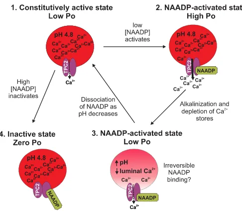
Fig. 9 incorporates our biophysical data into a predictive model of the regulation of TPC2 as the Ca2⫹release channel of
lysosome-like acidic Ca2⫹stores. In the absence of activating
ligands, TPC2 channels gate with very occasional brief open-ings (Po⬍0.0009). We term this gating state of TPC2 the “
con-stitutively active state.” Low concentrations of NAADP in-crease TPC2Po, but this is characterized by (i) a delay in onset, (ii) frequent episodes of synchronized gating of multiple chan-nels, and (iii) irreversible activation at pH 7.2 (on the timescale of a single-channel experiment). We term this gating state of TPC2 the “NAADP-activated state.” Thus, the rate of associa-tion of NAADP to TPC2 is slow, and the rate of dissociaassocia-tion of NAADP from TPC2 is even slower. The association rate does not appear to increase with either increases in luminal [Ca2⫹]
or a change in pH but is concentration-dependent, being faster at 10MNAADP than at 10 nMNAADP as expected for a
ligand-receptor interaction. At acidic pH (4.8), the rate of
dis-sociation of NAADP from TPC2 is increased (NAADP binding to TPC2 is no longer irreversible). In contrast to the effects of low concentrations of NAADP, high concentrations of NAADP (1 mM) abolish even constitutive activity, and we term this state
of TPC2 the “inactivated state.” The effects of high [NAADP] appear to be of rapid onset, but this is difficult to quantify because constitutive activity is so low.
We have reconstituted immunopurified TPC2 into artificial membranes. How do our single-channel currents compare with the currents measured from whole lysosomes isolated from HEK293 cells overexpressing TPC2 (13)? We demonstrate a Ca2⫹/K⫹permeability ratio (PCa2⫹/PK⫹) of 2.6 and show, in
many single-channel traces, that TPC2 is permeable to K⫹. In contrast, Schiederet al.(13) conclude that TPC2 is imperme-able to K⫹. However, it is possible that these authors were unable to accurately measure PCa2⫹/PK⫹ due to a gating
mechanism that inactivated the channels (13). With whole lysosome current recordings, it is difficult to determine whether a rectifying current is obtained because the channels are open but impermeable to K⫹or whether the channels are shut due to a gating mechanism (inactivation or block). We have demonstrated that the luminal environment of TPC2 plays an important regulatory role. Perhaps there are additional luminal factors present within a lysosome that can affect TPC2 gating.
In summary, purified TPC2 behaves as a functional ion channel and, crucially, possesses the conduction properties required of an intracellular Ca2⫹release channel. TPC2 also exhibits a number
of unusual gating characteristics that provide an underlying explanation for many of the peculiarities of NAADP-induced Ca2⫹release in cellular systems (9, 46) including extreme
sen-FIGURE 8.Effects of NAADP on RyR.Histograms showing the effect of NAADP on RyR1 and RyR2 with Ca2⫹as the permeant ion (mean⫾S.D.;nⱖ3) are displayed. The Ca2⫹sensitivity of RyR1 was checked at the end of the experiment by lowering the cytosolic [Ca2⫹] to subactivating levels (⬍1 nM
[image:8.603.306.549.54.266.2]free Ca2⫹) with 10 mMcytosolic EGTA. All channel openings were abolished (Po⫽0;n⫽3). In separate bilayer experiments with RyR2 channels from the same preparations as those used in the histogram, the Ca2⫹sensitivity was also confirmed with EGTA (10 mM).
FIGURE 9.Model summarizing NAADP modulation of TPC2 channel gat-ing.Without NAADP present, TPC2Pois very low, but it still gates with occa-sional brief openings, and therefore, it is in aconstitutively active state(1). Low [NAADP] increasesPoto optimum levels when Ca2⫹loading of the stores is high (2). Ca2⫹release depletes the stores of Ca2⫹, leading to a reducedPo, but causes alkalinization within the stores, which could lead to irreversible bind-ing of NAADP to TPC2 (3). Both state (2) and state (3) represent NAADP-acti-vated states. As the acidic pH is restored, NAADP dissociates from TPC2, and luminal Ca2⫹stores are replenished. High [NAADP] inactivates the channel (Po⫽0) in a reversible manner (4); we term this state theinactive state.
at Univ of St Andrews on December 16, 2013
http://www.jbc.org/
[image:8.603.55.279.54.158.2]sitivity to low concentrations of NAADP, inhibition by 1M
Ned-19 and by high [NAADP], and time-dependent NAADP actions (38). A key aspect of TPC2 regulation is that the sensitivity and reversibility of NAADP binding are con-trolled by the luminal pH and [Ca2⫹]. Further investigation into the detailed gating mechanisms by which NAADP and other agents can regulate TPC channels will be an exciting area for future research that will transform our understanding of cellular NAADP-mediated processes.
REFERENCES
1. Lee, H. C., and Aarhus, R. (1995)J. Biol. Chem.270,2152–2157 2. Lee, H. C. (2000)J. Membr. Biol.173,1– 8
3. Genazzani, A. A., and Galione, A. (1997)Trends Pharmacol. Sci. 18,
108 –110
4. Guse, A. H. (2009)Curr. Biol.19,R521–R523
5. Patel, S., Churchill, G. C., and Galione, A. (2001)Trends Biochem. Sci.26,
482– 489
6. Guse, A. H., and Lee, H. C. (2008)Sci. Signal.1,re10
7. Galione, A., Evans, A. M., Ma, J., Parrington, J., Arredouani, A., Cheng, X., and Zhu, M. X. (2009)Pflugers Arch.458,869 – 876
8. Churchill, G. C., Okada, Y., Thomas, J. M., Genazzani, A. A., Patel, S., and Galione, A. (2002)Cell111,703–708
9. Ruas, M., Rietdorf, K., Arredouani, A., Davis, L. C., Lloyd-Evans, E., Koe-gel, H., Funnell, T. M., Morgan, A. J., Ward, J. A., Watanabe, K., Cheng, X., Churchill, G. C., Zhu, M. X., Platt, F. M., Wessel, G. M., Parrington, J., and Galione, A. (2010)Curr. Biol.20,703–709
10. Calcraft, P. J., Ruas, M., Pan, Z., Cheng, X., Arredouani, A., Hao, X., Tang, J., Rietdorf, K., Teboul, L., Chuang, K. T., Lin, P., Xiao, R., Wang, C., Zhu, Y., Lin, Y., Wyatt, C. N., Parrington, J., Ma, J., Evans, A. M., Galione, A., and Zhu, M. X. (2009)Nature459,596 – 600
11. Brailoiu, E., Churamani, D., Cai, X., Schrlau, M. G., Brailoiu, G. C., Gao, X., Hooper, R., Boulware, M. J., Dun, N. J., Marchant, J. S., and Patel, S. (2009)
J. Cell Biol.186,201–209
12. Tugba Durlu-Kandilci, N., Ruas, M., Chuang, K. T., Brading, A., Par-rington, J., and Galione, A. (2010)J. Biol. Chem.285,24925–24932 13. Schieder, M., Ro¨tzer, K., Bru¨ggemann, A., Biel, M., and Wahl-Schott, C. A.
(2010)J. Biol. Chem.285,21219 –21222
14. Ishibashi, K., Suzuki, M., and Imai, M. (2000)Biochem. Biophys. Res. Com-mun.270,370 –376
15. Zong, X., Schieder, M., Cuny, H., Fenske, S., Gruner, C., Ro¨tzer, K., Gries-beck, O., Harz, H., Biel, M., and Wahl-Schott, C. (2009)Pflugers Arch.458,
891– 899
16. Clapham, D. E., and Garbers, D. L. (2005)Pharmacol. Rev.57,451– 454 17. Pitt, S. J., Funnell, T., Zhu, M. X., Sitsapesan, M., Venturi, E., Parrington, J.,
Ruas, M., Galione, A., and Sitsapesan, R. (2010)Biophys. J.98,682a– 683a 18. Sitsapesan, R., Montgomery, R. A., MacLeod, K. T., and Williams, A. J.
(1991)J. Physiol.434,469 – 488
19. Colquhoun, D., and Sigworth, F. J. (1983) inSingle-channel recording
(Sak-mann, B., and Neher, E., eds.) 2nd Ed., pp. 483–587, Plenum Publishing Corp., New York
20. Fatt, P., and Ginsborg, B. L. (1958)J. Physiol.142,516 –543
21. Naylor, E., Arredouani, A., Vasudevan, S. R., Lewis, A. M., Parkesh, R., Mizote, A., Rosen, D., Thomas, J. M., Izumi, M., Ganesan, A., Galione, A., and Churchill, G. C. (2009)Nat. Chem. Biol.5,220 –226
22. Lindsay, A. R., and Williams, A. J. (1991)Biochim. Biophys. Acta1064,
89 –102
23. Bezprozvanny, I., and Ehrlich, B. E. (1994)J. Gen. Physiol.104,821– 856 24. Tinker, A., and Williams, A. J. (1992)J. Gen. Physiol.100,479 – 493 25. Ehrlich, B. E., and Watras, J. (1988)Nature336,583–586
26. Churchill, G. C., and Galione, A. (2000)J. Biol. Chem.275,38687–38692 27. Kinnear, N. P., Boittin, F. X., Thomas, J. M., Galione, A., and Evans, A. M.
(2004)J. Biol. Chem.279,54319 –54326
28. Kinnear, N. P., Wyatt, C. N., Clark, J. H., Calcraft, P. J., Fleischer, S., Jeya-kumar, L. H., Nixon, G. F., and Evans, A. M. (2008)Cell Calcium44,
190 –201
29. Cancela, J. M., Churchill, G. C., and Galione, A. (1999)Nature398,74 –76 30. Morgan, A. J., and Galione, A. (2008)Methods46,194 –203
31. Sitsapesan, R., and Williams, A. J. (1994)J. Membr. Biol.137,215–226 32. Lukyanenko, V., Gyo¨rke, I., and Gyo¨rke, S. (1996)Pflugers Arch.432,
1047–1054
33. Lloyd-Evans, E., Morgan, A. J., He, X., Smith, D. A., Elliot-Smith, E., Sil-lence, D. J., Churchill, G. C., Schuchman, E. H., Galione, A., and Platt, F. M. (2008)Nat. Med.14,1247–1255
34. Churchill, G. C., and Galione, A. (2001)J. Biol. Chem.276,11223–11225 35. Morgan, A. J., and Galione, A. (2007)Biochem. J.402,301–310 36. Rosen, D., Lewis, A. M., Mizote, A., Thomas, J. M., Aley, P. K., Vasudevan,
S. R., Parkesh, R., Galione, A., Izumi, M., Ganesan, A., and Churchill, G. C. (2009)J. Biol. Chem.284,34930 –34934
37. Churamani, D., Dickinson, G. D., Ziegler, M., and Patel, S. (2006)Biochem. J.397,313–320
38. Genazzani, A. A., Empson, R. M., and Galione, A. (1996)J. Biol. Chem. 271,11599 –11602
39. Sutko, J. L., Airey, J. A., Welch, W., and Ruest, L. (1997)Pharmacol. Rev. 49,53–98
40. Sutko, J. L., and Airey, J. A. (1996)Physiol. Rev.76,1027–1071
41. Rousseau, E., Smith, J. S., and Meissner, G. (1987)Am. J. Physiol.253,
C364 –C368
42. Buck, E., Zimanyi, I., Abramson, J. J., and Pessah, I. N. (1992)J. Biol. Chem. 267,23560 –23567
43. Lindsay, A. R., Tinker, A., and Williams, A. J. (1994)J. Gen. Physiol.104,
425– 447
44. Chu, A., Díaz-Mun˜oz, M., Hawkes, M. J., Brush, K., and Hamilton, S. L. (1990)Mol. Pharmacol.37,735–741
45. Marx, S. O., Ondrias, K., and Marks, A. R. (1998)Science281,818 – 821 46. Bak, J., Billington, R. A., and Genazzani, A. A. (2002)Biochem. Biophys.
Res. Commun.295,806 – 811
47. Saftenku, E., Williams, A. J., and Sitsapesan, R. (2001) Biophys.J.80,
2727–2741
TPC2 Is an NAADP-sensitive Ca
2ⴙRelease Channel
at Univ of St Andrews on December 16, 2013
http://www.jbc.org/


![FIGURE 5. The slow onset of NAADP-induced TPC2 channel activation is�stationary or modal gating behavior is still operating at high [NAADP]](https://thumb-us.123doks.com/thumbv2/123dok_us/8696558.380790/6.603.339.527.54.276/figure-naadp-induced-channel-activation-stationary-behavior-operating.webp)