ISSN Online: 2163-0585 ISSN Print: 2163-0569
DOI: 10.4236/ojmn.2020.101004 Nov. 27, 2019 27 Open Journal of Modern Neurosurgery
Treatment of Pediatric Brain Tumors in
Brazzaville (Congo) about a Case Series
Hugues Brieux Ekouele Mbaki
1,2*, Léon Boukassa
1,2,
Olivier Brice Ngackosso
2, Sinclair Brice Kinata Bambino
2,
Gedeon Colin Thouassa
3, Rel Gerald Boukaka Kala
41Université Marien Ngouabi, Brazzaville, Republic of the Congo
2Service de Chirurgie Polyvalente, CHU de Brazzaville, Brazzaville, Republic of the Congo 3Université Cadi Ayyad, Marrakech, Morocco
4Université Cheikh Anta Diop, Dakar, Senegal
Abstract
Introduction: Tumors of the central nervous system are the most common group of solid neoplasm in children and account for 20% to 25%. They are common in Sub-Saharan countries, despite the insufficiency of histological diagnosis. No study has been performed concerning the pediatric brain tu-mors in the Republic of Congo. The aim of this study was to describe the conditions of neurosurgical management of pediatric tumors in Brazzaville. Materials and Methods: We performed a retrospective and descriptive study, from January 2014 to December 2017 (48 months), into the neurologi-cal unit of the surgineurologi-cal department of Brazzaville. We included all patients aged from 17 years old and below, hospitalized for a brain tumor. Results: We have identified 11 cases of brain tumors. The average age was 8.1 ± 4.3 years old, a sex ratio of 0.57. Ten out of the eleven patients of our series have intracranial hypertension. We found six cases of infratentorial tumors and five of supratentorial location. Only three cases had histology. Ten patients were operated, limited by ventriculoperitoneal shunt in 6 cases, surgical re-section in three cases, biopsy in one case. There were no possibilities of radi-otherapy and chemradi-otherapy during this period of study. Conclusion: A mul-tidisciplinary team must be organized to improve the management of pedia-tric brain tumors in our context. Histological diagnosis and possibilities of radiotherapy are imperatively needed.
Keywords
Pediatric Brain Tumor, Posterior Fossa Tumors, Surgical Resection, Brazzaville
How to cite this paper: Mbaki, H.B.E., Boukassa, L., Ngackosso, O.B., Bambino, S.B.K., Thouassa, G.C. and Kala, R.G.B. (2020) Treatment of Pediatric Brain Tumors in Brazzaville (Congo) about a Case Series. Open Journal of Modern Neurosurgery, 10, 27-35.
https://doi.org/10.4236/ojmn.2020.101004
Received: September 24, 2019 Accepted: November 24, 2019 Published: November 27, 2019
Copyright © 2020 by author(s) and Scientific Research Publishing Inc. This work is licensed under the Creative Commons Attribution International License (CC BY 4.0).
DOI: 10.4236/ojmn.2020.101004 28 Open Journal of Modern Neurosurgery
1. Introduction
Tumors of the central nervous system are the most common group of solid tu-mors in children and account for 20% to 25% of childhood neoplasms [1] [2] [3]. In the United States, mortality rates among children with brain tumors ex-ceed those in children with acute lymphoblastic leukemia [1]. Magnetic Radi-ologic Imaging (MRI) is the required non-invasive investigation for the diagno-sis of brain tumors. Accurate diagnodiagno-sis is important in selecting the optimal therapy for a child with a brain tumor. Surgery is the initial treatment in the majority of cases, with extensive resection for the best long-term survival. Im-portant factors that have contributed to improving the long-term survival of children with brain tumors include advances in neuroimaging, histopathology, neurosurgery, and radiotherapy, associated with chemotherapy [3]. There are different types with variable survival rates depending on the histology, biology, age, and degree of spread. Some types of brain tumors may be completely re-sected, many cannot be removed without a considerable degree of morbidity [1]. Pediatric brain tumors are common in Sub-Saharan countries, despite the in-sufficiency of histological diagnosis, because brain computed-tomography scan (CT-Scan) helps to evoke this diagnosis and to carry out a treatment [4]. Fur-thermore, conditions for identifying childhood brain tumors are difficult be-cause of economic conditions; no insurance health system is associated with a difficulty to have access to an equipped center with a multidisciplinary team in-cluding neurosurgeons [5]. So far, no study has been performed concerning pe-diatric brain tumors in the Republic of Congo.
This work stands as one of a kind and will aim to describe the conditions of neurosurgical management of pediatric tumors in Brazzaville (Republic of Congo).
2. Material and Methods
We performed a retrospective and descriptive study, from January 2014 to De-cember 2017 (48 months) into the neurological unit of the surgical department of the University Hospital of Brazzaville. This unit is the national reference cen-ter for the management of neurosurgical diseases, including pediatric brain tu-mors, with four neurosurgeons.
The University Hospital is equipped with CT-scan and MRI, an operative room dedicated to neurosurgery, with an operative microscope, a Mayfield head clamp, and bipolar coagulation among others.
We selected all patients aged from 17 years old and below, hospitalized in the neurological unit of the surgical department for a brain tumor.
DOI: 10.4236/ojmn.2020.101004 29 Open Journal of Modern Neurosurgery
care unit or multipurpose intensive care unit if ventilatory assistance was needed. When the patient was stable, with good clinical presentation, postopera-tive care could be performed directly in the neurological unit of the surgical de-partment. Postoperative control was performed by CT-scan 24 hours after sur-gery, and or in case of clinical aggravation. Depending on the histology, a patient could be oriented to oncology for further management. Concerning the histolo-gy, pathological material was sent to an anatomopathological laboratory of our hospital or to Kinshasa (Democratic Republic of Congo).
Funding for treatment was directly assured by the child’s parents including the radio-imaging as well as all the operative prescriptions, hospital stay, anato-mopathological examination, and medical treatment.
The parameters evaluated were the diagnosis, therapeutic and evolutionary. Collection of data was performed from a hospital register and parent of pa-tient’s phone information. Statistical values were obtained using the software Numbers for Macintosh 5.3 2008-2018 Apple Inc.
3. Results
3.1. Studied Population and Anthropometric Characteristics
We have identified 11 cases of brain tumors. The average age was 8.1 ± 4.3 years old, a sex ratio of 0.57.
3.2. Diagnostic, Therapeutic Aspects and Evolution
Median delay for admission in the hospital was 5.9 ± 3.7 months, with extremes ranging from 1 to 12 months. Intracranial hypertension was the most common clinical presentation in our case series. We found six cases of infratentorial tu-mors and five of supratentorial location. About cases of brainstem tutu-mors, bi-opsy was not discussed. All cases of hydrocephalus needed a ventriculoperito-neal shunt. The parents of the patient n˚2 refused the surgery for lack of finan-cial means. It was about decompressive surgery with at least one diagnostic bi-opsy, or even an excision of the cerebellar lesion. All clinical parameters, the to-pography of the lesion at neuroimaging, modalities of treatment, histology when it was possible and evolutionary are summarized in Table 1 below.
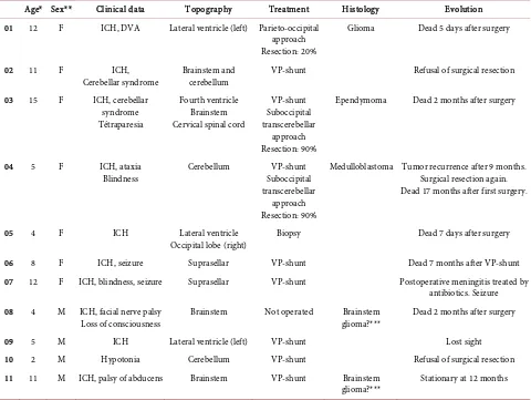
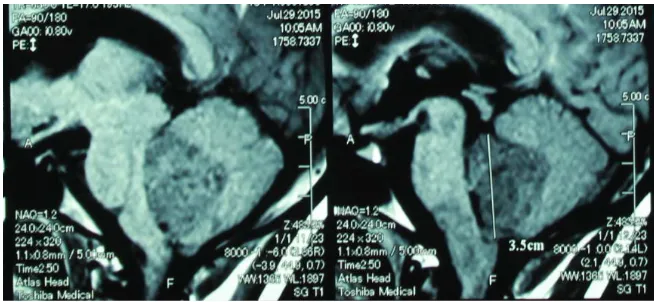
Figure 1 and Figure 2 below indicate the radiological aspects of brain tumors for 2 cases: in the first one, there is a left intraventricular lesion with moderate hydrocephalus; the lesion is tissue and heterogeneous in Flair sequence of MRI. In the second, there is a lesion located in fourth ventricle, and another in me-dulla oblongata, with hyposignal in T1 sequence.
4. Discussion
Epidemiologyhisto-DOI: 10.4236/ojmn.2020.101004 30 Open Journal of Modern Neurosurgery
[image:4.595.59.539.146.507.2]logic type [6]. The epidemiological profile of pediatric brain tumors has been poorly described in Africa. In the region of Marrakesh (South Morocco), on one hundred and thirty-six patients, the average age of patients was 8.28 years, with
Table 1. Characteristics of the series.
Age* Sex** Clinical data Topography Treatment Histology Evolution 01 12 F ICH, DVA Lateral ventricle (left) Parieto-occipital
approach Resection: 20%
Glioma Dead 5 days after surgery
02 11 F ICH,
Cerebellar syndrome Brainstem and cerebellum VP-shunt Refusal of surgical resection 03 15 F ICH, cerebellar
syndrome Tétraparesia
Fourth ventricle Brainstem Cervical spinal cord
VP-shunt Suboccipital transcerebellar
approach Resection: 90%
Ependymoma Dead 2 months after surgery
04 5 F ICH, ataxia
Blindness Cerebellum Suboccipital VP-shunt transcerebellar
approach Resection: 90%
Medulloblastoma Tumor recurrence after 9 months. Surgical resection again. Dead 17 months after first surgery.
05 4 F ICH Lateral ventricle
Occipital lobe (right) Biopsy Dead 7 days after surgery
06 8 F ICH, seizure Suprasellar VP-shunt Dead 7 months after VP-shunt
07 12 F ICH, blindness, seizure Suprasellar VP-shunt Postoperative meningitis treated by antibiotics. Seizure 08 4 M ICH, facial nerve palsy
Loss of consciousness Brainstem Not operated glioma?*** Brainstem Dead 2 months after surgery
09 5 M ICH Lateral ventricle (left) VP-shunt Lost sight
10 2 M Hypotonia Cerebellum VP-shunt Refusal of surgical resection
11 11 M ICH, palsy of abducens Brainstem VP-shunt Brainstem
glioma?*** Stationary at 12 months
*Age (years). **Sex: M = male. F = female. Brainstem glioma***: diagnosis evocated on radiological aspect, not confirmed by histology. ICH: Intracranial hypertension. DVA: decreased visual acuity. VP-Shunt: Ventriculoperitoneal shunt.
[image:4.595.208.539.541.707.2]DOI: 10.4236/ojmn.2020.101004 31 Open Journal of Modern Neurosurgery
Figure 2. MRI (sagittal view, T1) of case n˚3. Tumor located in fourth ventricle and
me-dulla oblongata.
a sex ratio of 1.6 [7]. But in another Moroccan study about two cities, Rabat and Casablanca, the authors found on 542 patients that 51.8% were males and 48.2% were females [8]. In Cameroon (Center of Africa) cerebral tumors in children represented 35.29% of all cerebral tumors. The average age was 9 years, and they found 47.62% of males against 52.38% of females [4]. Broalet et al. (Ivory Coast) found a frequency of 15.74% with a sex ratio of 0.5 [5]. In our case series, we found 7 females against 4 males.
Clinical aspects
Ten on eleven patients of our series have intracranial hypertension (Table 1). Mbonda, E. et al. [4] in Cameroon found intracranial hypertension in 88% of cases. Ndubuisi et al. [9] in Nigeria found in a series of 54 cases from 2006 to 2007 (one year) that a significant proportion of cases presented an advanced disease. They found that 48% of patients had an alteration of the level of con-sciousness and more than 70% with a definite focal deficit.
Topography
About location of the lesion, Barnholtz-Sloan et al. [2] in a study about pedia-tric brain tumors in Non-Hispanics, Hispanics, African Americans and Asians; the majority of the tumors were infratentorial in each racial group (57% of Non-Hispanics, 47% of Hispanics, 55% of African Americans and 53% of Asians). Hazmiri et al. [7] in Morocco found the same results (infratentorial tumors in 61.53% of cases). Some authors like Broalet et al. (Ivory Coast) found a majority of supratentorial tumors, 54.38% [5]. In our case series, we found six cases of infratentorial tumors and five of supratentorial location. Globally, there is no difference between these locations in the literature, but the difference is found depending on the age of patients: before three years old, supratentorial tumors are predominant and between three and eleven years old, tumors of posterior fossa are predominant; after this step, there is no difference about loca-tion [10] [11].
Histology
Barn-DOI: 10.4236/ojmn.2020.101004 32 Open Journal of Modern Neurosurgery
holtz-Sloan et al. found that the Hispanics and Asians had the highest propor-tions of these tumors. Hispanics, African Americans, and Asians had higher proportions of astrocytoma, high-grade tumors compared to Non-Hispanics [2].
In Morocco (North of African continent), astrocytoma and medulloblastoma accounted for 46.32% (29.41% and 16.91%, respectively) in the study of Hazmiri
et al. [7]. In another study, also in Morocco, Karkouri et al. [8] found 34.5% for
medulloblastoma, followed by pilocytic astrocytoma (17.3%) and diffuse astro-cytoma grade 2 (12.5%). The difference between these two studies can be ex-plained by methodology about the selection of patients (cases ranged until 19 years old in the first group and until 15 years old in the second). Near the Re-public of Congo, in Cameroon; astrocytoma was the most frequent type in his-tology, in 45.23% of cases [4]. The same result was found in the Ivory Coast (as-trocytoma) for 19% of cases [5]. In our study, only three cases had histology.
Treatment
Surgery represents the initial treatment for the majority of pediatric brain tu-mors. The surgical resection that is as extensive as possible is important for long-term survival with most tumors [3].
Pilocytic astrocytoma is generally circumscribed and slow-growing tumors. They are commonly located within the cerebellar hemispheres. When complete tumor excision is achieved, there is no need for another therapy, and complete surgical resection is curative. When total resection is not possible without dam-age to eloquent structures of the brain, chemotherapy or radiotherapy can be used for the residual and recurrent tumors [3] [12] [13]. With the introduction of neuro-navigation systems, functional brain mapping and cortical mapping, the lesions located in eloquent brain areas become more accessible for surgical resection with minimal morbidity [3] [13].
Medulloblastomas are undifferentiated embryonal neuroepithelial tumors of the cerebellum. Complete resection should be performed at 74.6%. Brainstem infiltration could be one of the major reasons for the high incidence of subtotal excision. Standard therapy consists of total surgical resection followed by radia-tion to the entire craniospinal axis and or chemotherapy [3] [13] [14]. Some pa-tients might require a ventricular shunt or third ventriculostomy prior to the re-section of the tumor, and the majority of them will have a resolution of the hy-drocephalus after tumor resection. Cerebellar mutism syndrome is one surgical complication characteristically developing after surgery of post-erior fossa [13].
DOI: 10.4236/ojmn.2020.101004 33 Open Journal of Modern Neurosurgery
In our context of work, total resection of the tumor can be performed for ce-rebellar pilocytic astrocytoma. But the difficulty of to manage the surgical treat-ment of pediatric brain tumors is characterized by poor working conditions re-garding possibilities of brain mapping because the neuro-navigation system is not available. Also, there was no possibility of radiotherapy in Congo, during the period of study. This situation contributes to explain a case of recurrence of medulloblastoma after a surgical resection estimated at 90%.
Evolutionary
About some sub-Saharan series, postoperative with complementary treatment follow up is characterized by satisfying evolution in 15% of cases, global mortal-ity around 22% and, postoperative mortalmortal-ity estimated at 7%, survival at 1 and 5 years were 56% and 47% respectively [5] [17]. Also, pediatric brain tumors sur-vivors at 5 years have an increased endocrine disease, psychiatric disorders, cog-nitive and developmental disorders [18] [19]. Most of this morbidity can be at-tributed to direct neurological damage to the developing brain caused by the tumor, surgery, toxicity of chemotherapy and effects of irradiation. Generally, children treated at a young age and those who receive the most intensive therapy are more likely to develop late effects [20].
Limitation of the study
This was a retrospective study, limited in the collection of data from perioper-ative aspects and evolutionary during hospitalization (blood loss, duration of in-tervention, anesthetics parameters). The size of this case series was limited at pa-tients admitted in the neurological unit of the surgical department. It was im-portant to verify if all cases of brain tumors admitted in the pediatric department were addressed for neurosurgical management.
5. Conclusion
The epidemiological profile of pediatric brain tumors has been poorly described in Sub-Saharan countries. Their diagnosis can be improved by more access to CT-scan and MRI. Intracranial hypertension is the most common clinical pres-entation. The majority of these tumors are infratentorial. Histological data are dominated by low-grade astrocytoma and medulloblastoma. Surgical resection that is as extensive as possible is important for long-term survival, but the quali-ty of life after surgery depends on postoperative morbidiquali-ty, chemotherapy and radiotherapy toxicities. In our context, to organize a multidisciplinary team to manage these cases, including pediatrician, radiologist, neurosurgeon, and on-cologist is needed to improve the management of pediatric brain tumors. Histo-logical data and the possibilities of chemotherapy and radiotherapy are impera-tively needed.
Conflicts of Interest
DOI: 10.4236/ojmn.2020.101004 34 Open Journal of Modern Neurosurgery
References
[1] Vidyasagar, R., Abernethy, L., Pizer, B., Avula, S. and Parkes, L.M. (2016) Quantita-tive Measurement of Blood Flow in Paediatric Brain Tumours: A ComparaQuantita-tive Study of Dynamic Susceptibility Contrast and Multi Time-Point Arterial Spin La-belled MRI. British Journal of Radiology, 89, Article ID: 20150624.
https://doi.org/10.1259/bjr.20150624
[2] Barnholtz-Sloan, J.S., Severson, R.K., Stanton, B., Hamre, M. and Sloan, A.E. (2005) Pediatric Brain Tumors in Non-Hispanics, Hispanics, African Americans and Asians: Differences in Survival after Diagnosis. Cancer Causes Control, 16, 587-592.
https://doi.org/10.1007/s10552-004-7843-2
[3] Robertson, P.L. (2006) Advances in Treatment of Pediatric Brain Tumors. NeuroRx, 3, 276-291.https://doi.org/10.1016/j.nurx.2006.01.001
[4] Mbonda, E., Lele Siaka, C., Djientcheu, V.P., Nguefack, S., Mbonda Chimi, P.C., Chiabi, A., Mbassi Awa, H., Sando, Z. and Gonsu Fotsin, J. (2011) Aspects cliniques, scanographiques et histologiques des tumeurs cérébrales de l’enfant à Yaoundé, Cameroun. Schweizer Archiv für Neurologie und Psychiatrie, 162, 284-287.
https://doi.org/10.4414/sanp.2011.02312
[5] Broalet, E., Haidara, A., Zunon-Kipre, Y., N’dri Oka, D., N’da, H., Jibia, A., Kakou, M., Varlet, G. and Bazeze, V. (2007) Approche Diagnostique des tumeurs cérébrales chez l’enfant—expérience du service de neurochirurgie du CHU de Yopougon Ab-idjan. African Journal of Neurological Sciences, 26, 27-38.
https://doi.org/10.4314/ajns.v26i2.7596
[6] Johnson, K.J., Cullen, J., Barnholtz-Sloan, J.S., Ostrom, Q.T., Langer, C.E., Turner, M.C., McKean-Cowdin, R., Fisher, J.L., Lupo, P.J., Partap, S., Schwartzbaum, J.A. and Scheurer, M.E. (2014) Childhood Brain Tumor Epidemiology: A Brain Tumor Epidemiology Consortium Review. Cancer Epidemiology, Biomarkers & Preven-tion, 23, 2716-2736.https://doi.org/10.1158/1055-9965.EPI-14-0207
[7] Hazmiri, F.-Z., Boukis, F., Ait Benali, S., El Ganouni, N.C.I. and Rais, H. (2018) Tumeurs cérébrales de l’enfant: A propos de 136 cas. Case series. Pan African Med-ical Journal, 30, 291.https://doi.org/10.11604/pamj.2018.30.291.13208
[8] Karkouri, M., Zafad, S., Khattab, M., Benjaafar, N., El Kacemi, H., Sefiani, S., Ketta-ni, F., Dey, S. and Soliman, A.S. (2010) Epidemiologic Profile of Pediatric Brain Tumors in Morocco. Child’s Nervous System, 26, 1021-1027.
https://doi.org/10.1007/s00381-010-1097-y
[9] Ndubuisi, C.A., Ohaegbulam, S.C. and Ejembi, G.O. (2018) Paediatric Brain Tu-mours Managed in Enugu, Southeast Nigeria: Review of One Centre Experience.
Nigerian Postgraduate Medical Journal, 25, 186-190.
https://doi.org/10.4103/npmj.npmj_132_18
[10] Garcia-Santos, J.M., Torres del Rio, S., Sanchez, A. and Martinez-Lage, J.F. (2002) Basal Ganglia and Thalamic Tumours: An Imaging Approximation. Child’s Nerv-ous System, 18, 412-425.https://doi.org/10.1007/s00381-002-0606-z
[11] Koob, M. and Girard, N. (2014) Cerebral Tumors: Specific Features in Children.
Diagnostic and Interventional Imaging, 95, 965-983.
https://doi.org/10.1016/j.diii.2014.06.017
[12] Karajannis, M., Allen, J.C. and Newcomb, E.W. (2008) Treatment of Pediatric Brain Tumors. Journal of Cellular Physiology, 217, 584-589.
https://doi.org/10.1002/jcp.21544
DOI: 10.4236/ojmn.2020.101004 35 Open Journal of Modern Neurosurgery https://doi.org/10.1016/j.nurt.2009.04.006
[14] Kumar, L.P., Deepa, S.J., Moinca, I., Suresh, P. and Naidu, K. (2015) Medulloblas-toma: A Common Pediatric Tumor: Prognostic Factors and Predictors of Outcome.
Asian Journal of Neurosurgery, 10, 5051.
https://doi.org/10.4103/1793-5482.151516
[15] Nageswara Rao, A.A., Do, J.S., Wells, E.M. and Packer, R.J. (2012) Biologically Tar-geted Therapeutics in Pediatric Brain Tumors. Pediatric Neurology, 46, 203-211.
https://doi.org/10.1016/j.pediatrneurol.2012.02.005
[16] Rogers, H.A., Estranero, J., Gudka, K. and Grundy, R.G. (2017) The Therapeutic Potential of Targeting the PI3K Pathway in Pediatric Brain Tumors. Oncotarget, 8, 2083-2095.https://doi.org/10.18632/oncotarget.13781
[17] Uche, E.O., Shokunbi, M.T., Malomo, A.O., Akang, E.E.U., Lagunju, I. and Ama-nor-Boadu, S.D. (2013) Pediatric Brain Tumors in Nigeria: Clinical Profile, Man-agement Strategies, and Outcome. Child’s Nervous System, 29, 1131-1135.
https://doi.org/10.1007/s00381-013-2105-9
[18] Gunn, M.E., Lähdesmäki, T., Malila, N., Arola, M., Grönroos, M., Matomäki, J. and Lähteenmäki, P.M. (2015) Late Morbidity in Long-Term Survivors of Childhood Brain Tumors: A Nationwide Registry-Based Study in Finland. Neuro-Oncology, 17, 747-756.https://doi.org/10.1093/neuonc/nou321
[19] Mostoufi-Moab, S. and Grimberg, A. (2010) Pediatric Brain Tumor Treatment: Growth Consequences and Their Management. Pediatric Endocrinology Reviews, 8, 6-17.
[20] Fischer, C., Petriccionne, M., Donzelli, M. and Pottenger, E. (2016) Improving Care in Pediatric Neuro-Oncology Patients: An Overview of the Unique Needs of Child-ren with Brain Tumors. Journal of Child Neurology, 31, 488-505.