REVIEW ARTICLE
The Modic Vertebral Endplate and Marrow
Changes: Pathologic Significance and Relation to
Low Back Pain and Segmental Instability of the
Lumbar Spine
R. Rahme R. Moussa
SUMMARY:Two decades following their description, the significance of Modic vertebral endplate and marrow changes remains a matter of debate. These changes are closely related to the normal degenerative process affecting the lumbar spine, and their prevalence increases with age. However, the exact pathogenesis underlying these changes and their relation to segmental instability of the lumbar spine and to low back pain remain unclear. In this paper, we review the literature relevant to this topic and discuss the currently available evidence regarding the pathologic and clinical significance of Modic changes.
D
egenerative vertebral endplate and subchondral bone marrow changes were first noted on MR imaging by de Roos et al in 1987.1A formal classification was subsequentlyprovided by Modic et al in 1988,2 based on a study of 474
patients, most of whom had chronic low back pain (LBP). These authors described 2 types of endplate and marrow changes:
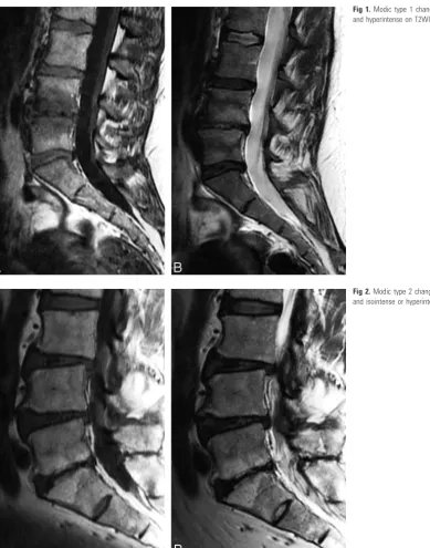
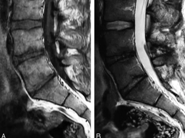
Type 1 changes (Fig 1) were hypointense on T1-weighted imaging (T1WI) and hyperintense on T2-weighted imaging (T2WI) and were shown to represent bone marrow edema and inflammation.
Type 2 changes (Fig 2) were hyperintense on T1WI and isointense or slightly hyperintense on T2WI and were associ-ated with conversion of normal red hemopoietic bone marrow into yellow fatty marrow as a result of marrow ischemia.1,2
Modic type 3 changes (Fig 3) were subsequently described as hypointense on both T1WI and T2WI and were thought to represent subchondral bone sclerosis.3Mixed-type 1/2 and 2/3 Modic changes have also been reported, suggesting that these changes can convert from one type to another and that they all present different stages of the same pathologic process.4The
absence of Modic changes, a normal anatomic appearance, has often been designated Modic type 0.5
Epidemiology
The prevalence of Modic changes among patients with degen-erative disk disease (DDD) of the lumbar spine varies between 19% and 59%, with type 1 and 2 changes being the most com-mon and type 3 and mixed-type changes being relatively rare.1-4,6-14There is disagreement as to whether Modic type 1 or 2 changes are most prevalent in this patient population. Although several series, including the original study of Modic et al,2have shown that type 2 changes are the most frequent
and may account for up to 90% of Modic changes,1-3,9,10,12,13 other studies have suggested that type 1 changes may be more common and may constitute up to 68% of Modic changes in these patients.4,7,11Such differences in the quoted prevalence
of Modic changes and the relative frequency of each Modic type are most likely the result of sampling errors and variations among the studied populations. Modic changes are most com-mon at L4-L5 and L5-S14,10,12,13and are associated with
in-creasing age.10,12These changes usually occur adjacent to
de-generated or herniated intervertebral disks.1-3,9,10,15
Modic changes are uncommon in asymptomatic individu-als without DDD.6,16,17In the series of Toyone et al,6only 9.6%
of patients without DDD had such changes. Weishaupt et al16
reported a prevalence of 3%–10% among 60 asymptomatic volunteers 20 –50 years of age. In particular, type 1 changes were only seen by 1 radiologist in 1 volunteer in 1 of 300 lumbar intervertebral spaces. In a population-based sample of four hundred twelve 40-year old Danes, Kjaer et al17observed Modic changes in the lumbar spines of 9.6% of subjects with-out DDD and 34.1% of those with DDD.
Differential Diagnosis
Intervertebral disk space infections typically give rise to verte-bral marrow edema, manifesting as areas of low signal inten-sity on T1WI and high signal inteninten-sity on T2WI, thereby mim-icking type 1 Modic changes.18,19 Moreover, contrast
enhancement in the disk and endplates may occur in both conditions.18-21However, because of desiccation and
dehy-dration, the disk often appears normal or hypointense on T2WI in DDD, whereas its T2WI signal intensity is typically increased in spondylodiskitis.18,19,21Also, the vertebral
end-plates are usually preserved in DDD rather than eroded or destroyed as seen in disk space infection.18,21Finally, the pres-ence of paraspinal or epidural inflammation and/or collection should orient the diagnosis toward an infectious pro-cess.18,20,21In addition to these imaging considerations, the
clinical presentation and context and the results of laboratory tests such as erythrocyte sedimentation rate and C-reactive protein (CRP) can help differentiate between the 2 entities.18
In particular, the CRP appears to be a very reliable indicator of disk space infection, being raised in up to 100% of patients at the time of diagnosis.18
Pathology and Pathogenesis
In their original study, Modic et al2analyzed histopathologic
sections from 3 patients with type 1 changes and 3 patients with type 2 changes. The authors found that type 1 changes
From the Department of Neurosurgery, Saint-Joseph University and Hoˆtel-Dieu de France, Beirut, Lebanon.
Please address correspondence to Ralph Rahme, MD, Department of Neurosurgery, Hoˆtel-Dieu de France, Ashrafieh, Beirut, Lebanon; e-mail: [email protected]
were associated with disruption and fissuring of endplates and formation of a fibrovascular granulation tissue. In contrast, type 2 changes were associated with fatty degeneration of the red marrow and its replacement by yellow marrow. They con-cluded that type 1 changes correspond to the inflammatory stage of DDD and indicate an ongoing active degenerative process, whereas type 2 changes represent the fatty stage of DDD and are related to a more stable and chronic process. These authors later postulated that type 3 changes represent the sclerotic stage of DDD.3
According to Modic,15the altered signal intensity detected
by MR imaging is not, in and of itself, the causal pathologic process but rather a reflection of the causal process, which is some type of biomechanical stress or instability. Karchevsky et al10concluded that these changes likely represent a response of
the bone marrow to the degenerative process involving the
disk. In fact, type 1 changes have been shown to develop in 8% of patients following diskectomy and 40% following chemo-nucleolysis, which may be viewed as models of accelerated disk degeneration.15Kokkonen et al22observed a strong positive
correlation between Modic changes and disk degeneration and proposed that endplate degeneration is more likely to be a sequel in the process of disk degeneration than a factor con-tributing to disk damage.
Crock23suggested that repeated trauma to intervertebral disks results in the production of inflammatory mediators in the nucleus pulposus and that diffusion of such toxic chemi-cals through vertebral endplates could result in a local inflam-matory reaction resulting in LBP. Brown et al24studied spec-imens of intervertebral disks, vertebral endplates, and adjacent cancellous bone they obtained during anterior diskectomy and fusion from patients with chronic LBP and DDD. They
Fig 1.Modic type 1 changes are hypointense on T1WI (A) and hyperintense on T2WI (B).
Fig 2.Modic type 2 changes are hyperintense on T1WI (A) and isointense or hyperintense on T2WI (B).
REVIEW
[image:2.594.63.453.38.534.2]observed cracks and defects in the vertebral endplates, with increases in vascular density and the number of sensory nerve fibers, and hypothesized that such changes could represent a means of increasing disk nourishment and could be a source of LBP in patients with DDD. Burke et al25observed a greater
increase in proinflammatory mediators such as interleukin-6, interleukin-8, and prostaglandin E-2 in the disks of patients with type 1 Modic changes undergoing fusion for LBP than in those of patients undergoing diskectomy for sciatica. These authors proposed that the production of proinflammatory mediators within the nucleus pulposus may be a major factor in the genesis of diskogenic LBP. Vital et al26concluded that
Modic type 1 changes correspond to edema of vertebral end-plates and subchondral bone. This edema could correspond to microfractures of cancellous bone and endplate cracks accom-panied by an increased vascular density along with an increase in the number of nerve endings and in the levels of proinflam-matory chemical mediators, and these vascular and inflamma-tory changes would follow the initial mechanical phenomena. Schmid et al9 found a positive correlation between the
presence and extent of Modic changes and the amount of car-tilage in the extruded disk in patients undergoing lumbar mi-crodiskectomy and concluded that these changes may result from avulsion-type disk herniation. Ohtori et al27found that
the cartilaginous endplates of patients with Modic changes had more protein gene product (PGP) 9.5 immunoreactive nerve fibers and tumor necrosis factor (TNF) immunoreactive cells than those with normal endplates. PGP 9.5 immunoreac-tivity was seen exclusively in patients with diskogenic LBP, whereas TNF immunoreactivity was seen in both patients with LBP and healthy controls. In addition, the number of TNF immunoreactive cells in endplates with Modic type 1 changes was higher than those with type 2 changes. The authors con-cluded that inflammatory cytokines and nerve ingrowth into vertebral endplates may be a cause of diskogenic LBP and that type 1 changes, representing more active inflammation, seem to be mediated by proinflammatory cytokines, whereas type 2 and 3 changes could be more quiescent stages of the process.
In a randomized controlled trial, Korhonen et al28found
that infliximab, a monoclonal antibody against TNF-␣, was no more effective than placebo in the treatment of disk hernia-tion–induced sciatica. However, the authors noted a trend to-ward better results in the infliximab group when a Modic change was colocalized at the symptomatic level. Fayad et al29
found that patients with chronic LBP and predominantly type 1 inflammatory Modic changes had better short-term relief of symptoms following intradiskal steroid injection than those with predominantly type 2 changes, which further supports the inflammatory nature of Modic type 1 changes and the role of inflammation in the generation of LBP.
Modic Changes and LBP
Kjaer et al17suggested that Modic changes constitute the cru-cial element in the degenerative process around the disk in relation to LBP and clinical findings. They demonstrated that DDD on its own was a fairly quiet disorder, whereas DDD with Modic changes was much more frequently associated with clinical symptoms. Most authors agree that, among Modic changes, type 1 changes are the ones most strongly associated with LBP.6,8,13-15In a study of 74 patients with DDD, Toyone
et al6observed that 73% of patients with type 1 changes had LBP as opposed to only 11% of those with type 2 changes. Mitra et al8found a positive trend between the evolution of
type 1 Modic changes into type 2 changes and the improve-ment of symptoms. In addition, they observed that patients in whom type 1 changes increased were clinically worsened. Al-bert and Manniche14reported a strong association between
Modic changes and LBP as 60% of patients with Modic changes but only 20% of those without such changes had LBP. These authors also showed that type 1 changes were more strongly associated with LBP than type 2 changes. In a study of 228 Finnish middle-aged male workers, Kuisma et al13found
that Modic changes at L5-S1, especially type 1 changes and extensive lesions, were strongly associated with pain symp-toms and LBP.
The relationship between Modic changes and diskogenic
[image:3.594.54.376.44.283.2]LBP remains a matter of debate.4,7,22,30Braithwaite et al4and Weishaupt et al7showed that Modic changes have a very high
specificity (96%–96.8%) and positive predictive value (88%– 91.3%) for pain reproduction during diskography in patients with chronic LBP. In contrast, other MR imaging findings such as advanced disk degeneration and high-intensity zones were found to be much less specific for diskogenic LBP. Weishaupt et al7further demonstrated that moderate and
se-vere Modic changes (ie, those extending over 25% or more of the vertebral height) have a specificity and positive predictive value of 100% for a concordant pain response on diskography. The findings of these authors have been challenged by those of Sandhu et al30and Kokkonen et al,22who failed to demon-strate any significant association between the presence of Modic changes and pain provocation during diskography in patients with chronic LBP. Given the limited sample sizes in these studies and their conflicting results, no conclusions can be drawn at this time with certainty regarding the relationship between Modic changes and diskogenic LBP.
Modic Changes and Segmental Instability
In the study of Toyone et al,670% of patients with type 1
Modic changes but only 16% of those with type 2 changes were found to have segmental hypermobility, defined as a sagittal translation of 3 mm or more on dynamic flexion-extension films. The authors concluded that patients with chronic LBP and type 1 Modic changes had more frequent instability quiring arthrodesis than those with type 2 changes. Their re-sults were challenged by those of Bra¨m et al,31who did not find any relationship between type 1 Modic changes and lumbar instability by using the same radiologic criteria. A major lim-itation of these studies is their reliance on a radiologic defini-tion of lumbar instability for which a consensus has to exist.32 The relationship between Modic type 1 changes and seg-mental instability is mostly supported by indirect evidence coming from outcome studies following lumbar fusion.26,33-36
In a study assessing osseous union following lumbar fusion in 33 patients, Lang et al33found that all 19 patients with solid
fusion had type 2 Modic changes, whereas 10 of 14 patients with nonunion had type 1 changes. They concluded that the persistence of type 1 Modic changes after fusion suggests pseudarthrosis. Buttermann et al34 similarly observed that
nonfusion was associated predominantly with the persistence of type 1 Modic changes. In a study of 56 patients treated with anterior lumbar interbody fusion for LBP, Chataigner et al35 found that patients with type 1 Modic changes had much bet-ter outcomes than those with isolated DDD and those with type 2 changes, in whom the results were generally poor. Vital et al26assessed clinical and radiologic outcomes following in-strumented posterolateral fusion in 17 patients with chronic LBP and type 1 Modic changes. Six months later, all type 1 changes had converted, 76.5% into type 2 changes and 23.5% back to normal, and clinical improvement was seen in all pa-tients. They concluded that fusion accelerates the course of type 1 Modic changes probably by correcting the mechanical instability and that these changes appear to be a good indicator of satisfactory surgical outcome after arthrodesis. In a study of 60 patients with severe chronic LBP and single-level DDD treated with instrumented fusion, Esposito et al36showed that
patients with type 1 Modic changes had excellent results and
improved much better than patients with type 2 changes whose clinical outcome was poor.
Natural History
Braithwaite et al4suggested that Modic changes can convert
from one type to another and that they all present different stages of the same pathologic process. Mixed-type changes are assumed to develop before conversion to one of the true Modic types.4,26In their original study, Modic et al2followed 16 patients longitudinally. Among the 6 patients with type 1 Modic changes, 5 patients showed at least a partial conversion into type 2 within 14 months to 3 years, whereas the remaining patient demonstrated reverse transformation into Modic type 0. In contrast, none of the 10 patients with type 2 Modic changes showed a change during the 2- to 3-year follow-up period. The authors concluded that type 2 changes are stable and unchangeable with time, whereas type 1 changes are un-stable. This view was supported by a greater prevalence of type 2 changes in their study.
It is generally agreed that type 1 changes are unstable le-sions.2,8,15,26Vital et al26demonstrated that all type 1 changes
converted into either type 2 changes or back to normal within 6 months following lumbar fusion, which paralleled clinical improvement in all patients. In a longitudinal study of 44 non-operated patients with LBP and sciatica followed for 12–72 months, Mitra et al8found that 92% of type 1 changes either converted wholly or partially into type 2 changes (52%) or became more extensive (40%) and that only 8% of these changes remained the same. They concluded that type 1 Modic changes are dynamic lesions that, in most cases, either increase in size or convert into type 2 changes.
The stability of type 2 changes has been recently questioned by several authors.5,12,14 Kuisma et al12 studied the natural
history of Modic changes in 60 nonoperated patients with sci-atica. They found that 14% of Modic changes evolved into another type within 3 years, 80% of the conversions being from type 2 to either type 1 or mixed type 1/2. They also found that nonconverted Modic changes increased in size and that new Modic changes developed adjacent to degenerated disks in 6% of patients, 77% of these new changes being either type 1 or mixed type 1/2. They concluded that type 2 changes may be less stable than previously assumed and speculated that an acute ongoing inflammatory process in some type 2 changes causes conversion of yellow to red marrow, which suggests superimposed changes such as continued or accelerated de-generation, a view shared by Modic.15In a study of 166
non-operated patients with sciatica undergoing repeat MR imaging at 14 months, Albert and Manniche14found that the
preva-lence of Modic changes increased from 9% at baseline to 29% at follow-up, all new changes developing at the level of the previous disk herniation. In addition, many patients with pre-existent Modic changes developed additional type 1 changes. They concluded that lumbar disk herniation was a strong risk factor for developing Modic changes, especially type 1, during the following year. Marshman et al5reported on 2 patients
and 2 are instead interchangeable and equipotent in symp-tom-generating capacity.
Conclusion
From this review, it appears that Modic changes are dynamic markers of the normal age-related degenerative process affect-ing the lumbar spine. These lesions can convert from one type to another with time, with mixed-type changes probably rep-resenting the intermediate stages in this conversion. Type 1 changes are likely to be inflammatory in origin and seem to be strongly associated with active low back symptoms and seg-mental instability, thus reflecting a state of active degeneration and biomechanical instability of the lumbar spine. Accord-ingly, these changes appear to predict an excellent outcome following lumbar fusion. In contrast, type 2 changes are less clearly associated with LBP and seem to indicate a more bio-mechanically stable state, though superimposed stress may oc-casionally cause their reverse conversion into type 1 changes. Finally, the exact nature and pathogenetic significance of type 3 changes remains largely unknown.
References
1. de Roos A, Kressel H, Spritzer C, et al.MR imaging of marrow changes adjacent to end plates in degenerative lumbar disk disease.AJR Am J Roentgenol
1987;149:531–34
2. Modic MT, Steinberg PM, Ross JS, et al.Degenerative disk disease: assessment of changes in vertebral body marrow with MR imaging. Radiology
1988;166:193–99
3. Modic MT, Masaryk TJ, Ross JS, et al.Imaging of degenerative disk disease.
Radiology1988;168:177– 86
4. Braithwaite I, White J, Saifuddin A, et al.Vertebral end-plate (Modic) changes on lumbar spine MRI: correlation with pain reproduction at lumbar discog-raphy.Eur Spine J1998;7:363– 68
5. Marshman LA, Trewhella M, Friesem T, et al.Reverse transformation of Modic type 2 changes to Modic type 1 changes during sustained chronic low-back pain severity: report of two cases and review of the literature.J Neurosurg Spine
2007;6:152–55
6. Toyone T, Takahashi K, Kitahara H, et al.Vertebral bone-marrow changes in degenerative lumbar disc disease: an MRI study of 74 patients with low back pain.J Bone Joint Surg Br1994;76:757– 64
7. Weishaupt D, Zanetti M, Hodler J, et al.Painful lumbar disk derangement: relevance of endplate abnormalities at MR imaging. Radiology
2001;218:420 –27
8. Mitra D, Cassar-Pullicino VN, McCall IW.Longitudinal study of vertebral type-1 end-plate changes on MR of the lumbar spine. Eur Radiol
2004;14:1574 – 81
9. Schmid G, Witteler A, Willburger R, et al.Lumbar disk herniation: correlation of histologic findings with marrow signal intensity changes in vertebral end-plates at MR imaging.Radiology2004;231:352–58
10. Karchevsky M, Schweitzer ME, Carrino JA, et al.Reactive endplate marrow changes: a systematic morphologic and epidemiologic evaluation.Skeletal Ra-diol2005;34:125–29
11. Kjaer P, Leboeuf-Yde C, Korsholm L, et al.Magnetic resonance imaging and low back pain in adults: a diagnostic imaging study of 40-year-old men and women.Spine2005;30:1173– 80
12. Kuisma M, Karppinen J, Niinimaki J, et al.A three-year follow-up of lumbar spine endplate (Modic) changes.Spine2006;31:1714 –18
13. Kuisma M, Karppinen J, Niinima¨ki J, et al.Modic changes in endplates of lumbar vertebral bodies: prevalence and association with low back and sciatic pain among middle-aged male workers.Spine2007;32:1116 –22
14. Albert HB, Manniche C.Modic changes following lumbar disc herniation.Eur Spine J2007;16:977– 82
15. Modic MT.Modic type 1 and type 2 changes.J Neurosurg Spine2007;6:150 –51 16. Weishaupt D, Zanetti M, Hodler J, et al.MR imaging of the lumbar spine: prevalence of intervertebral disk extrusion and sequestration, nerve root compression, end plate abnormalities, and osteoarthritis of the facet joints in asymptomatic volunteers.Radiology1998;209:661– 66
17. Kjaer P, Korsholm L, Bendix T, et al.Modic changes and their associations with clinical findings.Eur Spine J2006;15:1312–19
18. James SL, Davies AM.Imaging of infectious spinal disorders in children and adults.Eur J Radiol2006;58:27– 40
19. Ross JS, Modic MT.Current assessment of spinal degenerative disease with magnetic resonance imaging.Clin Orthop Relat Res1992;279:68 – 81 20. Van Goethem JW, Parizel PM, van den Hauwe L, et al.The value of MRI in the
diagnosis of postoperative spondylodiskitis.Neuroradiology2000;42:580 – 85 21. Ledermann HP, Schweitzer ME, Morrison WB, et al.MR imaging findings in
spinal infections: rules or myths?Radiology2003;228:506 –14
22. Kokkonen SM, Kurunlahti M, Tervonen O, et al.Endplate degeneration ob-served on magnetic resonance imaging of the lumbar spine: correlation with pain provocation and disc changes observed on computed tomography dis-kography.Spine2002;27:2274 –78
23. Crock HV.Internal disc disruption: a challenge to disc prolapse fifty years on.
Spine1986;11:650 –53
24. Brown MF, Hukkanen MV, McCarthy ID, et al.Sensory and sympathetic in-nervation of the vertebral endplate in patients with degenerative disc disease.
J Bone Joint Surg Br1997;79:147–53
25. Burke JG, Watson RW, McCormack D, et al.Intervertebral discs which cause low back pain secrete high levels of proinflammatory mediators.J Bone Joint Surg Br2002;84:196 –201
26. Vital JM, Gille O, Pointillart V, et al.Course of Modic 1 six months after lumbar posterior osteosynthesis.Spine2003;28:715–21
27. Ohtori S, Inoue G, Ito T, et al.Tumor necrosis factor-immunoreactive cells and PGP 9.5-immunoreactive nerve fibers in vertebral endplates of patients with discogenic low back pain and Modic type 1 or type 2 changes on MRI.
Spine2006;31:1026 –31
28. Korhonen T, Karppinen J, Paimela L, et al.The treatment of disc-herniation-induced sciatica with infliximab: one-year follow-up results of FIRST II, a randomized controlled trial.Spine2006;31:2759 – 66
29. Fayad F, Lefevre-Colau MM, Rannou F, et al.Relation of inflammatory Modic changes to intradiscal steroid injection outcome in chronic low back pain.Eur Spine J2007;16:925–31
30. Sandhu HS, Sanchez-Caso LP, Parvataneni HK, et al.Association between find-ings of provocative discography and vertebral endplate signal changes as seen on MRI.J Spinal Disord2000;13:438 – 43
31. Bra¨m J, Zanetti M, Min K, et al.MR abnormalities of the intervertebral disks and adjacent bone marrow as predictors of segmental instability of the lum-bar spine.Acta Radiol1998;39:18 –23
32. Nizard RS, Wybier M, Laredo JD.Radiologic assessment of lumbar interver-tebral instability and degenerative spondylolisthesis.Radiol Clin North Am
2001;39:55–71
33. Lang P, Chafetz N, Genant HK, et al.Lumbar spinal fusion: assessment of functional stability with magnetic resonance imaging.Spine1990;15:581– 88 34. Buttermann GR, Heithoff KB, Ogilvie JW, et al.Vertebral body MRI related to
lumbar fusion results.Eur Spine J1997;6:115–20
35. Chataigner H, Onimus M, Polette A.Surgery for degenerative lumbar disc disease: should the black disc be grafted[in French]?Rev Chir Orthop Reparatrice Appar Mot1998;84:583– 89