w
w
-3 polyunsaturated fatty acids ameliorate type
1 diabetes and autoimmunity
Xinyun Bi, … , Xiaoxi Li, Allan Zijian Zhao
J Clin Invest.
2017;
127(5)
:1757-1771.
https://doi.org/10.1172/JCI87388
.
Despite the benefit of insulin, blockade of autoimmune attack and regeneration of
pancreatic islets are ultimate goals for the complete cure of type 1 diabetes (T1D).
Long-term consumption of w-3 polyunsaturated fatty acids (PUFAs) is known to suppress
inflammatory processes, making these fatty acids candidates for the prevention and
amelioration of autoimmune diseases. Here, we explored the preventative and therapeutic
effects of w-3 PUFAs on T1D. In NOD mice, dietary intervention with w-3 PUFAs sharply
reduced the incidence of T1D, modulated the differentiation of Th cells and Tregs, and
decreased the levels of IFN-g, IL-17, IL-6, and TNF-a. w-3 PUFAs exerted similar effects on
the differentiation of CD4
+T cells isolated from human peripheral blood mononuclear cells.
The regulation of CD4
+T cell differentiation was mediated at least in part through w-3
PUFA eicosanoid derivatives and by mTOR complex 1 (mTORC1) inhibition. Importantly,
therapeutic intervention in NOD mice through nutritional supplementation or
lentivirus-mediated expression of an w-3 fatty acid desaturase, m
fat-
1, normalized blood glucose and
insulin levels for at least 182 days, blocked the development of autoimmunity, prevented
lymphocyte infiltration into regenerated islets, and sharply elevated the expression of the b
cell markers pancreatic and duodenal homeobox 1 (
Pdx1
) and paired box 4 (
Pax4
). The
findings suggest that w-3 PUFAs could potentially serve as a therapeutic modality for T1D.
Research Article
Autoimmunity
Endocrinology
Find the latest version:
Introduction
Type 1 diabetes (T1D) is a polygenic and organ-specific autoim-mune disease, in which a certain subclass of T lymphocytes is involved in executing autoimmune attacks that lead to the destruc-tion of pancreatic β cells (1, 2). Although the current prevailing treatment of T1D is lifelong daily injection of recombinant insulin, the exogenously delivered insulin does not carry C-peptide, which has been known to provide protective effects on the microvascula-ture (3) and on neuronal (4) and renal (5) function. Even with prop-er blood glucose control, T1D patients are prone to complications such as retinopathy, neuropathy, and cardiovascular disease (6, 7). Thus far, there is no effective method of reversing autoimmuni-ty once a patient enters the course of T1D. Research in the past decade has been focused on islet allografting (8) and stem cell– derived β cells (9), as well as immunotherapies using mAbs target-ing CD3 (10), glutamic acid decarboxylase-65 Ig (GAD65-Ig) (11), and cytotoxic T lymphocyte–associated antigen-4–Ig (CTLA-4– Ig) (12). Although promising phase I and II trials were reported in patients with early onset of T1D, phase III trials of anti-CD3 and GAD65-Ig immunotherapy did not meet the primary endpoints (10, 11, 13). Similarly, CTLA4-Ig preserved C-peptide concentra-tions for only 9 months, despite continuous i.v. administration
for 2 years (12). The limitations of islet transplantation include a shortage of donors, fibrosis of transplanted islets, and various side effects of immunosuppressive agents, making it unlikely to be a primary therapeutic approach for T1D (8, 14). Indeed, either a sin-gle immunosuppressive agent or islet allografting alone appears to be insufficient to completely control or reverse the autoimmune attack on β cells. A new paradigm that targets several pathogenic pathways is therefore sorely needed for a complete cure of T1D.
Both genetic susceptibility and environmental factors can trigger the onset of T1D. In particular, environmental factors such as viral infection and nutritional imbalance can cause dysregula-tion of CD4+ T cells (15), leading to the infiltration of autoreactive
CD8+ T cells into pancreatic islets and, ultimately, the killing of β cells. Eventually, a subsequent wave of β cell killing results in a severe reduction of β cell function and mass (1, 15). Numerous studies have shown that alterations of multiple immune cells play major roles in the etiology of T1D, chief among which Th1, Th2, and Th17 cells and Tregs have been extensively elucidated (16). Th1 cells evoke cell-mediated immunity and phagocyte-depen-dent inflammation, whereas Th2 cells can cause strong antibody responses but inhibit phagocyte-independent inflammation (17, 18). The Th1-dominated responses dictate the pathogenesis of organ-specific autoimmune disorders (19). Th1 and Th17 cells mainly secrete inflammatory factors such as IFN-γ and IL-17, which, by acting alone or synergistically, can aggravate the pro-gression of T1D (20). In contrast, Th2 cells and Tregs can antag-onize autoimmune responses through cytokines such as IL-4 and
Despite the benefit of insulin, blockade of autoimmune attack and regeneration of pancreatic islets are ultimate goals for the complete cure of type 1 diabetes (T1D). Long-term consumption of ω-3 polyunsaturated fatty acids (PUFAs) is known to suppress inflammatory processes, making these fatty acids candidates for the prevention and amelioration of autoimmune diseases. Here, we explored the preventative and therapeutic effects of ω-3 PUFAs on T1D. In NOD mice, dietary intervention with ω-3 PUFAs sharply reduced the incidence of T1D, modulated the differentiation of Th cells and Tregs, and decreased the levels of IFN-γ, IL-17, IL-6, and TNF-α. ω-3 PUFAs exerted similar effects on the differentiation of CD4+ T cells isolated
from human peripheral blood mononuclear cells. The regulation of CD4+ T cell differentiation was mediated at least in part
through ω-3 PUFA eicosanoid derivatives and by mTOR complex 1 (mTORC1) inhibition. Importantly, therapeutic intervention in NOD mice through nutritional supplementation or lentivirus-mediated expression of an ω-3 fatty acid desaturase, mfat-1, normalized blood glucose and insulin levels for at least 182 days, blocked the development of autoimmunity, prevented lymphocyte infiltration into regenerated islets, and sharply elevated the expression of the β cell markers pancreatic and duodenal homeobox 1 (Pdx1) and paired box 4 (Pax4). The findings suggest that ω-3 PUFAs could potentially serve as a therapeutic modality for T1D.
ω
-3 polyunsaturated fatty acids ameliorate type 1
diabetes and autoimmunity
Xinyun Bi,1,2 Fanghong Li,1 Shanshan Liu,3 Yan Jin,4 Xin Zhang,3 Tao Yang,5 Yifan Dai,3 Xiaoxi Li,3 and Allan Zijian Zhao1 1Collaborative Innovation Center for Cancer Medicine, Institute of Biomedical and Pharmaceutical Sciences, Guangdong University of Technology, Guangzhou, Guangdong Province, China. 2The Center of
Immunological Genetics and HLA Typing, First Affiliated Hospital of Nanjing Medical University, Nanjing, Jiangsu Province, China. 3The Center of Metabolic Disease Research, Nanjing Medical University,
Nanjing, Jiangsu Province, China. 4Department of Pathology, Wuxi Maternity and Child Health Care Hospital Affiliated to Nanjing Medical University, Wuxi, Jiangsu Province, China. 5Department of
Endocrinology, First Affiliated Hospital of Nanjing Medical University, Nanjing, Jiangsu Province, China.
Authorship note: X. Li and A.Z. Zhao are co-senior authors.
Conflict of interest: The authors have declared that no conflict of interest exists.
Submitted: March 10, 2016; Accepted: February 2, 2017.
jects also resulted in a much-reduced incidence of childhood-onset T1D (23). Recently, a multicenter, randomized, double-blind trial of docosahexaenoic acid (DHA) supplementation in infants at a high genetic risk of T1D, beginning either in the last trimester of pregnan-cy or in the first 5 months after birth, revealed significantly reduced inflam-mation as reflected in the lower levels of high-sensitivity C-reactive protein (24). Apart from this, our earlier studies showed that stable cellular production of ω-3 PUFAs could enhance insulin secretion and confer strong resistance to cytokine-induced β cell destruction (25). These studies indicated that an
ω-3 PUFA–enriched-diet can have a strong influence on immune responses as well as on pancreatic islet function and prompted us to investigate the strategy of using ω-3 PUFAs as a preven-tative, or even therapeutic, modality to deter T1D. Specifically, we improvised a nutritional as well as a gene therapy approach to elevate circulating eicos-apentaenoic acid (EPA) and DHA levels, with the goals of correcting the dysregu-lation of T cell differentiation, silencing the immune attack against pancreatic
β cells, and regenerating β cells.
Results
Intervention with dietary ω-3 PUFAs reduces the incidence of T1D. We designed
a dietary intervention study, in which female NOD mice were fed a diet con-taining enriched EPA/DHA for 35 weeks, starting at 5 weeks of age. In addition to a control group fed a regular diet, a sepa-rate group of animals was fed a diet con-taining equal levels of arachidonic acid (AA, an ω-6 PUFA). The dietary inter-vention resulted in significant changes in fatty acid composition in serum and tis-sue samples (Supplemental Tables 1–3; supplemental material avail-able online with this article; https://doi.org/10.1172/JCI87388DS1). The EPA/DHA-enriched diet resulted in a substantial increase in ω-3 PUFAs that was accompanied by a significant decrease (P < 0.0001) in ω-6 PUFAs, leading to sharply reduced ratios of ω-6/ω-3 PUFAs relative to ratios in AA diet–fed mice. Nonfasting blood glucose con-centrations were used to monitor the incidence of diabetes, which was diagnosed by the presence of glucose concentrations of greater than 11.11 mmol/l for 2 consecutive weeks. No spontaneous reversal to less than 11.11 mmol/l was observed in any of the control groups (n = 15 per group). Consistent with results reported by others (26, 27), we found that 80% of female NOD mice on a regular diet devel-IL-10 (21). The intervention of dysregulated Th cells is therefore
critical to stopping autoimmune progression and the inflammato-ry attack against β cells.
Mounting evidence suggests that dietary supplementation of fish oil starting from infancy has the beneficial effect of alleviating autoimmune progress and T1D. A longitudinal DAISY (Diabetes Autoimmunity Study in the Young) study showed that long-term dietary intake of ω-3 polyunsaturated fatty acids (PUFAs) starting from 1 year of age is associated with a reduced risk of islet auto-immunity in children with familial T1D (22). A Norwegian case- control study in which cod liver oil was given, starting from 1 year of age, to infants with a high risk of T1D and to population control
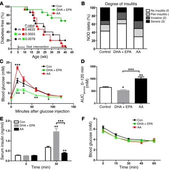
sub-Figure 1. ω-3 PUFAs ameliorate the development of T1D and normalize glucose metabolism in NOD mice. (A) Blood glucose concentrations in 3 groups of NOD mice on varied diets were monitored weekly until 40 weeks of age. Sustained hyperglycemia for 2 consecutive weeks (>11.11 mmol/l) marked the onset of disease, which was used to create a life table to determine the incidence of diabetes (n = 15/group). Statistical calculation was done using a Mantel-Cox log-rank test. (B) Sections (4-μm-thick) of pancreas from 20-week-old NOD mice were formaldehyde fixed, paraffin embedded, and stained with H&E (n =7/ group). Islets were sorted into the following 4 categories on the basis of the relative degree of immune infiltration: no insulitis (0), peri-insulitis (1), invasive insulitis (2), or severe insulitis (3). Representative pancreatic sections are shown in Supplemental Figure 1. The differences in severe insulitis between DHA plus EPA group and the control group (P < 0.0001) and between the DHA plus EPA group and the AA group (P = 0.0008) were significant. The finding of no insulitis in the DHA plus EPA group was increased compared with the control (P = 0.02) and AA (P < 0.0001) groups. Statistical calculation was done using Pearson’s χ2
[image:3.585.38.374.55.394.2]were no different among the 3 groups, suggesting that ω-3 PUFAs could not prevent the initiation of lymphocyte infiltration. Taken together, these results indicated that ω-3 PUFA supplementation in NOD mice sharply reduced the percentage of islets showing the most severe insulitis (Figure 1B).
Intervention with ω-3 PUFA can normalize glucose metabolism in NOD mice. To study the function of β cells and the homeostasis of blood glucose, we evaluated the impact of ω-3 and ω-6 PUFAs on glucose homeostasis in NOD mice on different diets. Fasting blood glucose concentrations were indistinguishable among the 3 groups. However, during the i.p. glucose tolerance test, the NOD mice maintained on an EPA/DHA-enriched diet showed signifi-cantly lower blood glucose concentrations at all time points than did mice in the other 2 groups (Figure 1, C and D). The improved glucose tolerance in the EPA/DHA-fed mice was primarily attributed to the elevated glucose-induced insulin secretion (Fig-ure 1E), as the insulin tolerance tests revealed no significant differ-ence among the 3 groups of NOD mice (Figure 1F).
Modulation of CD4+ T cell differentiation by ω-3 and ω-6 PUFAs.
The interplay among CD4+ T cells, particularly Th1, Th2, Th17,
oped diabetes by the age of 40 weeks. In contrast, only 33% of the mice fed an EPA/DHA-enriched diet were diabetic, which was sig-nificantly different (P = 0.0076) according to a Mantel-Cox log-rank test. Interestingly, 93% of NOD mice on the diet containing compa-rable levels of AA developed diabetes at the same age, although there was no significant difference between the AA intervention group and the control diet group (Figure 1A). Thus, long-term supplementation of dietary EPA/DHA reduced the incidence of T1D and delayed its onset in female NOD mice.
Intervention with ω-3 PUFAs blocks the progression of immune infiltration in NOD mice. The progression of peri-insulitis and
[image:4.585.42.523.55.475.2]insu-litis occurs between the initiation and detection of hyperglycemia in NOD mice (28). We used H&E-stained pancreatic sections to evaluate the extent of lymphocyte infiltration into pancreatic islets isolated from 20-week-old NOD mice (16 weeks after different dietary intervention) (29) (Supplemental Figure 1). By the age of 20 weeks, the islets from the EPA/DHA-fed mice had a signifi-cantly reduced incidence of severe insulitis compared with those from mice maintained on an AA-enriched diet or a regular diet. The percentages of peri-insulitis and invasive insulitis incidence
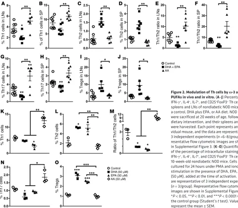
Figure 2.Modulation of Th cells by ω-3 and ω-6 PUFAsin vivo and in vitro. (A–J) Percentage of IFN-γ+, IL-4+, IL-7+, and CD25+FoxP3+ Th cells in
spleens and LNs of nondiabetic NOD mice fed a control, DHA plus EPA, or AA diet. NOD mice were sacrificed at 20 weeks of age, following dietary intervention, and their spleens and LNs were harvested. Each point represents an indi-vidual mouse, and the data are representative of 3 independent experiments (n =6–8/group). Rep-resentative flow cytometric images are shown in Supplemental Figure 3. (K–O) Quantification of the percentage of intracellular staining of IFN-γ+, IL-4+, IL-7+, and CD25+FoxP3+ Th cells from
IL-4 secreted from CD8+ T cells were too low to be detected by
FACS. Thus, CD8+ T cells were probably not the primary targets of
autoimmune regulation by ω-3 PUFAs. Taken together, interven-tion with ω-3 PUFAs normalized the ratio of Th1/Th2 cells and had an inhibitory effect on Th1 and Th17 cells, but a stimulatory effect on Tregs in NOD mice.
Dietary ω-3 PUFAs restore cytokine secretion profiles in NOD mice. Activated T cells produce a series of cytokines that exert many
actions on T cell themselves and on other immune cells. Measure-ment of pro- and antiinflammatory cytokines in the blood of NOD mice showed that an EPA/DHA-enriched diet had no significant impact on proinflammatory factors such as IL-17A and TNF-α and decreased the secretion of IFN-γ and IL-6, but increased the levels of IL-4 and IL-10, which are cytokines with known immunosup-pressive characteristics. In contrast, the AA-enriched diet promot-ed the secretion of IFN-γ, IL-17A, IL-6, and TNF-α, while it had no effect on IL-4 and IL-10 secretion (Supplemental Table 4), which was consistent with the proinflammatory nature of AA.
Direct effects of ω-3 PUFAs on CD4+ T cell differentiation in
vitro. The results described above led us to examine the direct
impact of ω-3 PUFAs on CD4+ T cell differentiation in vitro. CD4+
T cells were purified from the spleens and lymph nodes (LNs) of 10-week-old female NOD mice and treated with DHA, EPA, or AA, separately. Treatment with EPA or DHA, both of which are
ω-3 PUFAs, reduced the population of Th1 cells, increased Th2 and Tregs, as well as their secreted cytokines (30, 31) plays crucial
roles in priming an immune response against pancreatic β cells (19, 32–35). The analysis of Th1 and Th2 cells in NOD mice revealed that Th1 cells significantly increased from 5.03% to 14.8%, while Th2 cells decreased from 5.65% to 2.24% after the onset of T1D in the mice fed a regular diet. Consequently, the Th1/Th2 ratio was significantly disturbed and polarized toward Th1 cells (Supplemen-tal Figure 2, A and B). In contrast, intervention with an EPA/DHA- enriched diet reduced Th1 cell percentages and elevated the popu-lation of Th2 cells, thus rebalancing the ratio of Th1/Th2 cells (Fig-ure 2, A–F and Supplemental Fig(Fig-ure 3). Consistent with its role as a proinflammatory fatty acid, dietary addition of AA further boosted the population of Th1 cells, resulting in a drastically exacerbated ratio of Th1/Th2 cells (Figure 2, A–F and Supplemental Figure 3).
Dietary EPA/DHA not only corrected the imbalance of effec-tive Th1 and Th2 cells but also reduced the proportion of Th17 cells and increased the population of Tregs. In contrast, an AA- enriched diet increased the numbers of Th17 cells and had no sig-nificant effect on Treg numbers (Figure 2, G–J and Supplemental Figure 3). We also performed a parallel analysis of some cytokines secreted by CD8+ T cells following intervention with an ω-3 or ω-6
[image:5.585.43.539.54.343.2]PUFA–enriched diet. Although an ω-6 PUFA–enriched diet also markedly elevated the secretion of IFN-γ, an ω-3 PUFA–enriched diet did not induce significant changes in IFN-γ secretion from CD8+ T cells (Supplemental Figure 4). Furthermore, the levels of
Figure 3. Diverse metabolic production of ω-3 PUFAs regulates Th cell differentiation. (A–F) Presence of different eicosanoids from ω-3 or ω-6 PUFAs in pancreas samples from NOD mice fed a control, DHA plus EPA, or AA diet (n = 6/group). (G–K) Quantification of the percentage of intracellular staining of IFN-γ+, IL-4+, IL-7+, and CD25+ FoxP3+ Th cells from 10-week-old nondiabetic NOD mice. Cells were cultured for 24 hours under PMA and ionomycin
promoting Th2 cell and Treg populations. In contrast, 15-HETE significantly elevated Th17 cell numbers (Figure 3, G–K and Sup-plemental Figure 7). The results suggested that some of the EPA- and DHA-derived metabolites were at least partly responsible for balancing CD4+ T cell differentiation.
ω-3 PUFA–regulated CD4+ T cell differentiation via mTOR
complex 1 activity. Our previous studies showed that ω-6 and ω-3 PUFAs had opposite effects on mTOR kinase (38), a critical reg-ulator of Th cell activation and fate decision (39, 40). To test the involvement of this pathway in the regulation of T cell differenti-ation by PUFAs, we treated the isolated CD4+ T cells from spleens
of NOD mice with AA (ω-6), DHA, or EPA (ω-3) for 24 hours. The phosphorylated form of S6 (Ser235/236) and dephosphorylated form of 4E-BP1 are indicative of the activation of mTOR complex 1 (mTORC1) (41). The treatment with AA alone strongly elevated phosphorylated S6 (p-S6) levels. However, EPA or DHA alone (or in combination) only had a small or marginal effect on S6 phos-phorylation (Figure 4A). To further investigate the cross-regula-tion of mTORC1 activity by different PUFAs, we treated CD4+ T
cells with different combinations of AA plus EPA or DHA for 24 hours. Although AA elevated p-S6 levels, the addition of EPA or DHA to the AA-containing medium ameliorated the AA-induced increase in p-S6. Consistent with this observation, p–4E-BP1 lev-els were higher in the medium containing AA plus EPA/DHA than the medium containing AA alone, indicative of mTORC1 inhibi-tion (Figure 4B). The phosphorylainhibi-tion of PKC and AKT (Ser473), however, remained unchanged by incubation with ω-3 PUFAs (Figure 4B). Thus, ω-6 PUFAs could activate mTORC1, but not mTORC2, in CD4+ T cells, and such activation could be reversed
by the addition of ω-3 PUFAs. As an additional confirmation of the importance of mTORC1 in PUFA-mediated CD4+ T cell
differen-tiation, coincubation with rapamycin completely blocked AA-pro-moted differentiation into Th1 cells (Figure 4, C and D). Thus, the counterregulation of mTORC1 activity by ω-3 and ω-6 PUFAs plays a critical role in the differentiation of CD4+ T cells.
and Treg numbers, and consequently balanced the Th1/Th2 ratio. However, addition of AA to the culture significantly increased Th1 and Th17 cell populations, but did not affect Th2 cell or Treg pop-ulations (Figure 2, K–O, and Supplemental Figure 5), suggesting that, although ω-6 PUFAs favored the activation of Th1 and Th17,
ω-3 PUFAs could counteract the excessive production of Th1 by increasing the population of Th2 cells and Tregs. We observed similar results when we analyzed secreted cytokines in the culture media. Accordingly, EPA and DHA suppressed the production of IL-17A and significantly increased IL-4 and IL-10 secretion (Supplemental Table 5). DHA also decreased the secreted levels of IFN-γ. Incubation with AA generated the opposite effect by increasing secreted IL-17A, IFN-γ, and IL-6, while significant-ly reducing the amount of IL-4. Thus, ω-3 PUFAs promoted the secretion of antiinflammatory cytokines and inhibited the produc-tion of proinflammatory cytokines.
Regulation of CD4+ T cell differentiation by ω-3 PUFA metabolites.
Several prominent ω-3 and ω-6 PUFA–derived eicosanoids such as prostaglandins (PGs), thromboxanes (TXs), leukotrienes (LTs), and hydroxyeicosatetraenoic acids (HETEs) are synthesized via COX and lipoxygenase (LOX) activity and can have multiple bio-logical effects on inflammation (36, 37). Metabolomic analysis of
ω-3 and ω-6 PUFA metabolites in the pancreatic samples revealed that the levels of some DHA and EPA metabolites such as resol-vin D1 (RvD1); 16,17-epoxy docosapentaenoicacid (16,17-EDP); prostaglandin D3 (PGD3); and 17,18-dihydroxy-5Z,8Z,11Z,14Z- eicosatetraenoic acid (17,18-DiHETE) were significantly higher (P < 0.05) than those in mice fed a regular diet or an AA-enriched diet. On the other hand, the levels of the AA-derived metabolites 15-HETE, 20-HETE, and 15-oxo-5Z,8Z,11Z,13E-eicosatetraenoic acid (15-oxo-ETE) were higher in the samples from mice fed an AA-enriched diet (Figure 3, A–F and Supplemental Figure 6). Eval-uation of the biological effects of these metabolites on CD4+ T cell
[image:6.585.46.381.56.291.2]differentiation indicated that DHA-derived RvD1 and EPA-derived PGD3 sharply inhibited the differentiation into Th1 cells, while
Figure 4.ω-3 PUFAs regulate Th cell differ-entiation through the inhibition of mTORC1.
(A and B) Immunoblot analysis of mTOR activation in lysates of naive CD4+ T cells from
nondiabetic NOD mice. Cell lysates were stimu-lated for 24 hours with anti-CD3 and anti-CD28 Abs plus various doses of AA, DHA, and EPA (top lanes) in serum-containing medium. (C) Representative flow cytometric images, with the numbers in quadrants indicating the per-centage of IFN-γ+ Th cells in splenocytes from
10-week-old nondiabetic NOD mice. Spleno-cytes were cultured under PMA and ionomycin stimulation in the presence of AA (50 μM) plus rapamycin (10 nM) or AA alone. (D) Quantifi-cation of Th1 cell percentages (n = 3/group). **P < 0.01 versus the AA group (Student’s t test). Data are representative of 3 independent experiments, and flow cytometric samples were gated on CD4+ T cells (CD3+CD8–). All values
Reversal of diabetes development by gene therapy or nutritional supplementation of ω-3 PUFAs. Intervention with an ω-3 PUFA– enriched diet before hyperglycemia constituted only a preventive method against T1D development in NOD mice. We speculated that there would also be a therapeutic benefit of such a modality for diabetic NOD mice. To this end, we designed a lentiviral vec-tor carrying a modified Caenorhabditis elegans (C. elegans) cDNA, mfat-1 (referred to herein as lenti-mfat-1), that encodes an ω-3 fat-ty acid desaturase (42). By adding a double bond at the ω-3 posi-tion, the mFAT-1 enzyme can specifically convert ω-6 PUFAs into
ω-3 PUFAs, thereby elevating endogenous levels of ω-3 PUFAs with a concomitant decrease in ω-6 PUFAs. In parallel, we also set up a group of female NOD mice that were fed an EPA/DHA- enriched diet after they became diabetic.
Delivery (i.v.) of lenti-mfat-1 (109 transducing units per
kilo-gram [TU/kg]) into NOD mice increased blood concentrations of ω-3 PUFAs, primarily EPA and DHA. Because of the activity of mFAT-1, the concentrations of ω-6 PUFAs, mainly AA, were sig-nificantly reduced, and the ratio of ω-6/ω-3 was decreased from 4.2 to 1.7 (Supplemental Table 6). Approximately 3 to 4 weeks after lenti-mfat-1 treatment, nonfasting blood glucose levels had grad-ually dropped to 8 mmol/l or lower in 7 of 10 diabetic NOD mice. The euglycemic condition was sustained for at least 9 weeks after virus delivery. Similarly, in the dietary intervention group, 7 of 11 diabetic mice regained normal glucose concentrations 3 weeks after starting an EPA/DHA-enriched diet (Figure 5A). The treat-ed mice maintaintreat-ed a euglycemic condition and survivtreat-ed for at least 182 days. Concomitant with the normalization of blood glu-cose levels, serum insulin levels in the lenti-mfat-1–treated group and the EPA/DHA-enriched diet group were completely restored, reaching 0.5 ng/ml and 0.4 ng/ml, respectively (Figure 5B). These insulin levels were similar to those observed in the prediabetic
NOD mice and in sharp contrast to the almost undetectable insu-lin levels in the control lentivirus–treated (lenti-con–treated) dia-betic NOD mice (Figure 5B).
Interestingly, either treatment method could only rescue those diabetic NOD mice with nonfasting blood glucose levels below 20 mmol/l. A ketone test of β-hydroxybutyrate (BHOB) revealed that mice bearing blood glucose levels above 20 mmol/l had very severe diabetic ketoacidosis (DKA), with BHOB levels reaching 7.0 mmol/l, which was at least 10-fold higher than the levels seen in NOD mice with blood glucose levels below 20 mmol/l (Figure 5C).
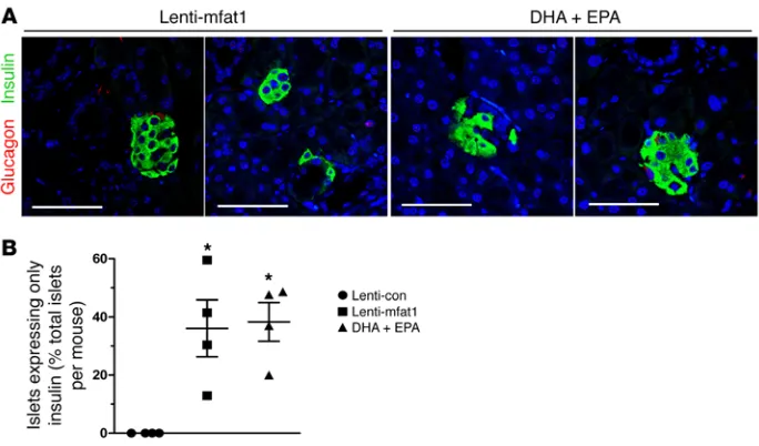
Therapeutic supplementation of ω-3 PUFAs promotes islet and
β cell regeneration. At the end of 9 weeks of therapy with the
len-tiviral gene or the EPA/DHA-enriched diet, confocal imaging revealed newly formed insulin-staining islets with a diameter of at least 50 μm that were located adjacent to pancreatic ducts (Fig-ure 6, A and B). Importantly, lymphocyte infiltration of neopan-creatic islets in the lenti-mfat-1–treated or EPA/DHA-enriched dietary group was much lower compared with that observed in the lenti-con–treated groups (Figure 6, C and D), suggesting that both therapeutic approaches deterred the immune attack on the pancre-as. Interestingly, we found that a high percentage of regenerated islets (~40%) had essentially all β cells, with very few α cells (Fig-ure 7, A and B). Confocal imaging revealed multiple cells with both insulin and glucagon staining (indicated by yellow fluorescence) within the regenerated islets near pancreatic ducts after either interventional approach (Figure 8, A–E), which was indicative of transdifferentiation from α cells into β cells. Consistent with these observations, blood glucagon levels in the nonfasted ω-3 PUFA intervention group were much lower than those in the nontreated diabetic mice as well as in the lenti-con–treated NOD mice (Figure 9A). Also, compared with the nontreated diabetic and lenti-con– treated groups, the transcription levels of Pdx1, Pax4, and
arista-Figure 5.ω-3 PUFAs exert a therapeutic effect on hyperglycemia in diabetic NOD mice. (A) Nonfasting blood glucose levels in diabetic NOD mice (nonfasting blood glucose levels for 2 consecutive weeks = 11.1–20 mmol/l) after i.v. tail-vein injection of lenti-con (black, n = 7) or lenti-mfat-1 (green, n = 7), or DHA plus EPA dietary intervention (purple, n = 7). ***P < 0.0001 versus the lenti-con group (Student t test). (B and
[image:7.585.45.404.55.334.2]less-related homeobox (Arx) were drastically increased following treatment with either ω-3 PUFA interventional approach (Figure 9, B–D). Taken together, our data showed that the lenti-mfat-1 virus could regenerate pancreatic β cells, at least some of which came from transdifferentiated α cells.
Therapeutic supplementation of ω-3 PUFAs altered CD4+ T cell
differentiation profiles in diabetic NOD mice. Implementation of
either ω-3 PUFA dietary intervention had an inhibitory effect on the population of Th1 and Th17 cells but strongly promoted Th2 and Treg differentiation and activation (Figure 9, E–N and Supple-mental Figure 8). Interestingly, the stimulatory effect on Th2 cells achieved through the gene therapy method was even more effec-tive than that of dietary intervention. Thus, either ω-3 PUFA sup-plementation method could restore the differentiation profiles of CD4+ T cells and therefore stall the progression of autoimmunity.
Modulation of human CD4+ T cell differentiation by ω-3 PUFAs.
The data presented above led us to further explore whether ω-3 PUFAs have a similar impact on human CD4+ T cell
differentia-tion. Human peripheral blood mononuclear cells (PBMCs) were isolated from 4 T1D patients and 5 nondiabetic donors (patients’ information, including A1C and autoantibodies, is provided in Supplemental Table 7) and treated with 100 μM DHA, EPA, and AA, separately, for 24 hours. CD4+ T cell differentiation was
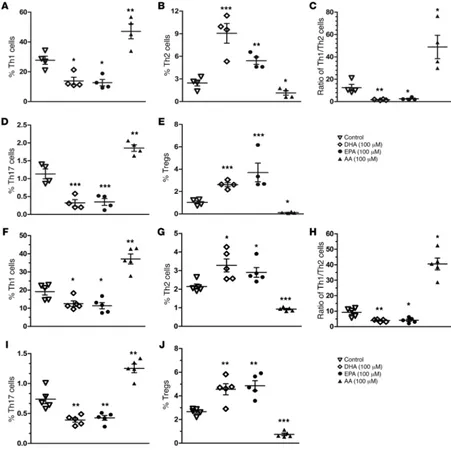
[image:8.585.76.522.54.430.2]ana-lyzed by flow cytometry. Consistent with the findings in NOD mice, we found that EPA and DHA essentially rebalanced the Th1/Th2 ratio from approximately 12 to approximately 1 by both increasing Th2 cell populations and decreasing Th1 cell popula-tions in the samples from T1D patients (Figure 10, A–E and Sup-plemental Figure 9). Meanwhile, we also observed a significant reduction of Th17 cells and an elevation of Tregs. In contrast, AA,
ed by B cells change dynamically or even become undetectable in some patients (43). In addition, the strong association between T1D and the HLA class II locus can trigger the activation of CD4+ T
cells (44). Thus, our studies have primarily focused on autoimmune responses driven by CD4+ T cells in NOD mice. Elevated
popula-tions of Th1 cells, the main effectors of diabetes in NOD mice, are a hallmark of T cell–mediated autoimmunity that in turn can further stimulate CD8+ T cells and macrophages to promote inflammatory
and immune attacks on pancreatic β cells (45). CD4+ T cells
prefer-entially differentiate into Th1 cells, with a concomitant decrease in Th2 cells in NOD mice, as indicated by the elevated ratio of IFN-γ/ IL-4 (46, 47). In addition to dysregulated Th1/Th2 ratios, T1D patients have an elevated percentage of activated Th17 cells and decreased numbers of Tregs. Specific anti–IL-17 antibodies have been shown to significantly increase the proportion of Tregs (48). Mesenchymal stem cell–based cellular therapy has already shown promise in elevating Tregs, which then suppress immune attack in a T1D model (49). In our study, supplementation of ω-3 PUFAs, provided as either a preventative or therapeutic modality, signifi-cantly suppressed Th1 and Th17 cells, reduced secreted IFN-γ
and IL-17 levels, and increased Th2 cells and Tregs. Meanwhile, the regulatory effects of ω-3 and ω-6 PUFAs on IFN-γ secretion by CD8+ T cells were consistent but somewhat minor when
com-pared with the effects on IFN-γ secretion by CD4+ T cells, and IL-4
secretion by CD8+ T cells was undetectable. Thus, CD8+ T cells
par-ticipate in the pathogenesis of T1D mainly through their cytotoxic effects on islet cells, but not through cytokine secretion (50, 51). In further extending this concept to application in humans, we found that DHA and EPA had the same effects on the differentiation of CD4+ T cells isolated from T1D donors as those observed in NOD
mice. Thus, ω-3 PUFA–initiated global changes in CD4+ T cells are
likely the primary cellular mechanisms underlying the blockade of autoimmune development.
Multiple mechanisms may have contributed to the effects of
ω-3 PUFAs on CD4+ T cell differentiation. EPA/DHA-derived
eico-sanoids, synthesized through the activities of enzymes such as LOX and COX, tend to be antiinflammatory, whereas those derived from
ω-6 PUFAs are proinflammatory (37). For example, AA-derived an ω-6 PUFA generally considered to be proinflammatory, exerted
effects on CD4+ T cell differentiation that were completely
oppo-site to those induced by EPA or DHA. We also obtained similar results in the T cell samples from nondiabetic donors (Figure 10, F–J and Supplemental Figure 10). To further confirm the impact of ω-3 and ω-6 PUFAs on human CD4+ T cells, we also used flow
cytometry to follow the expression of transcription factors such as T-bet, GATA3, and RORγT in human PBMCs following treat-ment with AA, DHA, or EPA. The results were clearly consistent with the staining panels of cytokines from the nondiabetic donors (Supplemental Figure 11).
Discussion
The deterrence of autoimmune attack and regeneration of pan-creatic β cells remain daunting tasks in the pursuit of a complete cure for T1D. The study described here suggested the clinical potential of gene therapy or nutritional supplementation of ω-3 PUFAs — EPA and DHA in particular — in preventing and revers-ing the development of autoimmunity and T1D. Both approaches alleviated T cell–mediated autoimmunity by rebalancing the ratio of Th1 and Th2 cells, decreasing Th17 cells, and increasing Tregs, while reducing circulating levels of inflammatory cytokines. Importantly, we observed a full recovery of pancreatic islet func-tion and a near-complete absence of any lymphocyte infiltrafunc-tion into regenerated islets in the diseased NOD mice following the nutritional or gene therapeutic intervention. To our knowledge, this represents the first report of a single treatment method that can both stall autoimmunity and, concomitantly, fully restore pancreatic β cell function. Three recent clinical trials in inde-pendent cohorts have convincingly demonstrated that long-term dietary intake of ω-3 PUFAs, starting from infancy, could signifi-cantly decrease the incidence of islet autoimmunity and T1D (22– 24). The results from our studies are entirely consistent with the findings revealed in these clinical studies.
A targeted autoimmune attack on islets involves dysregulation and dysfunction of both T cells and B cells (16). T cell–mediated immunity is primarily responsible for the direct damage of β cells and inflammatory injury (16). The titers of autoantibodies
secret-Figure 7.Islet and β cell regeneration in diabetic NOD mice treated with ω-3 PUFAs. Pancreases were harvested from 9-week-old mice that had received lentivirus treatment and DHA plus EPA dietary intervention. Confocal images (A) and quantification (B) of islets expressing only insulin, without α
[image:9.585.37.379.55.256.2]molecules such as CD28 (53, 54). T cell–specific mutation of mTOR suppresses differentiation of Th17 cells (54) and promotes naive CD4+ T cell differentiation into Tregs (54). In our studies, EPA/
DHA strongly inhibited the activation of mTORC1 by AA. Similar to the effect of EPA and DHA, rapamycin also blocked the Th1 dif-ferentiation induced by AA. Such results are also reminiscent of the anti-diabetes and autoimmunity prevention effects of rapamycin in NOD mice that were reported by several groups (55–59).
Increased intake of ω-6 PUFAs has been found to amplify immune response and the inflammatory events in mice with aller-gic diseases (60). Moreover, oleic acid and linoleic acid diets have been reported to elevate the development of autoimmunity and intestinal inflammation in mice (61, 62). Although the conclusions from these studies were generally consistent with our findings, one study also found the beneficial effect of an ω-6 PUFA–rich diet in alleviating inflammation in drug-induced mucosal damage (63). Thus, the effects of ω-6 PUFAs on inflammatory diseases should be carefully considered in different immune disease models. eicosanoids play a role in the development of arthritis, lupus, and
asthma (52). Some of the eicosanoids that showed drastic chang-es in our metabolomics analysis of pancreatic samplchang-es played dis-tinct roles in CD4+ T cell differentiation. For example, PGD3, an
EPA-derived metabolite, had a strong inhibitory effect on Th1 and Th17 cell differentiation and elevated Th2 and Treg populations. On the other hand, 17,18-DiHETE, another EPA-derived metab-olite, decreased only Th17 cell populations. RvD1, a DHA-derived metabolite, had a strong inhibitory effect on Th1 cell differentiation and strongly promoted Th2 and Treg differentiation. As one might expect, AA-derived proinflammatory eicosanoids, such as 15-HETE and 20-HETE, had effects on naive CD4+ T cell differentiation that
were exactly opposite to those of EPA/DHA-derived metabolites, further confirming the specificity of the effects of these eicosanoids.
Regulation of mTOR activity plays a critical role in the differ-entiation of CD4+ T cells (40). Application of rapamycin can inhibit
[image:10.585.104.486.56.427.2]the proliferation of T cells and the differentiation into Th1 and Th17 cells, even in the presence of T cell receptor (TCR) costimulatory
cyte infiltration into regenerated islets. Second, both interventions regenerated pancreatic islets and fully restored blood insulin lev-els. In the absence of a meaningful number of preexisting β cells (as is the case in diabetic NOD mice), neogenesis can be achieved via either differentiation of progenitors within the ductal epitheli-um (64) or transdifferentiation of acinar (65) or α cells (66) into β
cells. Although the underlying mechanisms remain to be defined, neogenesis is probably the primary event, as we found expression levels of Pdx1 (67) and Pax4 (68) to be sharply elevated. We also Arguably, the most striking finding from this study is the
reversal of autoimmune development and stable normalization of blood glucose levels following gene therapeutic or nutritional supplementation of ω-3 PUFAs in diabetic NOD mice. We believe these findings have strong clinical implications for the treatment of T1D. First, both intervention methods successfully readjusted the differentiation of CD4+ T cells, including the rebalancing of
the Th1/Th2 ratio, the reduction of Th17 cells, and the elevation of Treg numbers, thus causing a nearly complete absence of
lympho-Figure 9.β Cell regeneration andmodulation of Th cell subsets after lentivirus and dietary therapy with ω-3 PUFAs in diabetic NOD mice. Mice were sacrificed at 9 weeks of age following lentivirus treatment and DHA plus EPA dietary intervention, and pancreases were harvested. (A) Concentrations of nonfasting serum glucagon levels in nondiabetic mice; diabetic NOD mice (nonfasting blood glucose level for 2 consecutive weeks <20 mmol/l) before treatment; and diabetic NOD mice (nonfasting blood glucose level for 2 consecutive weeks >11.1 mmol/l) after ω-3 PUFA therapy (n = 5–7/group). *P < 0.05, **P < 0.01, and ***P < 0.0001 versus the nondiabetic group (Student’s t test). Data are representative of 2 independent experiments. (B–D) mRNA expression of Pdx1, Pax4, and Arx measured by RT-PCR in pancreases from NOD mice that received ω-3 PUFA therapy. *P < 0.05, **P < 0.01, and ***P < 0.0001 compared with the lenti-con group (n = 3 per group) (Student’s t test). Data are representative of 3 independent experiments. (E–N) Quan-tification (n = 4–10 per group) of the percentage of intracellular staining of IFN-γ+, IL-4+, IL-7+, and CD25+FoxP3+ Th cells in LNs and spleens of diabetic NOD
[image:11.585.46.532.51.498.2]with T1D (69, 70). Part of this failure may reside in the complexi-ty of the pathogenesis of T1D. On this note, we were encouraged to observe the same effects of EPA/DHA on human CD4+ T cells
as those on murine T cells. In addition, the results from our stud-ies are entirely consistent with the conclusions derived from the DAISY and Norwegian clinical studies (22, 23).
In this study, the daily intake of EPA/DHA applied to NOD mice was approximately 3.6 g/kg BW, which is high by clinical standards. Such a dose could very well be a saturating amount in these mice, and a dose well below this level may only be needed to achieve a thera-peutic effect. We do not yet know whether the same dose for mice discovered the phenomenon of α cell transdifferentiation, as a
sig-nificant percentage of newly generated islets often had very few
α cells and glucagon/insulin double staining following the thera-peutic interventions. For reasons that are not clear, we were only able to rescue a subset of diabetic mice with our therapeutic inter-ventions, particularly those mice without overt conditions (non-fasting blood glucose ≤20 mmol/l and only mild ketoacidosis), suggesting an irreversible damage to the neogenesis mechanisms after prolonged exposure to the severe DKA condition.
[image:12.585.73.524.57.508.2]Most of the preventative and therapeutic modalities devel-oped in NOD mice have not been successfully realized in humans
Figure 10.ω-3 and ω-6 PUFAs readjust CD4+ T cell differentiation in PBMCs from T1D patients and nondiabetic donors in vitro. Quantification of the
percentage of intracellular staining of IFN-γ+, IL-4+, IL-7+, and CD25+FoxP3+ Th cells in PBMCs from 4 T1D patients (A–E) and 5 nondiabetic donors (F–J).
glucometer (Accu-Chek; Roche). For insulin tolerance tests, mice were fasted for a 4-hour period during the light cycle before i.p. injec-tion of regular human insulin (0.8 U/kg; Novo Nordisk) diluted in ster-ile saline. Blood glucose levels were measured before insulin admin-istration and then every 15 minutes afterwards, for 60 minutes (26).
Histological studies and insulitis scoring. After the mice were
sac-rificed, pancreases were harvested and fixed with 4% paraformal-dehyde overnight at 4°C. The pancreases were paraffin embedded and sectioned (4 μm thickness). To assess insulitis, islets from H& E-stained sections were scored using standard methods and placed into 1 of 4 categories: 1) no insulitis; 2) peri-insulitis (leukocytes in the periphery of the islet); 3) invasive insulitis (25%–50% coverage of the islet); and 4) severe insulitis (>50% infiltration). Paraffin sections of mouse pancreases were collected at intervals of 100 μM. Mouse islets (20–50 per pancreata) were scored for the evaluation of insulitis. This procedure was used for 1 paraffin cross section of pancreatic tissue placed on individual slides from each of the 7 animals included in the histopathological study, and all slides were evaluated by a pathologist and scored for degree of perivascular and islet-associated monocyte infiltration. In addition, for each section included, a standard H& E-stained slide was prepared and submitted for pathological review.
Confocal microscopy and immunofluorescence. For
immunofluores-cence staining, 5-μm-thick paraffin sections were assayed with DAPI as a counterstain. The primary antibodies used were: rabbit anti-mouse insulin (1:100; Cell Signaling Technology) and monoclonal anti-mouse glucagon (1:1,000; Sigma-Aldrich). The secondary antibodies (1:1,000; Molecular Probes, Thermo Fisher Scientific) used were: Alexa Fluor 555 donkey anti-mouse and Alexa Fluor 488 donkey anti-rabbit. Sec-tions were examined under a Zeiss LSM 710 confocal microscope.
Serum analysis. Blood was collected when the NOD mice were
sacrificed. Serum insulin levels were measured using an insulin ELISA Kit (EMD Millipore). Plasma glucagon and BHOB levels were measured using an ELISA Kit (Mercodia) and a Colorimetric Assay Kit (Cayman Chemical).
Human PBMC isolation and flow cytometry. Human blood samples
were obtained from T1D donors. The resulting PBMCs were prepared by differential density gradient separation (Lymphoprep; AXIS-SHIELD). The following anti-human surface antibodies were purchased from BD Biosciences: CD3 (UCHT1) and CD8 (RPA-T8). The following anti-mouse surface antibodies were purchased: CD3 (145-2C11; BD Biosci-ences) and CD8 (53-6.7; eBioscience). For intracellular cytokine FACS, we stimulated cells with PMA (50 ng/ml; Sigma-Aldrich); ionomycin (500 ng/ml; Sigma-Aldrich); and brefeldin A (5 μg/ml; Sigma-Aldrich) for 5 hours. For the detection of CD4+ T cells, CD3+ cells were gated as
whole T lymphocytes with CD3-PerCP Cy5.5 mAb. In this gated CD3+
cell population, CD8 was labeled with CD8-FITC mAb. The CD8– cells
were selected as the targeted CD4+ T cells (see Figure 4C and
Supple-mental Figures 2, 3, and 5 and 7–11). Cells were fixed and permeabi-lized with Fixation/Permeabilization Diluent (eBioscience) and then stained for IFN-γ, IL-4, and IL-17 (Miltenyi Biotec for mouse samples; BD Biosciences for human samples). For Treg staining, we used human and mouse Treg-staining kits (catalogs 88-8999 and 88-8111; eBiosci-ence) according to the manufacturer’s protocol. Cells (105 per sample)
were collected on an Accuri-C6 (BD Biosciences) and analyzed using FlowJo software (Tree Star). Each test was repeated 3 times. Analysis of mouse Th1, Th2, and Th17 cytokines in culture supernatants was per-formed with a BD Cytometric Bead Array.
can simply be mathematically translated into a dose for humans to achieve the same results, given the very different metabolic profiles of mice and humans. However, the DAISY study showed that daily supplementation of a mere 150-mg dose of EPA/DHA in infants would have a strong preventative effect in children (22). For infants, starting at 1 year of age, the Norwegian study suggested that daily supplementation (1.0 g DHA/EPA) could reduce their risk of devel-oping T1D (23). These EPA/DHA levels were far below those applied here in the NOD mice, further raising the hope of using such an approach to alleviate T1D in the clinic. Unlike the nutritional supple-mentation approach, gene therapy led not only to an increase of ω-3 PUFAs but also a significant decrease of ω-6 PUFAs, particularly the proinflammatory AA and its derivatives, which might have also con-tributed to the suppression of autoimmunity and the restoration of blood glucose levels. The dose of EPA/DHA needed to reverse T1D in the clinic will require a very thorough trial.
Our observations may also offer clinical guidance, in that those patients who are either at the early-onset stage of T1D or have con-sistently had good management of their blood glucose levels may benefit the most from these interventions. These treatment modal-ities, if cleared in safety evaluations, may potentially be helpful in the treatment of other types of autoimmune diseases as well.
Methods
Mice and diets. The NOD (J001976) mouse colony, which was the same
colony used in most of the other published studies (71–73), was pur-chased from The Jackson Laboratory and maintained in a strict specif-ic pathogen–free (SPF) barrier facility at the Model Animal Research Center of Nanjing University. Both male and female NOD mice were tested regularly (at least every 3 months) for microbiological screening when housed at the barrier facility. All microbial screening test results were negative. Mice had ad libitum access to water and food. The 3 groups of mice in this study were fed a semi-purified control diet or the same control diet supplemented with either 10% AA or 10% EPA/DHA (by weight). The diets met National Research Council (NRC) nutrition requirements for mice and varied only in PUFA lipid content (74). The source of ω-3 PUFAs was fish oil (ShangHai HOPE Industry Co., Ltd.). Analysis of other fatty acids and vitamine D3 is shown in Supplemental Table 8. The SPF-level regular mouse diet and the PUFA-enriched diet were manufactured by XIETONG Bioengineering Co., Ltd. Details of the sources of protein, other fats, and other additives are provided in Supplemental Table 9. The distribution of fatty acid species in the fish oil and DHA/EPA diets was analyzed by gas chromatography–mass spectrometry (GC-MS) and is detailed in Supplemental Table 8. All PUFA-enriched diets were vacuum sealed and stored at –20°C before thawing. The PUFA-enriched diets were replenished for the mice every 48 hours. The same lot of PUFA-enriched diets was used throughout this study. Diabetes was confirmed by the presence of blood glucose concentrations above 11.11 mmol/l for 2 consecutive weeks, with the first week of hyperglycemia considered the age of disease onset.
Glucose tolerance tests and insulin tolerance tests. All 3 groups of mice
olites were quantified using a 5500 QTRAP hybrid triple quadrupole linear ion trap mass spectrometer (SCIEX) equipped with a turbo ion spray electrospray ionization (ESI) source.
Statistics. All data are presented as the mean ± SEM. P values
were calculated using a 2-tailed Student’s t test or 1-way ANOVA with a Tukey’s post-hoc multiple comparisons test. A P value of less than 0.05 was considered statistically significant. Survival plot analyses were used to evaluate the difference in the incidence of diabetes onset between the different groups of NOD mice, with the differences being determined using a Mantel-Cox log-rank test. All statistic analyses (Student’s t tests, ANOVA, and survival analyses) were performed using GraphPad Prism 5.0 software (GraphPad Software).
Study approval. All experimental protocols were approved by the
Research Ethics Committee of Nanjing Medical University, and all experiments were conducted in compliance with guidelines for the care and use of laboratory animals and approved by the IACUC of Nanjing Medical University. PBMCs from T1D patients and nondia-betic donors were provided with prior informed consent, and blood samples were collected with IRB approval of the First Affiliated Hospi-tal of Nanjing Medical University.
Author contributions
XB, SL, XZ, YD, and XL designed and performed experiments, analyzed data, and contributed to the writing of the first draft of the manuscript. FL and YJ supervised the histopathological stud-ies and revised the manuscript. XB and TY recruited patients and performed experiments with human PBMCs. AZZ designed experiments, supervised the work, and wrote and revised the manuscript. All authors reviewed the manuscript.
Acknowledgments
We thank Yu Jiang (Department of Pharmacology and Chemical Biology, University of Pittsburgh School of Medicine, Pittsburgh, Pennsylvania, USA) and Xiaochun Bai (Department of Cell Biolo-gy, School of Basic Medical Sciences, Southern Medical Universi-ty, Guangzhou, China) for their helpful scientific discussions and technical assistance. This work was supported by grants from the National Program on Key Basic Research Project of China (973 Program, 2013CB945202, to AZZ and FL); the National Natural Science Foundation of China (NSFC) (81170780 and 81630021, to AZZ; 81372798, to FL; 81200570, to XL); the Natural Science Foundation of Jiangsu Province of China (JSNFC) (BK20130059, to AZZ; 2011766, to XL); and the Key University Science Research Project of Jiangsu Province (12KJB310006, to XL).
Address correspondence to: Allan Zijian Zhao, No. 100 Waihuan Xi Road, Guangzhou Higher Education Mega Center, Panyu Dis-trict, Guangzhou, Peoples Republic of China, 510006. Phone: 86.18027330698; E-mail: azzhao@gdut.edu.cn. Or to: Xiaoxi Li, 101 Longmian Avenue, Jiangning District, Nanjing 211166, Peoples Republic of China. Phone: 86.17715230828; E-mail: lxx@njmu.edu.cn.
CD4+ T cell isolation, cell culture, and Western blot analysis. Naive CD4+ T cells from spleens were obtained by negative selection using the
Miltenyi Biotec CD4+ Purification Kit II, according to the
manufactur-er’s recommendations. RPMI 1640 (Gibco, Thermo Fisher Scientific) supplemented with 10% FBS (HyClone; GE Healthcare), 50 mM β -mer-captoethanol, 100 U/ml penicillin, 100 mg/ml streptomycin, nonessen-tial amino acids, 1 mM sodium pyruvate, and 10 mM HEPES was used as a complete culture medium. Magnetic-activated cell sorter–purified (MACS-purified) CD4+ T cells (purity >96%) were cultured in 12-well
plates (2.0 × 10⁶ cells per well) and stimulated with 2 μg/ml soluble
anti-CD3 Ab and 2 μg/ml soluble anti-CD28 Ab (both from BD Biosciences). For each analysis, 8 × 10⁶ CD4+ T cells were lysed and the aliquots
ana-lyzed by Western blotting using phospho-specific Abs or total protein (Cell Signaling Technology), with β-actin (Sigma-Aldrich) as the control.
RNA isolation, cDNA synthesis, and real-time PCR. RNA from NOD
mice pancreas was prepared using an RNeasy Micro Kit (QIAGEN). One microgram of RNA was used for the preparation of single-stranded cDNA using a QuantiTect Reverse Transcription Kit (QIAGEN). Real-time PCRs were performed using the QuantiNova SYBR Green PCR Kit (QIAGEN) on Applied Biosystems StepOne equipment (Applied Bio-systems). Reactions were performed at least in triplicate, and specifici-ty of the amplified products was determined by melting peak analysis. Quantification for each gene of interest was performed using the 2–ΔΔCt
method. Quantified values were normalized against the housekeeping gene Actb. The primers used are listed in Supplemental Table 10.
Gas chromatographic analysis of fatty acids. Lipids were extracted from
mouse food and whole-blood samples collected from mouse tail veins according to the method of Bligh and Dyer (75). Gas chromatography was performed on a Model 6890 N Network Gas Chromatograph (Agilent Technologies). Identification of components was done by comparison of retention times with those of authentic standards (Sigma-Aldrich).
Lentiviral vector design, production, and administration. In this
study, the lentiviral transfer vector pLJM1-EGFP (Addgene) containing mfat-1 cDNA encoding a ω-3 fatty acid desaturase under the control of the CMV promoter was designed on the basis of a design that was previously detailed and proven successful for converting ω-6 PUFAs into the corresponding ω-3 forms in transgenic mice (25). Vesicular stomatitis virus glycoprotein–pseudotyped lentiviral supernatant was produced by transient transfection of HEK293FT cells (Life Technol-ogies, Thermo Fisher Scientific) with the 3-plasmid system, including the lentiviral transfer vector pLJM1-EGFP, and the packaging plas-mids psPAX2 and pMD2.G. The lentiviral vectors were produced in 293FT cells and concentrated by ultracentrifugation to 108 TU/ml,
as described previously (76). Transducing units per milliliter (TU/ml) were determined for each vector stock by assessing EGFP+ cells, using
limiting dilution on 293FT cells. Both lenti-mfat-1 and lenti-con at 109
TU/kg were injected into the tail vein of diabetic NOD mice.
Metabolomic analysis. Pancreatic tissues from female NOD
mice fed different PUFA-enriched diets were subject to eicosanoid extraction according to a previously reported method (77). Chromato-graphic separation involved an ACQUITY UPLC BEH C18 Column (Waters) consisting of ethylene-bridged hybrid particles. The
1. Wållberg M, Cooke A. Immune mecha-nisms in type 1 diabetes. Trends Immunol. 2013;34(12):583–591.
2. Atkinson MA, et al. How does type 1 diabetes
develop?: the notion of homicide or β-cell suicide revisited. Diabetes. 2011;60(5):1370–1379. 3. Panero F, et al. Fasting plasma C-peptide and
micro- and macrovascular complications in
a large clinic-based cohort of type 1 diabetic patients. Diabetes Care. 2009;32(2):301–305. 4. Sima AA, et al. C-peptide prevents and improves
BB/Wor rat. Diabetologia. 2001;44(7):889–897. 5. Johansson BL, Sjöberg S, Wahren J. The influence
of human C-peptide on renal function and glu-cose utilization in type 1 (insulin-dependent) dia-betic patients. Diabetologia. 1992;35(2):121–128. 6. Johansson BL, Linde B, Wahren J. Effects of
C-peptide on blood flow, capillary diffusion capacity and glucose utilization in the exercising forearm of type 1 (insulin-dependent) diabetic patients. Diabetologia. 1992;35(12):1151–1158. 7. Bhatt MP, Lim YC, Kim YM, Ha KS. C-peptide
activates AMPKα and prevents ROS-mediated mitochondrial fission and endothelial apoptosis in diabetes. Diabetes. 2013;62(11):3851–3862. 8. Shapiro AM, et al. Islet transplantation in seven
patients with type 1 diabetes mellitus using a glu-cocorticoid-free immunosuppressive regimen.
N Engl J Med. 2000;343(4):230–238.
9. Kroon E, et al. Pancreatic endoderm derived from human embryonic stem cells generates glucose-responsive insulin-secreting cells in vivo.
Nat Biotechnol. 2008;26(4):443–452.
10. Bach JF. Anti-CD3 antibodies for type 1 diabetes: beyond expectations. Lancet. 2011;378(9790):459–460.
11. Ludvigsson J, et al. GAD65 antigen therapy in recently diagnosed type 1 diabetes mellitus.
N Engl J Med. 2012;366(5):433–442.
12. Orban T, et al. Co-stimulation modulation with abatacept in patients with recent-onset type 1 diabetes: a randomised, double-blind, placebo- controlled trial. Lancet. 2011;378(9789):412–419. 13. Wherrett DK, et al. Antigen-based therapy with
glutamic acid decarboxylase (GAD) vaccine in patients with recent-onset type 1 diabe-tes: a randomised double-blind trial. Lancet. 2011;378(9788):319–327.
14. Shapiro AM, et al. International trial of the Edmonton protocol for islet transplantation.
N Engl J Med. 2006;355(13):1318–1330.
15. van Belle TL, Coppieters KT, von Herrath MG. Type 1 diabetes: etiology, immunology, and ther-apeutic strategies. Physiol Rev. 2011;91(1):79–118. 16. Lehuen A, Diana J, Zaccone P, Cooke A. Immune cell crosstalk in type 1 diabetes. Nat Rev Immunol. 2010;10(7):501–513.
17. Mosmann TR, Cherwinski H, Bond MW, Giedlin MA, Coffman RL. Two types of murine helper T cell clone. I. Definition according to profiles of lymphokine activities and secreted proteins.
J Immunol. 1986;136(7):2348–2357.
18. Abbas AK, Murphy KM, Sher A. Functional diversity of helper T lymphocytes. Nature. 1996;383(6603):787–793.
19. Stechova K, et al. High T-helper-1 cytokines but low T-helper-3 cytokines, inflammatory cyto-kines and chemocyto-kines in children with high risk of developing type 1 diabetes. Diabetes Metab Res
Rev. 2007;23(6):462–471.
20. Luger D, et al. Either a Th17 or a Th1 effector response can drive autoimmunity: conditions of disease induction affect dominant effector cate-gory. J Exp Med. 2008;205(4):799–810. 21. Tang Q, et al. Visualizing regulatory T cell control
of autoimmune responses in nonobese diabetic mice. Nat Immunol. 2006;7(1):83–92. 22. Norris JM, et al. Omega-3 polyunsaturated fatty
acid intake and islet autoimmunity in children
at increased risk for type 1 diabetes. JAMA. 2007;298(12):1420–1428.
23. Stene LC, Joner G, Norwegian Childhood Diabe-tes Study Group. Use of cod liver oil during the first year of life is associated with lower risk of childhood-onset type 1 diabetes: a large, popu-lation-based, case-control study. Am J Clin Nutr. 2003;78(6):1128–1134.
24. Chase HP, et al. Effect of docosahexaenoic acid supplementation on inflammatory cytokine lev-els in infants at high genetic risk for type 1 diabe-tes. Pediatr Diabediabe-tes. 2015;16(4):271–279. 25. Wei D, et al. Cellular production of n-3 PUFAs and
reduction of n-6-to-n-3 ratios in the pancreatic beta-cells and islets enhance insulin secretion and confer protection against cytokine-induced cell death. Diabetes. 2010;59(2):471–478. 26. Norquay LD, et al. Insulin receptor substrate-2 in
beta-cells decreases diabetes in nonobese diabet-ic mdiabet-ice. Endocrinology. 2009;150(10):4531–4540. 27. Ferris ST, Carrero JA, Mohan JF, Calderon B,
Murphy KM, Unanue ER. A minor subset of Batf3-dependent antigen-presenting cells in islets of Langerhans is essential for the devel-opment of autoimmune diabetes. Immunity. 2014;41(4):657–669.
28. Anderson MS, Bluestone JA. The NOD mouse: a model of immune dysregulation. Annu Rev
Immunol. 2005;23:447–485.
29. Fox CJ, Danska JS. Independent genetic reg-ulation of T-cell and antigen-presenting cell participation in autoimmune islet inflammation.
Diabetes. 1998;47(3):331–338.
30. Makhlouf L, et al. Depleting anti-CD4 monoclo-nal antibody cures new-onset diabetes, prevents recurrent autoimmune diabetes, and delays allograft rejection in nonobese diabetic mice.
Transplantation. 2004;77(7):990–997.
31. Pfleger C, Meierhoff G, Kolb H, Schloot NC, p520/521 Study Group. Association of T-cell reactivity with beta-cell function in recent onset type 1 diabetes patients. J Autoimmun. 2010;34(2):127–135.
32. Feuerer M, Shen Y, Littman DR, Benoist C, Mathis D. How punctual ablation of regulatory T cells unleashes an autoimmune lesion within the pancreatic islets. Immunity. 2009;31(4):654–664. 33. Lindley S, Dayan CM, Bishop A, Roep BO,
Peak-man M, Tree TI. Defective suppressor function in CD4(+)CD25(+) T-cells from patients with type 1 diabetes. Diabetes. 2005;54(1):92–99.
34. Park H, et al. A distinct lineage of CD4 T cells regulates tissue inflammation by producing inter-leukin 17. Nat Immunol. 2005;6(11):1133–1141. 35. Emamaullee JA, et al. Inhibition of Th17 cells regulates autoimmune diabetes in NOD mice.
Diabetes. 2009;58(6):1302–1311.
36. Weylandt KH, Chiu CY, Gomolka B, Waechter SF, Wiedenmann B. Omega-3 fatty acids and their lipid mediators: towards an understanding of resolvin and protectin formation. Prostaglandins
Other Lipid Mediat. 2012;97(3–4):73–82.
37. Schmitz G, Ecker J. The opposing effects of n-3 and n-6 fatty acids. Prog Lipid Res. 2008;47(2):147–155.
38. Chen Z, et al. mTORC1/2 targeted by n-3 poly-unsaturated fatty acids in the prevention of mammary tumorigenesis and tumor progression.
Oncogene. 2014;33(37):4548–4557.
39. Yang K, et al. T cell exit from quiescence and differentiation into Th2 cells depend on Raptor- mTORC1-mediated metabolic reprogramming.
Immunity. 2013;39(6):1043–1056.
40. Delgoffe GM, et al. The kinase mTOR regulates the differentiation of helper T cells through the selective activation of signaling by mTORC1 and mTORC2. Nat Immunol. 2011;12(4):295–303. 41. Chi H. Regulation and function of mTOR
signal-ling in T cell fate decisions. Nat Rev Immunol. 2012;12(5):325–338.
42. Spychalla JP, Kinney AJ, Browse J. Identification of an animal omega-3 fatty acid desaturase by heterologous expression in Arabidopsis. Proc Natl
Acad Sci U S A. 1997;94(4):1142–1147.
43. Steck AK, et al. Predictors of Progression From the Appearance of Islet Autoantibodies to Early Childhood Diabetes: The Environmental Deter-minants of Diabetes in the Young (TEDDY).
Diabetes Care. 2015;38(5):808–813.
44. Nepom GT. Class II antigens and disease suscep-tibility. Annu Rev Med. 1995;46:17–25.
45. Boitard C. T-lymphocyte recognition of beta cells in type 1 diabetes: clinical perspectives. Diabetes
Metab. 2013;39(6):459–466.
46. Charlton B, Lafferty KJ. The Th1/Th2 bal-ance in autoimmunity. Curr Opin Immunol. 1995;7(6):793–798.
47. Delovitch TL, Singh B. The nonobese diabetic mouse as a model of autoimmune diabetes: immune dysregulation gets the NOD. Immunity. 1997;7(6):727–738.
48. Ferraro A, et al. Expansion of Th17 cells and func-tional defects in T regulatory cells are key features of the pancreatic lymph nodes in patients with type 1 diabetes. Diabetes. 2011;60(11):2903–2913. 49. Vija L, et al. Mesenchymal stem cells: Stem cell
therapy perspectives for type 1 diabetes. Diabetes
Metab. 2009;35(2):85–93.
50. Skowera A, et al. β-cell-specific CD8 T cell phe-notype in type 1 diabetes reflects chronic autoan-tigen exposure. Diabetes. 2015;64(3):916–925. 51. Jörns A, et al. Islet infiltration, cytokine
expres-sion and beta cell death in the NOD mouse, BB rat, Komeda rat, LEW.1AR1-iddm rat and humans with type 1 diabetes. Diabetologia. 2014;57(3):512–521.
52. Patterson E, Wall R, Fitzgerald GF, Ross RP, Stanton C. Health implications of high dietary omega-6 polyunsaturated Fatty acids. J Nutr
Metab. 2012;2012:539426.
53. Lee K, et al. Mammalian target of rapamycin protein complex 2 regulates differentiation of Th1 and Th2 cell subsets via distinct signaling pathways. Immunity. 2010;32(6):743–753. 54. Delgoffe GM, et al. The mTOR kinase
differential-ly regulates effector and regulatory T cell lineage commitment. Immunity. 2009;30(6):832–844. 55. Baeder WL, Sredy J, Sehgal SN, Chang JY, Adams
LM. Rapamycin prevents the onset of insulin- dependent diabetes mellitus (IDDM) in NOD mice. Clin Exp Immunol. 1992;89(2):174–178. 56. Rabinovitch A, Suarez-Pinzon WL, Shapiro AM,
57. Battaglia M, et al. Induction of tolerance in type 1 diabetes via both CD4+CD25+ T regulatory cells and T regulatory type 1 cells. Diabetes. 2006;55(6):1571–1580.
58. Battaglia M, Stabilini A, Migliavacca B, Hore-js-Hoeck J, Kaupper T, Roncarolo MG. Rapamy-cin promotes expansion of functional CD4+ CD25+FOXP3+ regulatory T cells of both healthy subjects and type 1 diabetic patients.
J Immunol. 2006;177(12):8338–8347.
59. Monti P, et al. Rapamycin monotherapy in patients with type 1 diabetes modifies CD4+ CD25+FOXP3+ regulatory T-cells. Diabetes. 2008;57(9):2341–2347.
60. van den Elsen LW, van Esch BC, Dingjan GM, Hofman GA, Garssen J, Willemsen LE. Increased intake of vegetable oil rich in n-6 PUFA enhances allergic symptoms and prevents oral tolerance induction in whey-allergic mice. Br J Nutr. 2015;114(4):577–585.
61. Pestka JJ, Vines LL, Bates MA, He K, Langohr I. Comparative effects of n-3, n-6 and n-9 unsatu-rated fatty acid-rich diet consumption on lupus nephritis, autoantibody production and CD4+ T cell-related gene responses in the autoimmune NZBWF1 mouse. PLoS ONE. 2014;9(6):e100255. 62. Ghosh S, Molcan E, DeCoffe D, Dai C, Gibson
DL. Diets rich in n-6 PUFA induce intestinal microbial dysbiosis in aged mice. Br J Nutr. 2013;110(3):515–523.
63. Ueda T, et al. Beneficial effect of an omega-6 PUFA-rich diet in non-steroidal anti-inflam-matory drug-induced mucosal damage in the murine small intestine. World J Gastroenterol. 2015;21(1):177–186.
64. Xu X, et al. Beta cells can be generated from endogenous progenitors in injured adult mouse pancreas. Cell. 2008;132(2):197–207. 65. Minami K, et al. Lineage tracing and
characteri-zation of insulin-secreting cells generated from adult pancreatic acinar cells. Proc Natl Acad Sci
U S A. 2005;102(42):15116–15121.
66. Thorel F, et al. Conversion of adult pancreatic alpha-cells to beta-cells after extreme beta-cell loss. Nature. 2010;464(7292):1149–1154. 67. Jonsson J, Carlsson L, Edlund T, Edlund
H. Insulin-promoter-factor 1 is required for pancreas development in mice. Nature. 1994;371(6498):606–609.
68. Sosa-Pineda B, Chowdhury K, Torres M, Oliver G, Gruss P. The Pax4 gene is essential for differentiation of insulin-producing beta cells in the mammalian pancreas. Nature. 1997;386(6623):399–402.
69. von Herrath M, Nepom GT. Animal mod-els of human type 1 diabetes. Nat Immunol. 2009;10(2):129–132.
70. Gill RG, et al. A preclinical consortium approach for assessing the efficacy of combined anti-CD3 plus IL-1 blockade in reversing new-onset
autoimmune diabetes in NOD mice. Diabetes. 2016;65(5):1310–1316.
71. Zhang L, et al. Monoclonal antibody blocking the recognition of an insulin peptide-MHC complex modulates type 1 diabetes. Proc Natl Acad Sci
U S A. 2014;111(7):2656–2661.
72. Karumuthil-Melethil S, Gudi R, Johnson BM, Perez N, Vasu C. Fungal β-glucan, a Dectin-1 ligand, promotes protection from type 1 diabetes by inducing regulatory innate immune response.
J Immunol. 2014;193(7):3308–3321.
73. Ablamunits V, et al. Synergistic reversal of type 1 diabetes in NOD mice with anti-CD3 and inter-leukin-1 blockade: evidence of improved immune regulation. Diabetes. 2012;61(1):145–154. 74. National Research Council (US)
Subcommit-tee on Laboratory Animal Nutrition. Nutrient Requirements of Laboratory Animals: Fourth Revised Edition, 1995. Washington (DC): National Academies Press (US); 1995. 75. Bligh EG, Dyer WJ. A rapid method of total lipid
extraction and purification. Can J Biochem
Physi-ol. 1959;37(8):911–917.
76. Tiscornia G, Singer O, Verma IM. Production and purification of lentiviral vectors. Nat Protoc. 2006;1(1):241–245.