Original Article
MiR-506-3p inhibits proliferation and invasion
by targeting EZH2 in glioblastomas
Yanyao Deng1, Le Xiao1, Chao Liu1, Yuan Li1, Ziqing Guo1, Bing Xie1, Ziqi Jin1, Zhicheng Lv2, Hongwei Zhu3,
Aimin Wang1
1Department of Neurology, The First Hospital of Changsha, Changsha, Hunan, China; 2Department of
Neurosurgery, The First Hospital of Chenzhou, Chenzhou, Hunan, China; 3Department of Gastroenterology, The Third Xiangya Hospital, Central South University, Changsha, Hunan, China
Received August 9, 2018; Accepted October 8, 2018; Epub April 15, 2019; Published April 30, 2019
Abstract: Background: Increasing evidence has indicated that microRNAs (miRNAs) play an important role in glio-blastoma cancer. Deregulation of miR-506-3p has been reported in several cancers. However, the expression and function of miR-506-3p in glioblastomas remain unclear. This study aimed to investigate the roles and underlying mechanisms of miR-506-3p in proliferation and invasion of glioblastoma cancer. Methods: Quantitative real-time PCR (qRT-PCR) and Western blot techniques were used to assess expression of miR-506-3p and enhancer of zeste homolog 2 (EZH2) in glioblastoma cell lines and tissues. Luciferase reporter assays were conducted to investigate the association between miR-506-3p and EZH2. MTT and Transwell invasion assays were performed to evaluate the effects of miR-506-3p on proliferation and invasion, respectively, in glioblastoma cells. Results: Data showed that levels of miR-506-3p were downregulated in glioblastoma tissues and cell lines. Overexpression of miR-506-3p repressed cell growth and suppressed cell invasion in glioblastoma cells, whereas knockdown of miR-506-3p pro-moted cell growth and increased cell invasion in glioblastoma cells. Moreover, EZH2 was a direct target of miR-506-3p in glioblastoma cells. Enforced expression of EZH2 and suppression of EZH2 alleviated effects of miR-506-miR-506-3p mimics and inhibitors on proliferation and invasion in vitro, respectively. Conclusion: Present results indicate that miR-506-3p plays a tumor suppressor gene role in human glioblastomas by regulating EZH2 genes.
Keywords: MiR-506-3p, glioblastoma, proliferation, invasion, EZH2
Introduction
Glioblastomas, derived from glial cells, have become the most common and most malig- nant tumors in China [1]. Despite multimodal therapies, such as surgery, radiation and me- dical therapies for treatment of glioblastoma, the average life expectancy for glioblastoma patients remains limited [2, 3]. Therefore, it is essential to find vital carcinogenic biomarkers and effective therapeutic strategies for gliobla- stomas.
MicroRNAs (miRNAs) are endogenous noncod-ing, 19-25 nucleotides RNAs, that negatively regulate a variety of genes expression by bind-ing to the 3’-untranslated region (UTR) of their target mRNAs involved in several cancers [4- 6]. Accumulating reports have suggested that miRNAs play important roles in many diverse
biological processes, including cell prolifera-tion, migraprolifera-tion, invasion, differentiaprolifera-tion, and apoptosis [7]. MiRNAs have been reported to serve a tumor-suppressor or oncogenic func-tion by targeting tumor-suppressor or onco-genes onco-genes, respectively [8-10]. It is notewor-thy that miRNAs have been implicated in tumor initiation and progression of glioblastomas [11, 12].
Materials and methods
Clinical samples and cell culture
Fresh glioblastoma tissues and adjacent nor-mal tissues were collected in the Department of Neurosurgery, the First Hospital of Chen- zhou. Histological features of all specimens were diagnosis by pathologists, according to WHO criteria. None of these patients received chemotherapy or radiotherapy before surgery. Normal human astrocytes (NHA) were pur-chased from ScienCell Research Laboratories (Corte Del Cedro Carlsbad, Canada) and cul-tured, according to manufacturer instructions. Human A172, LN229, U87, and U251 glioblas-toma cell lines were obtained from ATCC. All cells were maintained cultured in DMEM (Hy- clone, Logan, Utah, USA), supplemented with 10% fetal bovine serum (Hyclone) and incubat-ed in a humidifiincubat-ed atmosphere containing 5% CO2 at 37°C without antibiotics.
Quantitative miRNA and mRNA measurements
Total RNA was extracted using TRIzol Reagent (Ambion), according to manufacturer protocol. cDNA used to measure EZH2 were synthesized using PrimeScriptTM RT reagent kit (TaKaRa), according to manufacturer protocol. cDNA used to measure miR-506-3p were synthesized us- ing miRcute miRNA cDNA first strand synthesis kit (TIANGEN, Beijing), according to manufac-turer protocol. Expression of EZH2 was mea-sured using SYBR® Premix Ex TaqTM II (TaKa-
Ra) and GAPDH served as an internal referen- ce. Expression of miR-506-3p was measured using miRcute miRNA qPCR detection kit (TIAN- GEN, Beijing) and U6 served as an internal ref-erence. All experiments were performed in trip-licate. Results are represented as fold induc-tion using the 2-ΔΔCt method. Primers of
miR-transiently transfected at 70-80% confluence using the LipofectamineTM 2000 reagent (In- vitrogen, CA, USA), according to manufacturer instructions.
MTT assay
Cell proliferative impact was measured by MTT (3-(4,5-Dimethylthiazol-2-yl)-2,5-diphenyltetra- zolium bromide) assay. Cells were plated on 96-well plates (2-5×10³ cells/well). At
designat-ed time points, cells were incubatdesignat-ed with 2 mg/ mL of MTT solution for 2 hours. The resulting formazan product was dissolved in DMSO for O.D. measurement at 570 nm. The experiment was performed in triplicate.
Transwell invasion assay
Cell invasion was determined using 24-well Matrigel invasion chambers (Becton Dickin- son), according to manufacturer instructions. Cells (2×10⁴) were seeded per well in the up-
per well of the invasion chamber in DMEM with-out serum. The lower chamber well contained DMEM supplemented with 10% FBS to stimu-late cell invasion. After incubation for 48 hours, non-invading cells were removed from the top well with a cotton swab, while the bottom cells were stained with 0.05% crystal violet and pho-tographed in 5 independent fields for each well.
Western blotting analysis
[image:2.612.88.354.84.191.2]Total protein was extracted from cells. Cell ly- sates were prepared in lysis buffer and then separated by 10% SDS-PAGE (sodium dode- cyl sulfate-polyacrylamide gel Electrophoresis) and transferred to PVDF membranes (Millipore, Billerica, MA, USA). Membranes were blocked with 5% non-fat milk and then incubated over-night with the primary antibody to EZH2 (Cell
Table 1. Sequences for primers and siRNAs used in the study
Name Sequence
Primers for qRT-PCR
EZH2-F 5’-GACCTCTGTCTTACTTGTGGAGC-3’ EZH2-R 5’-CGTCAGATGGTGCCAGCAATAG-3’ GAPDH-F 5’-TTGGTATCGTGGAAGGACTCA-3’ GAPDH-R 5’-TGTCATCATATTTGGCAGGTT-3’ EZH2 siRNA 5’-AAGACTCTGAATGCAGTTGCTd(TT)-3’
5’-AGCAACUGCAUUCAGAGUCUUd(TT)-3’
F: forward primer, R: reverse primer.
506-3p and U6 were purchased from GenePharma (China). Primers used to detect expression of EZH2 are listed in Table 1.
miR-506-3p mimics, antisense, and transfection
Signaling Technology) or Actin (Santa Cruz Biotechnology, Santa Cruz, CA), followed by horseradish peroxidase-labeled secondary an- tibody incubation. Chemiluminescence signal was developed by ECL Plus Western Blotting Detection Reagents (GE Healthcare Life Sci- ences, Piscataway, NJ).
Luciferase activity assay
The EZH2 3’-UTR luciferase reporter construct was made by amplifying the EZH2 mRNA 3’- UTR sequence. Cells were co-transfected with pMIR/EZH2 vector or pMIR/EZH2/mut vector containing Firefly luciferase, along with 0.05 μg of the pRL-TK vector (Promega) containing Renilla luciferase and miR-506-3p mimic or scramble oligonucleotide. Luciferase activities were detected using the Dual-Luciferase Re- porter Assay System (Promega).
Statistical analysis
Statistical analysis was performed using SPSS 21.0 software (SPSS, USA). Data are expressed as the mean ± SD and differences between groups were analyzed using Student’s t test. Data indicates statistical significance when P < 0.05.
Results
Expression of miR-506-3p was downregulated in glioblastoma tissues and cell Lines
Expression of miR-506-3p was detected by qRT-PCR in 4 GBM cell lines (A172, LN229, U87, and U251) and NHAs. All 4 tested GBM
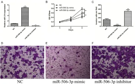
vitro. The efficiency of miR-506-3p mimics and inhibitors was confirmed by qRT-PCR. Compar- ed with the control miRNA, miR-506-3p expres-sion was significantly increased by the mimics while decreased by the inhibitors in U251 cell lines (Figure 2A).
MTT assay was performed to measure the effects of miR-506-3p on the proliferation of U251 cells. As shown in Figure 2B, miR-506-3p mimic transfection remarkably reduced the proliferation of U251 cells, compared with the control group. In contrast, the proliferation capacity of U251 cell lines was significantly enhanced by miR-506-3p repression induced by inhibitors. This study also confirmed the alteration of U251 cell invasion using Transwell assay. Results showed that the invasion of U251 cell lines was significantly reduced by miR-506-3p overexpression while enhanced by miR-506-3p silencing (Figure 2C). Present results indicate that low levels of endogenous miR-506-3p may play an important role in the development of glioblastomas by promoting proliferation and invasion.
EZH2 was a potential target of miR-506-3p in glioblastoma cells
[image:3.612.90.375.70.198.2]EZH2 has been considered a tumor oncogene in glioblastomas, according to previous studies [17, 18]. To further illuminate the underlying mechanisms of miR-506-3p regulating glio-blastoma cells, this study predicted the possi-ble targets of miR-506-3p using target predic-tion programs (TargetScan, PicTar, and miRan-da). Online analysis suggested that EZH2 was
Figure 1. Expression of miR-506-3p was downregulated in glioblastoma cell lines and tissues. A. Relative expression of miR-506-3p in glioblastoma cell lines (A172, LN229, U87, and U251) compared with the normal human as-trocytes cell line (NHAs). B. Comparison of the average expression levels of miR-506-3p between glioblastoma tissues and non-tumor tissues.
cell lines showed significantly lower miR-506-3p levels than those in the NHAs (Figure 1A). As shown in Figure 1B, miR-506-3p was downregulated in glioblastoma tissues, compar- ed with the normal tissues.
MiR-506-3p inhibits the pro-liferation and invasion in glio-blastoma cell lines
a potential direct target of miR-506-3p with a binding site in the 3’-UTR (Figure 3A). To further confirm whether this prediction was right,
lucif-erase reporter assay in U251 cells was per-formed. It was found that overexpression of miR-506-3p repressed the activity of
pMIR-Figure 3. EZH2 is a potential target of miR-506-3p in glioblastoma cells. A. Schematic representation of the putative binding sites in EZH2 mRNA 3’UTR for miR-506-3p. B. miR-506-3p mimic repressed luciferase activities controlled by wild-type EZH2-3’-UTR but did not affect luciferase activity controlled by mutant EZH2-3’-UTR. C. Relative mRNA expression levels of EZH2 were detected by real-time PCR. D. Western blot analysis was performed to evaluate ex-pression levels of EZH2 in the U251 cells, which was transfected with miR-506-3p mimic or scramble, respectively.
[image:4.612.89.520.72.341.2]β-actin was used as a loading control.
[image:4.612.92.522.414.593.2]WTEZH2-3’UTR plasmid in U251 cells, without changes in luciferase activity of pMIR-MUT- EZH2-3’UTR plasmid (Figure 3B). Moreover, miR-506-3p inhibited mRNA expression of EZ- H2 in U251 cells (Figure 3C). Ectopic expres-sion of miR-506-3p repressed protein levels of EZH2 in the U251 cells (Figure 3D). Present results indicate that miR-506-3p mediated re- gulation of EZH2 expression depended on its binding to a specific seed region in the EZH2 3’UTR.
Enforced expression of EZH2 alleviates effects of miR-506-3p on proliferation and invasion
The present study investigated whether overex-pression of EZH2 affects the role of miR-506-3p in U251 cells. pcDNA3.1-EZH2 was co-trans-fected with miR-506-3p, then the effects of proliferation and invasion of U251 cells were measured. pcDNA 3.1 empty vector served as control. Overexpression of EZH2 was confirmed by qRT-PCR and Western blot (Figure 4A, 4B). Present data showed U251 cells transfected with pcDNA3.1-EZH2 plus miR-506-3p mimics grew much faster (Figure 4C) and performed much stronger invasion (Figure 4D) ability than those transfected with pcDNA3.1 empty vector plus miR-506-3p mimics. Results indicate that
enforced expression of EZH2 alleviates effects of miR-506-3p on proliferation and invasion.
Silencing EZH2 alleviates effects of miR-506-3p inhibitor on proliferation and invasion
To further confirm the finding that EZH2 medi-ates the role of miR-506-3p in U251 cells, this study investigated if knockdown of EZH2 could affect the effects of miR-506-3p inhibitors on proliferation and invasion. Knockdown of EZH2 in U251 cells using siRNA targeted EZH2 was confirmed by qRT-PCR and Western blot (Figure 5A, 5B). Data showed that U251 cells, in which expression of EZH2 and miR-506-3p were both repressed, grew much slower (Figure 5C) and performed more weaker invasion (Figure 5D) ability than cells in which only expression of miR-506-3p was inhibited. Present results indi-cate that knockdown of EZH2 alleviates effects of miR-506-3p inhibitors on proliferation and invasion, supporting the hypothesis that miR-506-3p regulates proliferation and invasion of glioblastoma cells through regulating EZH2.
Discussion
Increasing evidence has suggested that miR-NAs play important roles in the development
and progression of tumors. The present study found that expression of miR-506-3p was downregulated in glioblastoma cell lines and tissues. Furthermore, in vitro experiments veri-fied that miR-506 inhibits the proliferation and invasion of glioblastoma cells. EZH2 was identi-fied as a direct target of miR-506-3p via the 3’UTR of EZH2. This study also confirmed that the miR-506-3p-EZH2 axis modulates prolifer-ation and invasion in glioblastoma cells. To date, the roles of miR-506-3p in cancer cells have not been well clarified. miR-506-3p plays contradictory roles in several cancers. In mela-noma, overexpression of miR-506-3p was criti-cal for promoting cancer growth, migration, and invasion [19], indicating that miR-506-3p ser- ves as an oncogene in melanomas. In contrast, in ovarian cancer, miR-506-3p, suppressing cell migration and invasion, was demonstrated as a key EMT inhibitor. In addition, miR-506-3p expression was positively relative with early FIGO stage and extended survival [20, 21]. Similar results have been found in cervical cer [22], liver cancer [23, 24], and breast can-cer [25], suggesting that miR-506-3p functions as a tumor suppressor gene in some tumors. The function of miR-506-3p in glioblastomas
remains poorly understood. In the present stu- dy, miR-506-3p was downregulated in glioblas-toma cell lines and tissues. Based on in vitro
experiments, it was verified that miR-506-3p serves as a tumor suppressor in glioblasto- mas.
MiRNAs typically perform their functions by re- pressing expression of target mRNAs. In ovari-an covari-ancer, miR-506-3p could inhibit prolifera-tion and promote senescence by directly target-ing the CDK4/6-FOXM1 axis [20]. Further stud-ies have shown that miR-506-3p could sup-press cervical cancer growth by directly target-ing the hedgehog pathway transcription factor Gli3 [22]. Moreover, recent profile studies dem-onstrated that miR-506-3p could regulate the biological behavior of cancer cells by targeting GATA6 and FLOT1 in oral squamous cell cancer [26] and renal cell cancer [27]. Present findings confirmed that miR-506-3p serves as a tumor suppressor in glioblastomas, but the underly- ing mechanisms remain still unclear. Therefore, TargetScan, PicTar, and miRanda databases were used to identify target genes of miR- 506-3p in glioblastomas. All three databases indicated that EZH2 may be a candidate miR-506-3p target gene. Moreover, previous
[image:6.612.92.519.71.308.2]ies have reported that EZH2 was a tumor onco-gene in several cancers [28, 29], including glio-blastomas [30, 31]. The present study conduct-ed a luciferase reporter assay to ensure whe- ther EZH2 is a direct target of miR-506-3p. Results suggested that regulation of EZH2 by miR-506-3p depended on its binding to the 3’UTR of EZH2. To establish whether the effects of miR-506-3p were exerted via direct inhibi-tion of EZH2, this study restored EZH2 expres-sion in miR-506-3p overexpressing cells and measured the proliferation and invasion of these cells. It was found that effects of prolif-eration and invasion were clearly increased. Moreover, this study also knock-downed EZH2 expression in miR-506-3p inhibiting cells and measured proliferation and invasion of these cells. It was found that effects of proliferation and invasion were clearly decreased. Results indicate that EZH2 is a mediator of miR-506- 3p function.
In conclusion, present results indicate that miR-506-3p, downregulated in glioblastoma cell lines and tissues, inhibits proliferation and invasion in vitro and that EZH2 is a di- rect target of miR-506-3p.
Acknowledgements
The study was supported by the Scientific Re- search Project of Health and Family Planning Commission of Hunan Province of China (B20- 17202).
Disclosure of conflict of interest
None.
Address correspondence to: Dr. Aimin Wang, De- partment of Neurology, The First Hospital of Ch- angsha, 311 Yinpan Road, Changsha 410005, Hunan, China. Tel: 17188613755012597; Fax: 073188618339; E-mail: 13755012597@163.com
References
[1] Chang L, Su J, Jia X and Ren H. Treating malig-nant glioma in chinese patients: update on te-mozolomide. Onco Targets Ther 2014; 7: 235-44.
[2] Kumthekar PU, Macrie BD, Singh SK, Kaur G, Chandler JP and Sejpal SV. A review of man-agement strategies of malignant gliomas in the elderly population. Am J Cancer Res 2014; 4: 436-44.
[3] Wang Y and Jiang T. Understanding high grade glioma: molecular mechanism, therapy and comprehensive management. Cancer Lett 2013; 331: 139-46.
[4] Ohdaira H, Sekiguchi M, Miyata K and Yoshida K. MicroRNA-494 suppresses cell proliferation and induces senescence in A549 lung cancer cells. Cell Prolif 2012; 45: 32-8.
[5] Wang LL, Wang L, Wang XY, Shang D, Yin SJ, Sun LL, Ji HB. MicroRNA-218 inhibits the prolif-eration, migration, and invasion and promotes apoptosis of gastric cancer cells by targeting LASP1. Tumour Biol 2016; 37: 15241-15252. [6] Yang S, Li Y, Gao J, Zhang T, Li S, Luo A, Chen
H, Ding F, Wang X, Liu Z. MicroRNA-34 sup-presses breast cancer invasion and metasta-sis by directly targeting Fra-1. Oncogene 2013; 32: 4294-303.
[7] Bushati N and Cohen SM. MicroRNA functions. Annu Rev Cell Dev Biol 2007; 23: 175-205. [8] Calin GA and Croce CM. MicroRNA signatures
in human cancers. Nat Rev Cancer 2006; 6: 857-66.
[9] Hwang HW and Mendell JT. MicroRNAs in cell proliferation, cell death, and tumorigenesis. Br J Cancer 2006; 94: 776-80.
[10] Volinia S, Calin GA, Liu CG, Ambs S, Cimmino A, Petrocca F, Visone R, Iorio M, Roldo C, Ferracin M, Prueitt RL, Yanaihara N, Lanza G, Scarpa A, Vecchione A, Negrini M, Harris CC, Croce CM. A microRNA expression signature of human solid
tumors defines cancer gene targets. Proc Natl
Acad Sci U S A 2006; 103: 2257-61.
[11] Ghasemi A, Fallah S and Ansari M. MicroRNA- 149 is epigenetically silenced tumor-suppres-sive microRNA, involved in cell proliferation and downregulation of AKT1 and cyclin D1 in human glioblastoma multiforme. Biochem Cell Biol 2016; 94: 569-576.
[12] Liu N, Zhang L, Wang Z, Cheng Y, Zhang P, Wang X, Wen W, Yang H, Liu H, Jin W, Zhang Y, Tu Y. MicroRNA-101 inhibits proliferation, mi-gration and invasion of human glioblastoma by targeting SOX9. Oncotarget 2017; 8: 19244-19254.
[13] Tong JL, Zhang CP, Nie F, Xu XT, Zhu MM, Xiao SD, Ran ZH. MicroRNA 506 regulates expres-sion of PPAR alpha in hydroxycamptothecin-re-sistant human colon cancer cells. FEBS Lett 2011; 585: 3560-8.
[14] Zhang Y, Lin C, Liao G, Liu S, Ding J, Tang F, Wang Z, Liang X, Li B, Wei Y, Huang Q, Li X, Tang B. MicroRNA-506 suppresses tumor pro-liferation and metastasis in colon cancer by directly targeting the oncogene EZH2. Onco- target 2015; 6: 32586-601.
[16] Deng J, Lei W, Xiang X, Zhang L, Yu F, Chen J, Feng M, Xiong J. MicroRNA-506 inhibits gastric cancer proliferation and invasion by directly targeting Yap1. Tumour Biol 2015; 36: 6823-31.
[17] Orzan F, Pellegatta S, Poliani PL, Pisati F, Caldera V, Menghi F, Kapetis D, Marras C, Schi- ffer D, Finocchiaro G. Enhancer of zeste 2 (EZH2) is up-regulated in malignant gliomas and in glioma stem-like cells. Neuropathol Appl Neurobiol 2011; 37: 381-94.
[18] Zhang Y, Yu X, Chen L, Zhang Z and Feng S. EZH2 overexpression is associated with poor prognosis in patients with glioma. Oncotarget 2017; 8: 565-573.
[19] Streicher KL, Zhu W, Lehmann KP, Georgantas RW, Morehouse CA, Brohawn P, Carrasco RA, Xiao Z, Tice DA, Higgs BW, Richman L, Jallal B, Ranade K, Yao Y. A novel oncogenic role for the miRNA-506-514 cluster in initiating mela-nocyte transformation and promoting melano-ma growth. Oncogene 2012; 31: 1558-70. [20] Liu G, Sun Y, Ji P, Li X, Cogdell D, Yang D, Parker
Kerrigan BC, Shmulevich I, Chen K, Sood AK, Xue F, Zhang W. MiR-506 suppresses prolifera-tion and induces senescence by directly target-ing the CDK4/6-FOXM1 axis in ovarian cancer. J Pathol 2014; 233: 308-18.
[21] Sun Y, Hu L, Zheng H, Bagnoli M, Guo Y, Rupai- moole R, Rodriguez-Aguayo C, Lopez-Berestein G, Ji P, Chen K, Sood AK, Mezzanzanica D, Liu J, Sun B, Zhang W. MiR-506 inhibits multiple targets in the epithelial-to-mesenchymal tran-sition network and is associated with good prognosis in epithelial ovarian cancer. J Pa- thol 2015; 235: 25-36.
[22] Wen SY, Lin Y, Yu YQ, Cao SJ, Zhang R, Yang XM, Li J, Zhang YL, Wang YH, Ma MZ, Sun WW, Lou XL, Wang JH, Teng YC, Zhang ZG. MiR-506 acts as a tumor suppressor by directly target-ing the hedgehog pathway transcription fac- tor Gli3 in human cervical cancer. Oncogene 2015; 34: 717-25.
[23] Dai W, Huang HL, Hu M, Wang SJ, He HJ, Chen NP, Li MY. MicroRNA-506 regulates prolifera-tion, migration and invasion in hepatocellular carcinoma by targeting F-spondin 1 (SPON1). Am J Cancer Res 2015; 5: 2697-707.
[24] Lu Z, Zhang W, Gao S, Jiang Q, Xiao Z, Ye L, Zhang X. MiR-506 suppresses liver cancer an-giogenesis through targeting sphingosine ki-nase 1 (SPHK1) mRNA. Biochem Biophys Res Commun 2015; 468: 8-13.
[25] Arora H, Qureshi R and Park WY. MiR-506 re- gulates epithelial mesenchymal transition in breast cancer cell lines. PLoS One 2013; 8: e64273.
[26] Deng L and Liu H. MicroRNA-506 suppresses growth and metastasis of oral squamous cell carcinoma via targeting GATA6. Int J Clin Exp Med 2015; 8: 1862-70.
[27] Yang FQ, Zhang HM, Chen SJ, Yan Y and Zheng JH. MiR-506 is down-regulated in clear cell renal cell carcinoma and inhibits cell growth and metastasis via targeting FLOT1. PLoS One 2015; 10: e0120258.
[28] Wang CG, Ye YJ, Yuan J, Liu FF, Zhang H and
Wang S. EZH2 and STAT6 expression profiles
are correlated with colorectal cancer stage and prognosis. World J Gastroenterol 2010; 16: 2421-7.
[29] Wang J, Yu L, Cai J, Jia J, Gao Y, Liang M, Wang Z. The role of EZH2 and DNA methylation in hMLH1 silencing in epithelial ovarian cancer. Biochem Biophys Res Commun 2013; 433: 470-6.
[30] Ahani N, Shirkoohi R, Rokouei M, Alipour Eskandani M and Nikravesh A. Overexpress- ion of enhancer of zeste human homolog 2 (EZH2) gene in human cytomegalovirus posi-tive glioblastoma multiforme tissues. Med Oncol 2014; 31: 252.