Abstract— Iris recognition algorithms, especially with the emergence of large-scale iris-based identification systems, must be tested for speed and accuracy and evaluated with a wide range of templates – large size, long-range, visible and different origins. This paper presents the acquisition of eye-iris images of dark-skinned subjects in Africa, a predominant case of very-dark-brown iris images, under near-infrared illumination. The peculiarity of these iris images is highlighted from the histogram and normal probability distribution of their grayscale image entropy (GiE) values, in comparison to Asian and Caucasian iris images. The acquisition of eye-images for the African iris dataset is ongoing and will be made publicly-available as soon as it is sufficiently populated.
Index Terms— Biometrics, Iris recognition systems, Iris dataset, Grayscale image entropy (GiE)
I.
INTRODUCTION
Human distinctive physiological and behavioural traits, called biometrics, have become profoundly useful in current identification systems. A lot of commercial systems with human-machine interface, access control, electronic voters’ and border control systems, deployed are enabled with fingerprint, iris, face, palm print, hand geometry or gait etc. Iris recognition is one of the most accurate means of personal identification. It has proved to be a fool-proof (or difficult to forge) biometric recognition technology because irises are highly distinctive; iris patterns of the eyes of the same individual and genetically identical twins are distinct [1].
The apparent richness of the iris textural patterns and improvement recorded with current algorithms has resulted in a lot of diverse applications, established in various research publications around the globe. This include improved recognition accuracy for large-scale iris-based identification systems[2], prediction of demographic attributes such as gender and ethnicity or relationships[3-8], development of user-friendly and less constrained commercial systems facilitated by long range and on-the-move acquisition[9-13].
Current publicly available iris datasets such as CASIA, ICE, UBIRIS etc, offer increased image template size with varying characteristics – short or long-range, near-infrared
Manuscript received July 19, 2012; revised August 12, 2012.
The authors are with the Electrical and Information Engineering Department of Covenant University, PMB1023 Ota, Nigeria (phone: 234-807-886-6304; e-mail: [email protected]).
or visible and different ethnic origins. This has greatly influenced the performance evaluation, especially in terms of speed and accuracy, of iris recognition and related algorithms. Notably, existing iris datasets consists majorly of Caucasian and Asians eye images captured under visible and near infrared lighting conditions. However, most dark-skinned individuals found in Africa, as compared to Caucasians or Asians, have very-dark-brown iris. There is a marked difference in the recognition rate of algorithms based on the use of irises of varying contrasts [14].
This paper introduces the peculiarity of eye-iris images of dark-skinned subjects in Africa, a predominant case of very-dark-brown iris images, under near-infrared illumination. This is highlighted from the histogram and normal probability distribution, in comparison to Asian and Caucasian iris images. The dataset is meant to further enhance the development of robust iris recognition algorithms and facilitate further research work in frontier areas of eye-iris research such as ethnicity/relationship prediction, gender prediction, ascertaining state of health and proper diagnosing eye defects.
A. The Human Iris
The human iris is the annular region of the eye bounded by the pupil and the sclera on either side. It is an internal though externally visible organ of the eye which is well protected from the environment and remains stable over time [15]. The complex iris texture is formed during foetal development and carries very distinctive visible information. ]. It comes in different colours – blue, gray, green, brown; indicating the colour of the eye [16].
B. Overview of Iris Recognition Systems
An iris recognition system consists of four main modules [1]:
1. Image acquisition module, in which a high-quality digitized eye-iris image of a subject, is captured by an iris scanner with a near-infrared illumination.
2. Image pre-processing module, in which iris localization (locating an iris in an eye image), image segmentation (detection and exclusion of occluding eyelids, eyelashes or reflections) and iris normalization (to obtain an iris image invariant to translation, scale or rotation) is carried out.
3. Feature extraction module, in which an encrypted and compact representation of the textural patterns of the iris is obtained in a way that its distinctiveness is made apparent. This representation, usually a feature vector or Iris code, is stored as a template in a database.
Development of
CUiris
:
A Dark-Skinned African Iris Dataset for
Enhancement of Image Analysis and Robust
Personal Recognition
4. Matching module, in which the feature vector obtained during recognition are compared against the stored templates to generate a matching score, a measure of similarity.
C. The Pupil-Iris Contrast
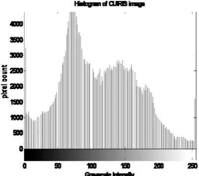
The pupil-iris contrast, one of the important factors in realising an accurate iris recognition algorithm, is dependent on the iris colour. The iris colour, a function of melanin pigmentation, is usually categorised as light or dark. Light eye-iris colours are blue, gray and green and are predominantly found among the Caucasians. Dark eye-iris colours are light-brown, dark-brown and very-dark-brown (or almost black); Asians usually have light-brown eye-iris colour while Africans usually have almost-black eye-iris colour. The very-dark-brown iris has a remarkable low contrast especially in the visible spectrum. The histogram, shown in fig.1, of an image is the function of the number of pixels against the gray levels, (i.e. 0 – 255 for an 8-bit image). It reflects the number of occurrence of each grayscale intensity, as a measure of contrast, in the image. Entropy is a statistical measure of randomness that can be used to characterize the texture of the input image. Grayscale image entropy, GiE, is defined as a measure of the amount of information contained in a grayscale image. The GiE value is formulated as the reduction of uncertainty about pixels classification as a result of observing the grayscale image [17]. It is defined as
∗ 2 1
,
[image:2.595.52.254.474.652.2]
It is usually used for image segmentation [17, 18]. The histogram and GiE values are used in this work to compare African/Asian/Caucasian images.
Fig 1: Histogram of a grayscale CUiris image
II. RELATEDWORK
Iris biometric research has witnessed great attention and improvement in the past decade resulting into a large number of publications and overall improvement. The major areas of contributions centre on the four major modules of iris recognition systems. The first module, Image acquisition, is extensively reviewed in this section.
There are currently seven publicly available iris image databases, hosted by academic and research institutions as summarized in Table 1 namely: Chinese Academy of Sciences – CASIA [19], University of Beira Interior – UBIRIS [9,11], ND-IRIS-0405 [20], a superset of Iris Challenge Evaluation, ICE 2005 & 2006 [21], University of Bath – BATH [22], Multimedia University – MMU [23], West Virginia University – WVU [247] and University of Olomuc – UPOL [25]. Apart from UPOL and UBIRIS, all of the databases contain near infrared, NIR images. Table 1 presents specifications of these databases.
Publications in this area address subjects’ position and movements, imaging distances, and lighting conditions. Images acquired under near-infrared illumination optimally reveal the richness of the iris structure and thus constitute over 70% of publicly available iris images. The images are used mainly for iris biometrics because it allows the iris texture of both “light” and “dark” eyes to be well visualized readily.
Recently, there is a shift to visible wavelength imaging as the more appropriate means to achieve “at a distance” and “on the move” imaging, based on images from the UBIRIS v2 dataset.. The dataset consist of visible-light, colour iris images, acquired with 4-8m distance between subject and sensor, and with subjects in motion. It represents 261 subjects, with over 11,000 iris images. The purpose of the dataset is to support research on visible-light iris images acquired under far from ideal imaging conditions [10, 11]. Also, experiments indicate the potential of using multispectral information to enhance the performance of iris recognition systems. This refers to the fusion of iris information from both near-IR and visible illumination [26, 27]. Another body of research shown that it is possible to acquire images while subjects walk at normal speed through an access control point [28,29] and at a distance of up to 10 meters with sufficient quality to support iris recognition [30, 31].
The second module is Image pre-processing which involves steps such as pupil and iris boundary detection, eyelid detection and removal and normalization. Popular approaches are Daugman algorithm [15,32,33], Wildes algorithm [34], Tisse algorithm [35] and Li Ma algorithm [36, 37, 38].
Recent publications collated in [12, 14, 39, 40] also listed a significant number of methods for segmenting the iris from the eye images. The significant difference in processing near infrared images as against visible-light images is that the pupillary boundary tends to be more distinct in near infrared whereas the limbic boundary appears to be more distinct in visible light.
The third module is Feature extraction. Broadly, there are four main approaches to iris representation (or feature extraction): phase-based methods [15, 32, 33], zero-crossing representation [41, 42], texture- analysis based methods [34, 37, 38] and intensity variation analysis [43]. Some of the features are x-y coordinates, radius, shape and size of the pupil, intensity values, orientation of the pupil ellipse and ratio between average intensity of two pupils. The features are encoded to a format suitable for recognition [14].
measures [36], neural networks [44] and normalized correlation measures [34].
Alongside these modules, some recent publications addressed the analysis and categorization of iris patterns for
determining the demographic attributes such as ethnicity and gender, of the subject. This categorization can also be used to create an indexing algorithm to speed search of an iris database and/or determining soft biometric traits of a person. Experiments conducted in [3, 4, 5, 7, 8, 45] reveal that the TABLE I:LIST OF PUBLICLY-AVAILABLE IRIS DATABASES AND THEIR SPECIFICATIONS
DATA BASE
VERSION ACQUISITION DEVICE
TOTAL IMAGES
SUBJECTS SUBJECT
ORIGIN
RESOLUTION IMAGE
FORMAT
DATASET PECULIARITY
CASIA [19]
V1 CASIA iris camera
756 108 Asian 320x280 bmp Pupil regions are edited to have constant intensity V2
CASIA-camera & OKI
2400 60 Asian 640x480 bmp Close-range images captured with two different sensors. V3:
Iris-Interval CASIA close-up iris camera
2,639 249 Asian 320x280 jpg Detailed texture features of iris images.
V3: Iris-Lamp
OKI IRISPASS-h camera
16,212 411 Asian 640x480 jpg Non-linear iris normalization and robust iris feature representation. V3:
Iris-Twins OKI IRISPASS-h camera
3,183 200 Asian 640x480 jpg Dissimilarity and similarity between iris images of twins.
Iris-Distance
CASIA long-range iris camera
2,567 142 Asian 2352x1728 jpg Long-range and high-quality iris images V4:
Iris-thousand IrisKing IKEMB-100 20,000 1,000 Asian 640x480 jpg Shows the uniqueness of iris features; for novel iris classification and indexing methods.
V4: Iris-Syn
CASIA image synthesis algorithm
10,000 1,000 (synthesized)
CASIA-IrisV1
640x480 jpg Synthesized iris images
UBIRIS [9,11]
V1 Nikon E5700 camera
1,877 241 Caucasian 400x300 jpg Images are coloured and noisy
V2 Canon EOS
5D camera
11,102 261 Caucasian 800x600 tiff Coloured images, captured on-the-move and at-a-distance
ND- IRIS-0405 [20]
Contains ICE 2005 & 2006[21]
IRIDIAN LG
EQU 2200 64,980 356 Caucasian 640x480 tiff Contains blur, off-axis and iris-occluded eye images.
BATH [22]
Iris DB
400 IrisGuard AD-100 dual-iris camera
8,000 200 Caucasian 1280x960 bmp
Images have notable eyelash and eyelid noise, on the iris portion. Iris DB
800 IrisGuard AD-100 dual-iris camera
16,000 400 Caucasian 1280x960 bmp
Iris DB 1600
IrisGuard AD-100 dual-iris camera
32,000 800 Caucasian 1280x960 bmp
MMU [23]
MMU1 LG IrisAccess 2200
450 100 Asian 320x280 bmp
Images includes minimal noise
MMU2 Panasonic BMET100US Authenticam
995 100 Asian 320x280 bmp
WVU [24]
- OKI IRISPASS-h camera
3,099 Caucasian Defocus blur, off-angle and heavily occluded images.
UPOL
[25] - SONY 950P 3CCD DXC- camera
local features of iris are unique to each subject while the global feature distributions of irises are similar for a specific race. The texture distribution differences of irises from different races seem to depend on the genes. The result reflects the Caucasian/Asian ethnicity of the person.
III. CUIRIS:THE DARK-SKINNED AFRICAN IRIS DATASET
The introductory version of the African iris dataset, called CUIRIS, is presented in this section.
A. Image Data Source and Data Description
The images were acquired from 81 subjects drawn from the community of Covenant University, Nigeria. The left and right irises of each subject were captured at a close range, in a single session, resulting in 162 eye-iris images in bitmap format. The images are in rectilinear format as specified by the ANSI/INCITS 379 and ISO/IEC 19794-6 Iris Image Interchange Format as shown in fig. 2. Images are named as either Left_ID or Right_ID where ID, the subject’s unique identification number. Other attributes of the subjects such as nationality, ethnic origin, age, use of contact lens or glasses were also collected.
B. Acquisition Device and Acquisition Method
The eye-iris images were acquired and digitized by the CrossMatch I Scan 2 dual iris scanner, shown in fig.3. The scanner captures the right and left irises concurrently. The specifications of the scanner are summarized in table 2. Proper quality assessment, integrated in the device SDK, is performed for each eye before the best image is stored. The iris scanner was connected to a PC via a USB port and operated in the automatic capture mode. Environmental condition, lighting was not controlled in order to simulate a real life scenario.
TABLE II:ISCAN2 DUAL-IRIS SCANNER SPECIFICATIONS
ATTRIBUTES SPECIFICATION
Iris Scans Dual optical system
Pixel Resolution 16.7 pixels/mm ( > 200 pixels over iris diameter of 9.5-13 mm)
Spatial Resolution 4.0 lp/mm at 60% or higher contrast
Biometric Data Interchange Formats
ANSI INCITS 379-2004; ISO/IEC 19794-6 Illuminating light Near infrared
IV. EXPERIMENTSANDRESULTS
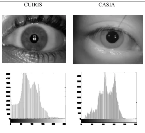
In this section, we compare images from the CUIRIS with images from two other popular datasets – UBIRIS v.1 and CASIA v.2, which contains Asian and Caucasian image sets respectively. Table 3 compares the histogram of a CUIRIS and a CASIA image; rich information area falls between 0 – 170 (darker) and 70 – 190 (lighter) gray levels for African and Asian respectively. Table 4 compares the histogram of a CUIRIS and a UBIRIS image; rich information area falls between 0 – 170 (darker) and 100 - 255 (lighter) gray levels for African and Caucasian respectively.
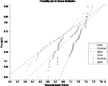
A predefined MATLAB code (m-file) was developed to: 1) randomly select a total of fifty samples from 50 subjects from each dataset; 2) compute the GiE values of the image histograms, as presented in Table 5; 3) compute the probability density function for the GiE values; and 4) plot the normal probability distribution of the GiE values.
TABLE III:HISTOGRAM OF CUIRIS AND CASIAIRIS IMAGES
CUIRIS CASIA
TABLE IV:HISTOGRAM CUIRIS AND UBIRIS IMAGES.
[image:4.595.310.547.278.495.2]CUIRIS UBIRIS
[image:4.595.57.286.481.567.2] [image:4.595.309.547.518.763.2] [image:4.595.43.272.628.737.2]TABLE V:SORTED GIE VALUES FOR FIFTY SAMPLES EACH FROM THE CUIRIS,CASIAIRIS AND UBIRIS DATATSETS
SAMPLE №
GIEVALUES
CUIRIS CASIA UBIRIS 1 5.7541 6.5099 6.2386 2 5.9515 6.5116 6.4642 3 5.9813 6.5186 6.5589 4 6.0027 6.5275 6.7479 5 6.0660 6.5420 6.7482 : : : : 27 6.6668 6.8440 7.2552 28 6.7142 6.8564 7.2764 29 6.7213 6.8603 7.2831 30 6.7817 6.8879 7.3093 31 6.8019 6.8938 7.3170 : : : : 45 7.1733 7.4605 7.4957 46 7.2341 7.4937 7.5043 47 7.2920 7.5317 7.5093 48 7.3990 7.5320 7.5149 49 7.4377 7.5344 7.5262 50 7.6557 7.5588 7.5325
The following deductions were made in the course of acquisition, visual inspection and analysis of the iris images: Visual inspection revealed that about 24.69% of the
subjects have a seemingly similar iris pattern; most of the subjects with this pattern have both parents from the same ethnic origin. This establishes the fact that individuals from the same ethnic origin have similar global features. Fig. 4 shows the normal distribution of the probabilities of
the GiEs for the 3 datasets. Africans
(GiE(min) = 5.75) are the most likely group with much darker contrast, possibly hiding away intricate features, than Asians (GiE(min) = 6.51) or Caucasians (GiE(min) = 6.24).
The lower GiE value of the Caucasian’s compared to Asian’s is apparently due to the lighting conditions during acquisition. The UBIRIS images were captured at visible wavelength and converted to grayscale images.
V. FUTUREWORK
The acquisition of eye-images for the African iris dataset is a work in progress. It consists of a significant number of images acquired using different sensors under both visible and near infrared illuminations. The dataset will be made publicly-available strictly for research purposes as soon as it is sufficiently populated. Subsequent versions will also contain other biometrics such as fingerprint, 2D and 3D face.
VI. CONCLUSION
[image:5.595.47.286.71.288.2]This paper established that individuals from the same ethnic origin have similar global features; Africans have a darker pupil-iris even in infrared imaging and images captured in the visible wavelength have notable contrast variations. A publicly-available iris dataset containing very-dark-brown irises of dark-skinned subjects of African origin will enhance the development of robust iris recognition algorithms especially for large-scale identification systems. The dataset will facilitate further research work in frontier areas of eye-iris research such as ethnicity prediction, gender prediction, ascertaining state of health and accurate diagnosis of eye defects.
[image:5.595.118.469.470.751.2]ACKNOWLEDGEMENT
We deeply appreciate the support of Prof. T.S. Ibiyemi, Covenant University students who willingly allowed us to collect their iris samples and the management of the University. We also thank Prof. Zhenan Sun (CASIA) and Hugo Proenca (UBIRIS) for providing us with iris images and useful ideas.
REFERENCES
[1] A. K. Jain, A. Ross and S. Prabhakar , “An Introduction to Biometric Recognition”, IEEE Transactions on Circuits and Systems for Video Technology, Special Issue on Image- and Video-Based Biometrics, Vol. 14, No. 1, pp 4-20, Jan. 2004.
[2] J. Daugman and I. Malhas, “Iris recognition border-crossing system in the UAE”, International Airport Review, Issue 2, 2004
[3] H. Zhang, Z. Sun, T. Tan and J. Wang, “Ethnic Classification Based on Iris Images”, CCBR 2011, LNCS 7098, pp. 82–90, 2011.
[4] X.C. Qiu, Z.A. Sun, and T.N. Tan. Learning appearance primitives of iris images for ethnic classification. In Int. Conf. on Image Processing, pages II: 405–408, 2007.
[5] X. Qiu, Z.Sun, T.Tan, Global texture analysis of iris images for ethnic classification, in: Springer LNCS 3832: International Conference on Biometrics, January 2006, pp. 411–418.
[6] V. Thomas, N. Chawla, K. Bowyer, P. Flynn, Learning to predict gender from iris images, in: Biometrics: Theory, Applications, and Systems, Sept 2007.
[7] S. Lagree and K.W. Bowyer, “Ethnicity prediction based on iris texture features”, 22nd Midwest Artificial Intellignece and Cognitive Science Conference (MAICS), April 2010.
[8] S. Lagree and K. W. Bowyer, “Predicting Ethnicity and Gender from Iris Texture”, Proc. IEEE International Conference on Technologies for Homeland Security (HST), pp. 440 – 445, Nov. 2011
[9] H. Proenc¸a and L.A. Alexandre, “UBIRIS: A Noisy Iris Image Database,” Proc. 13th Int’l Conf. Image Analysis and Processing, pp. 970-977, Sept. 2005.
[10]H. Proenca, “On the feasibility of the visible wavelength, at-a-distance and on-the-move iris recognition”. IEEE Workshop on Computational Intelligence in Biometrics: Theory, Algorithms, and Applications (CIB 2009), March 2009, 9-15.
[11]H. Proenca, S. Filipe, R. Santos, J. Oliveira and L. Alexandre, “The UBIRIS.v2: A Database of Visible Wavelength Images Captured On-The-Move and At-A-Distance”. IEEE Transactions on Pattern Analysis and Machine Intelligence 32 (8), August 2010, 1529-1535.
[12]H. Proenca, “Iris Recognition: On the Segmentation of Degraded Images Acquired in the Visible Wavelength”, IEEE Transactions on Pattern Analysis and Machine Intelligence, Vol. 32, no. 8, 2010, pp. 1502-1516
[13]H. Proenc¸a and L.A. Alexandre, “Iris Segmentation Methodology for Non-Cooperative Iris Recognition,” Proc. IEE Vision, Image, & Signal Processing, vol. 153, no. 2, pp. 199-205, 2006.
[14]S.V Sheela and P.A Vijaya, “Iris Recognition Methods - Survey”, International Journal of Computer Applications, Volume 3, No.5, June 2010
[15]J. Daugman, “The importance of being random: statistical principles of iris recognition,” Journal of Pattern Recognition, Vol. 36, No. 2, pp. 279–291, 2003.
[16]A.K. Jain, A. Ross, S. Pankanti, “Biometrics: A Tool for Information Security”, IEEE Transactions on Information Forensics and Security, Vol. 1, No. 2, June 2006.
[17]C.F. Sin and C.K. Leung, “Image Segmentation by Edge Pixel Classification with Maximum Entropy”, Proceedings of the International Symposium on Intelligent Multimedia, Video & Speech Processing, pp. 283-286, May 2-4, 2001.
[18]C.F. Sin and C.K. Leung, “Entropy-based Image Segmentation by Boundary Detection”, Proceedings of the International Symposium on Multimedia Information Processing, pp. 284-286, December 13-15, 2000
[19]Institute of Automation, Chinese Academy of Sciences, CASIA Iris Image Database, http://www.sinobiometrics.com , 2012.
[20]K. W. Bowyer and P. J. Flynn, “The ND-IRIS-0405 Iris Image Dataset”, http://www.nd.edu/˜cvrl/papers/ND-IRIS-0405.pdf
[21]National Institute of Standards and Technology, Iris Challenge Evaluation, http://iris.nist.gov/ICE/ , 2006
[22]Univ. of Bath, University of Bath Iris Image Database,
http://www.bath.ac.uk/elec-eng/pp./sipg/ , 2004.
[23]Multimedia University, MMU Iris Image Database,
http://pesona.mmu.edu.my/ccteo , 2004.
[24]A. Ross, S. Crihalmeanu, L. Hornak, and S. Schuckers, “A Centralized Web-Enabled Multimodal Biometric Database,” Proc. 2004 Biometric Consortium Conf., Sept. 2004.
[25]M. Dobes and L. Machala, UPOL Iris Image Database,
http://phoenix.inf.upol.cz/iris/ , 2004.
[26]C. Boyce, A. Ross, M. Monaco, L. Hornak, and X. Li, “Multispectral Iris Analysis: A Preliminary Study,” Proc. IEEE Conf. Computer Vision and Pattern Recognition Workshop Biometrics, pp. 51-59, June 2006.
[27]K. Smith, V.P. Pauca, A. Ross, T. Torgersen, and M. King, “Extended Evaluation of Simulated Wavefront Coding Technology in Iris Recognition,” Proc. First IEEE Int’l Conf. Biometrics: Theory, Applications, and Systems, pp. 1-7, Sept. 2007.
[28]J.R. Matey, D. Ackerman, J. Bergen, and M. Tinker, “Iris Recognition in Less Constrained Environments,” Advances in Biometrics: Sensors, Algorithms and Systems, pp. 107-131, Springer, Oct. 2007.
[29]K. Park and J. Kim, “A Real-Time Focusing Algorithm for Iris Recognition Camera,” IEEE Trans. Systems, Man, and Cybernetics, vol. 35, no. 3, pp. 441-444, Aug. 2005.
[30]C. Fancourt, L. Bogoni, K. Hanna, Y. Guo, R. Wildes, N. Takahashi, and U. Jain, “Iris Recognition at a Distance,” Proc. 2005 IAPR Conf. Audio and Video Based Biometric Person Authentication, pp. 1-13, July 2005.
[31]S. Yoon, K. Bae, K. Ryoung, and P. Kim, “Pan-Tilt-Zoom Based Iris Image Capturing System for Unconstrained User Environments at a Distance,” Lecture Notes in Computer Science, pp. 653-662, Springer, 2007.
[32]J. Daugman, “How iris recognition works,” IEEE Trans. Circuits System and Video Technology, Vol. 14, No. 1, pp. 21–30, Jan. 2004. [33]J. Daugman, “New methods in iris recognition “, IEEE transactions on
Systems, Man, and Cybernetics—part b: cybernetics, Vol. 37, No. 5, Oct. 2007.
[34]R. Wildes et al, “A machine-vision system for iris recognition,” Machine Vision and Application, Vol. 9, pp. 1–8, 1996.
[35]C. Tisse et al, “Person Identification Technique Using Human Iris Recognition,” Proc. Vision Interface, pp. 294-299, 2002.
[36]L. Ma et al “Personal identification based on iris texture analysis,” IEEE Transaction on Pattern Analysis Machine Intelligence, Vol. 25, No. 12, pp. 1519–1533, Dec. 2003.
[37]L. Ma, Y. Wang, and T. Tan, “Iris Recognition Based on Multichannel Gabor Filtering,” Proc. Fifth Asian Conf. Computer Vision, Vol. I, pp. 279-283, 2002.
[38]L. Ma, Y. Wang, and T. Tan, “Iris Recognition Using Circular Symmetric Filters,” Proc. Of the 16th International Conference on Pattern Recognition, Vol. II, pp. 414-417, 2002.
[39]K.W. Bowyer, K. Hollingsworth, and P. Flynn. “Image Understanding for Iris Biometrics: A Survey”. Computer Vision and Image Understanding 110 (2), pp. 281-307, 2008.
[40]K.W. Bowyer, K. Hollingsworth, and P. Flynn. “A survey of Iris Biometrics Research: 2008 -2010”, 2012
[41]W. Boles and B. Boashash, “A Human Identification Technique Using Images of the Iris and Wavelet Transform”, IEEE Trans. Signal Processing, Vol. 46, no. 4, pp. 1185-1188, 1998.
[42]C. Sanchez-Avila and R. Sanchez-Reillo, “Iris-based biometric recognition using dyadic wavelet transform”, IEEE Aerosp. Electron. Systems Magazine, Vol. 17, pp. 3–6, Oct. 2002.
[43]L. ma et al, “Local Intensity Variation Analysis for Iris Recognition”, Pattern recognition Journal, Vol. 37, Issue 6, pp. 1287-1298, June 2004.
[44]S. Lim, K. Lee, O. Byeon, and T. Kim, “Efficient Iris Recognition through Improvement of Feature Vector and Classifier”, ETRI Journal, Vol. 23, no. 2, pp. 61-70, 2001.
[45] L. Stark, K.W Bowyer and S. Siena, “Human perceptual categorization of iris texture patterns”, Biometrics Theory,Applications