Original Article
Safe regulable angle and optimum trajectory of the
second sacral alar iliac screw: a digital simulation study
Cong Li1, Xian Xu1, Jiayi Xu1, Jun Tan2, Wei Wu1
Departments of 1Trauma, 2Spinal Surgery, Shanghai East Hospital, School of Medicine, Tongji University, 150 JiMo
Road, Shanghai 200120, China
Received September 16, 2018; Accepted February 12, 2019; Epub May 15, 2019; Published May 30, 2019
Abstract: Objectives: The posterior second sacral alar iliac (S2AI) screw fixation technique has a wide range of clini
-cal applications. The optimum trajectory of S2AI screws has been reported by numerous studies. However, morpho
-logical and anatomical structures of the safe range of the S2AI screw fixation technique have rarely been studied. Methods: Three-dimensional reconstruction was performed on computed tomography (CT) data of 40 adult pelvises without relative lesions. One cylinder (diameter of 7.0 mm) was drawn to imitate the S2AI screw and CT imaging planes were rotated until they matched the ideal S2AI trajectory. Parameters of S2AI screw trajectory measure
-ments and the safe regulable angle were recorded. Results: Inward, outward, upward, and downward safe regulable angles, as well as TSV and SAG angles on the left side of females, were 4.7 ± 0.6°, 6.7 ± 0.4°, 8.1 ± 0.7°, 6.9 ± 0.6°, 11.5 ± 1.2°, and 15.5 ± 2.2°, respectively. Angles of the males and the right side of females were in accord with angles of the left side of females. Conclusion: This study recommends insertion of S2AI screws with approxi
-mately a 28° SAG angle in the sagittal plane (5° more for females) and a 34° TSV angle in the transverse plane (3° more for males). Insertion of the S2AI screws could be relatively close to the upward and outward sides, based on the optimum trajectory, because of less safe regulable angles at inward and downward sides.
Keywords: Safe regulable angle, insertion trajectory, sacral alar iliac screw, digital simulation
Introduction
After Sponseller and Kebaish devised the pos -terior second sacral alar iliac (S2AI) screw fixa -tion technique in 2007 [1, 2], it has been widely applied for surgical treatment of pelvic obliqui -ty, high-grade spondylolisthesis, lumbopelvic trauma, long spinal fusions, and other spinal deformities [3]. The S2AI screw fixation tech -nique has merits, compared to other methods of spinopelvic fixation, including the traditional iliac screw. The S2AI screw fixation technique has more fixed segments and stable biome -chanical torsion because both the ilium and the sacrum are anchored [4]. Compared to tradi -tional iliac screw fixation, the direction and length of S2AI screws could facilitate more reli -able stability [5-8]. Since minimally disturbed soft tissue completely covers the implants, the S2AI screw technique may minimize blood loss and infection rates [4, 9-11]. In contrast, the traditional iliac screw technique needs exten -sive surgical exposure of the posterior superior
iliac spine (PSIS). This could lead to minimal soft tissue coverage and contribute to a higher risk of bleeding and infection rates, reportedly as high as 4% [12]. Hence, the S2AI screw has been identified as a replacement for the iliac screw, due to those unfavorable factors [2].
S2AI screws traverse the sacroiliac joint and terminate in the ilium above the sciatic notch [1, 3, 4, 6, 14-18].
Typically, intraoperative fluoroscopy has been used to assess S2AI screw insertion, but most of the C-arm machines provide two-dimension -al images only. If a screw is very close to the surface of the inner and outer cortices of the sacral ala on lateral and frontal radiographs, it may have actually perforated the bone cortex, although it is still seen in the sacral ala in the radiographs. The presence of contrast, intesti -nal gas, and increased soft tissue density, due to obesity, can make it difficult to obtain and interpret appropriate intraoperative fluoroscop -ic images. These negative factors for S2AI screws also add to the insertion difficulty. Mal-positioned implants and excessive deflection of the S2AI screw could increase the risk of punc -turing the sacral or iliac cortex, damaging tis-sues proximal to the pelvis, such as the sciatic nerve, superior and inferior gluteal artery, veins and nerves, and other neurovascular tissues [19]. These factors could have catastrophic consequences [20-22].
Very few studies have reported the morphologi -cal and anatomi-cal structures for the safe range of the S2AI screw fixation technique, along with screw insertion in many lumbosa-cral-pelvic disorders. The current study attempt-ed to calculate safe regulable angles in four directions and the optimum trajectory of S2AI screws through digital simulation, thereby iden-tifying the presence of a stereoscopic safe
were routinely implemented before data pro -cessing. The study population consisted of 40 patients (20 males and 20 females), with a mean age of 34.2 years (range 18-57 years). Mean height of males and females was 171.68 ± 2.88 cm and 154.35 ± 4.67 cm, respectively. Mean weight of males was 62.57 ± 4.80 kg, while that of females was 55.35 ± 6.83 kg. DICOM image data of 40 adults were obtained by CT scanning the pelvic regions using a tube voltage of 120 kV, tube current of 200 mA, slice thickness of 1 mm, and interlayer spacing of 0.5 mm. For data consistency, the same inves-tigator, familiar with the anatomy of the pelvis, measured all parameters and recorded the average value of measurement results. Ex-periments were repeated three times. The fol -lowing steps were used for data analysis from each case.
Model construction
CT images were transformed into a three-dimensional (3D) pelvic reconstruction model via the interactive medical imaging control sys-tem Mimics 17.0 (Materialise, Leuven, Belgium). After 3D digital images were calculated and reconstructed, a cylinder (diameter of 7.0 mm) was drawn to imitate the S2AI screw [23].
Determination of the entry point
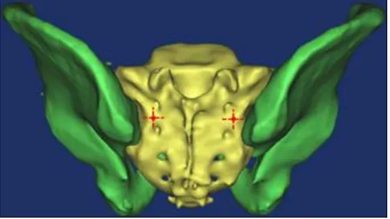
[image:2.612.91.375.73.233.2]Entry points of S2AI screws were chosen at the cross point of the lateral sacral crest and mid -line between the S1 and S2 dorsal foramen into a 3D pelvic reconstruction model (Figure 1). Figure 1. Entry points (red point) of S2AI screws were chosen at the cross
point of the lateral sacral crest and the midline between the S1 and S2 dor
-sal foramen in a three-dimensional pelvic reconstruction model.
range of S2AI screws. The aim is to help clinicians better understand the S2AI screw fix -ation technique and reduce ri-sks of perforation of the cor-tex.
Materials and methods
Figure 2. Optimum trajectory measurements of
transverse and sagittal plane images along S2AI trajectory. TSV angle (∠DAC), iliac width (Line EF), maximal length (Line AC), and sacral distance (Line AB) in transverse plane along S2AI trajectory (A). SAG angle (∠PAQ) in sagittal plane along S2AI trajectory
(B). The data of a patient (female, 28 years) on the left side; ∠DAC = 35.72°; line EF = 14.88 mm; line AC = 114.98; line AB = 25.31 mm; ∠PAQ = 34.20°.
Image plane adjustment
The entry point was specified for use at the cen -ter of rotation. CT imaging planes were rotated until they matched the ideal S2AI trajectory (greatest length and width of osseous channel) of the pelvis.
Parameter measurements
Optimum trajectory of S2AI screws: TSV angle is defined as lateral trajectory angulation in the transverse plane (Figure 2A, ∠DAC). SAG angle
is defined as caudal trajectory angulation in the sagittal plane (Figure 2B, ∠PAQ). Max-leng- th is defined as the maximal distance from the entry point to the anterior inferior iliac spine based on the optimum trajectory (Figure 2A, Line AC). Iliac width is defined as the narrowest iliac width measured between the outside corti-ces in the transverse plane (Figure 2A, Line EF).
Safe regulable angle of S2AI screw: As shown in Figure 3A, on the transverse plane, the axis of the optimal entry screw was crossed with the ilium cortex at A (the entry point). Point E, F’ (the narrowest spots of two margins) was used to make parallel lines (Line KG and Line IH) of the axis of optimum trajectory (Line AC), which pierced the vertebral body at points K and I. Through point I (the nearest entry point) and point A (the entry point) to make vertical lines (JI and GH), researchers formed the rectangular IJGH (the safe area of S2AI screws) and the rectangular LMNO (the ichnography of S2AI screw simulation). ∠JML and ∠INO were the safe regulable inward and outward angles of the screw transverse plane, respectively. As shown in Figure 3B, the quantitative measure -ment method of the safe area and safety adjustment on the sagittal plane was the same as on the transverse plane. Lastly, ∠UM’L’ and ∠TN’O’ are defined as upward and downward
safe regulable angles, respectively.
Statistical analysis
Statistical analysis was performed using SPSS statistical software program 22.0. Indepen-dent-samples t-test was used to detect differ -ences in gender and limb side of sacroiliac joints. For all statistical tests, P < 0.05 indi-cates significance.
Results
Tables 2 and 3 show the results of safe regula -ble angles. There were no significant differenc -es in gender and limb side of sacroiliac joints (P > 0.05). The TSV safe regulable angle is defined as the sum of inward and outward safe regulable angles in the transverse plane. Simi- larly, the SAG safe regulable angle is defined as the sum of upward and downward safe regula -ble angles in the sagittal plane. Inward, out-ward, upout-ward, downout-ward, TSV, and SAG safe regulable angles on the left side of females were 4.7 ± 0.6°, 6.7 ± 0.4°, 8.1 ± 0.7°, 6.9 ± 0.6°, 11.5 ± 1.2°, and 15.5 ± 2.2°, respective -ly. Angles of males and the right side of females were in accord with results of the left side of females.
Discussion
Despite significant advances and develop -ments in spinal instrumentation techniques, sacropelvic fixation remains a surgical chal -lenge, especially in neuromuscular scoliosis and severe adult scoliosis [24]. Poor bone qual -ity of the sacrum, complex anatomy, and tre -mendous biomechanical forces at the lumbo -sacral junction contribute to the high rates of instrumentation-related problems [9, 15, 25-27]. S2AI screws could provide immediate sta -bility and adequate biomechanical strength of constructs, based on discriminative features wherein both the ilium and the sacrum are anchored based on the direction and length of the screws. In addition, it could correct pelvic obliquity with lower implant profiles, with less soft tissue dissection, shorter operation times, and fewer complications [28, 29]. In recent years, the S2AI screw fixation technique has been widely applied for surgical treatment of lumbosacral-pelvic reconstruction and other disorders.
A previous study in a Chinese population rec-ommended average SAG and TSV angles of S2AI screw insertion of 30-40°, while the aver -age max-length was about 120 mm for males and 115 mm for females. The currents study recommends insertion of S2AI screws with a SAG angle of approximately 28° and a TSV angle of approximately 34°. The SAG angle of females should be approximately 5° more than males, consistent with previous studies [17]. The TSV angle of females should be about 3° less than males, based on current results. This Figure 3. Safe regulable angles of S2AI screws in
transverse and sagittal planes along the S2AI trajec-tory. The inward (∠JML) and outward (∠INO) safe regulable angles in transverse plane (A). The upward (∠UM’L’) and downward (∠TN’O’) safe regulable
angles in sagittal plane (B). The TSV safe regulable angle was defined as the sum of inward and outward safe regulable angles in the transverse plane. Simi
-larly, the SAG safe regulable angle was defined as the sum of upward and downward safe regulable angles in the sagittal plane. The safe angle data of a patient (female, 28 years) on the left side; ∠JML =
4.55°; ∠INO = 6.18°; ∠UM’L’ = 8.00°; ∠TN’O’ =
6.94°; TSV safe regulable angle = 10.73°; SAG safe regulable angle = 14.94°.
study recommends S2AI screws with the max-length of female 114 mm/male 121 mm, con -sistent with the findings of Kwan et al. [29]. This means that the max-length of ideal S2AI trajec -tory surpasses the length usually used in prac-tice, which is 80 mm [30]. Except for the inser -tion angle of S2AI screws, iliac width is a key indicator to decide whether S2AI screws suc-cessfully traverse the ideal iliac plane. Due to the complex anatomy of the pelvis, iliac width
Table 1. Parameters of S2AI screw trajectory measurements (Mean ± standard deviation)
Parameters Females Males
Left Right P Left Right P
Sag angle (°) 33.9 ± 6.5 34.8 ± 7.1 0.68 28.3 ± 7.7 29.0 ± 7.4 0.77
[image:5.612.90.384.239.343.2]Tsv angle (°) 34.1 ± 5.9 34.4 ± 5.1 0.86 37.8 ± 4.7 38.4 ± 5.3 0.71 Max-length (mm) 114.3 ± 9.5 114.2 ± 8.7 0.97 121.4 ± 9.3 121.2 ± 8.8 0.94 Iliac width (mm) 15.3 ± 2.6 15.5 ± 2.9 0.82 17.5 ± 3.4 17.8 ± 3.3 0.78 Sacral distance (mm) 24.7 ± 2.8 24.9 ± 2.5 0.81 24.4 ± 2.5 25.1 ± 2.2 0.35 For all statistical tests, the P value of less than 0.05 indicates statistical significance.
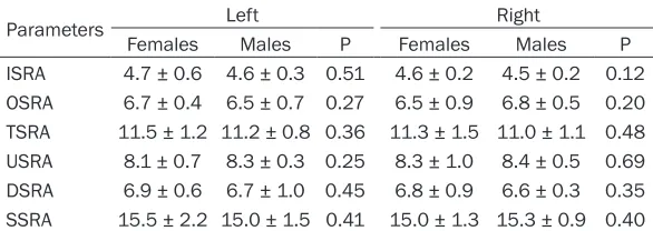
Table 2. The safe reguable angle of S2AI screws about the limb side of sacroiliac joint (Mean ± standard deviation)
Parameters Females Males
Left Right P Left Right P
ISRA 4.7 ± 0.6 4.6 ± 0.2 0.48 4.6 ± 0.3 4.5 ± 0.2 0.22 OSRA 6.7 ± 0.4 6.5 ± 0.9 0.37 6.5 ± 0.7 6.8 ± 0.5 0.12 TSRA 11.5 ± 1.2 11.3 ± 1.5 0.64 11.2 ± 0.8 11.0 ± 1.1 0.51 USRA 8.1 ± 0.7 8.3 ± 1.0 0.47 8.3 ± 0.3 8.4 ± 0.5 0.45 DSRA 6.9 ± 0.6 6.8 ± 0.9 0.68 6.7 ± 1.0 6.6 ± 0.3 0.67 SSRA 15.5 ± 2.2 15.0 ± 1.3 0.39 15.0 ± 1.5 15.3 ± 0.9 0.45 For all statistical tests, the P value of less than 0.05 indicates statistical significance. ISRA: the inward safe reguable angle; OSRA: the outward safe reguable angle; USRA: the upward safe reguable angle; DSRA: the downward safe reguable angle; TSRA: the TSV safe reguable angle; SSRA: the SAG safe reguable angle.
Table 3. The safe reguable angle of S2AI screws about the sexuality (Mean ± standard deviation)
Parameters Left Right
Females Males P Females Males P
ISRA 4.7 ± 0.6 4.6 ± 0.3 0.51 4.6 ± 0.2 4.5 ± 0.2 0.12 OSRA 6.7 ± 0.4 6.5 ± 0.7 0.27 6.5 ± 0.9 6.8 ± 0.5 0.20 TSRA 11.5 ± 1.2 11.2 ± 0.8 0.36 11.3 ± 1.5 11.0 ± 1.1 0.48 USRA 8.1 ± 0.7 8.3 ± 0.3 0.25 8.3 ± 1.0 8.4 ± 0.5 0.69 DSRA 6.9 ± 0.6 6.7 ± 1.0 0.45 6.8 ± 0.9 6.6 ± 0.3 0.35 SSRA 15.5 ± 2.2 15.0 ± 1.5 0.41 15.0 ± 1.3 15.3 ± 0.9 0.40 For all statistical tests, the P value of less than 0.05 indicates statistical significance. ISRA: the inward safe reguable angle; OSRA: the outward safe reguable angle; USRA: the upward safe reguable angle; DSRA: the downward safe reguable angle; TSRA: the TSV safe reguable angle; SSRA: the SAG safe reguable angle.
present study, iliac width of the determined transverse plane was L15.3 ± 2.6 mm/ R15.5 ± 2.9 mm in females and L17.5 ± 3.4 mm/R17.8 ± 3.3 mm in males. F. Zhu et al. examined the optimal trajectory and insertion ac- curacy of S2AI screws, in which the parameter of ili-ac width was included [17]. Their results of iliac width (L14.76 ± 2.46 mm/R14.94 ± 2.60 mm in females and L16.98 ± 3.52 mm/R17.00 ± 2.81 mm in males) were in accord with present results. Therefore, it is believed that the diameter of the screw will not constitute a limita-tion if the screw is in the safe regulable angle. This is because the current max-diameter of S2AI screw in clinical practice is 7.5 mm [30]. Thus, the safe regula -ble angle is critical for inser -tion of S2AI screws.
[image:5.612.90.383.435.539.2]-line, which can move about 8.0° upward and 6.5° downward when the entry point and screw angles remain constant on transverse plane. Similarly, it can move about 4.5° and 6.5° in-ward and outin-ward when the entry point and screw angles remain constant on sagittal pl- ane. Hence, the safe regulable angles of inward and downward sides of the screw are relatively small. It is obvious that, when bigger diameters and longer screws are used, the safe regulable angle will be smaller. In clinical cases, the posi-tion is also subjectively determined by sur-geons. The angle will slightly differ among dif -ferent surgeons. Therefore, to avoid penetrating the cortex of the pelvis, this study recommends that the insertion of S2AI screws be relatively close to the upward and outward sides, based on the optimum trajectory. This is due to the relatively less safe regulable angles at inward and downward sides.
The current study had some limitations, how-ever. First, the 3D imaging study did not inclu- de clinical cases or cadaveric experiments. Reference lines and data measurement were subjectively established. Second, this study was only based on a Chinese population. Thus, ethnic and gender differences may have impacted the parameters of S2AI screw inser -tion. Third, present data should be applied to guide S2AI screw insertion in patients without pelvic obliquity or pelvic asymmetry, since all measurements were from normal pelvises without deformities of the skeletal structure. Fourth, if bigger diameter screws are used, the safe regulable angle will be smaller. In prac -tice, there are many diameters of screws avail -able. However, only 7.0 mm screws were used in this study.
Conclusion
Given the complex anatomy of the pelvis, a S2AI screw could generate an optical illusion of the precise position on intraoperative fluoro -scopic radiographs but may have perforated the bone cortex of pedicle. Both optimal trajec -tories and safe regulable angles are advised to ensure the insertion accuracy of S2AI screws. This study recommends insertion of the S2AI screws with approximately 28° SAG angle in the sagittal plane (5° more for females) and about 34° TSV angle in the transverse plane (3° more for males). In addition, the TSV and SAG safe regulable angles are approximately
11.5° and 15.5°, respectively. It isrecommend -ed that the insertion of the S2AI screws be re-latively close to the upward and outward si- des, based on the optimum trajectory. This is because of the less safe regulable angles at inward and downward sides. To decrease per- foration risks and reduce iatrogenic damage, the current study suggests the generation of a three-dimensional pelvic model for every patient, ensuring the best selection of S2AI screws before surgery and decreasing compli -cations caused by improper screws.
Acknowledgements
Project name: Mechanism of ERCC1 gene mod -ifing DNA damage in pressure-induced calcifi -cation of intervertebral disc cartilage endplate. Project number: 2018KY813. Project location: China Pinghu Second People’s Hospital, 136 East Yashan Road, Zhapu Town, Pinghu City 314201, Zhejiang, China.
Disclosure of conflict of interest
None.
Address correspondence to: Wei Wu, The Departm-
ent of Trauma Surgery, Shanghai East Hospital, School of Medicine, Tongji University, 150 Jimo
Ro-ad, Shanghai 200120, China. Tel: +86 18964323- 671; E-mail: [email protected]
References
[1] Chang TL, Sponseller P, Kebaish K and Fish
-man E. Low profile pelvic fixation: anatomic parameters for sacral alar-iliac fixation versus traditional iliac fixation. Spine 2009; 8:
156S-157S.
[2] Martin CT, Witham TF and Kebaish KM. Sacro
-pelvic fixation: two case reports of a new per
-cutaneous technique. Spine 2011; 36:
E618-621.
[3] O’Brien JR, Yu WD, Rishi B, Paul S and Kebaish KM. An anatomic study of the S2 iliac tech
-nique for lumbopelvic screw placement. Spine
2009; 34: 439-442.
[4] Sponseller P, Zimmerman R, Ko P, Kebaish K,
Gunne AP, Mohammed A and Chang TL. 42.
Low profile pelvic fixation using S2 ALAR Iliac (S2AI) fixation in the pediatric population im
-proves results at 2-year minimum follow-up.
Spine J 2009; 9: 22S.
[5] Lebwohl NH, Cunningham BW, Anton D, Nor -imichi S, Lee G, Vince D, Oheneba BA and
-sacral fixation techniques in a calf spine
model. Spine 2002; 27: 2312-2320.
[6] Liu Z, Qiu Y, Yan H, Hu ZS, Zhu F, Qiao J, Xu LL, Wang B, Yu Y and Qian BP. S2 alar-iliac fixation: a powerful procedure for the treatment of ky -phoscoliosis. Orthop Surg 2016; 8: 81-84.
[7] García JM, Doblaré M, Seral B, Seral F, Palanca
D, Gracia L. Three-dimensional finite element analysis of several internal and external pelvis fixations. J Biomech Eng 2000; 122: 516-522. [8] Park JH, Hyun SJ, Kim KJ and Jahng TA. Free
hand insertion technique of S2 sacral alar-iliac screws for spino-pelvic fixation: technical note, acadaveric study. J Korean Neurosurg Soc
2015; 58: 578.
[9] Amit J, Hamid H, Strike SA, Menga EN, Spon
-seller PD and Kebaish KM. Pelvic fixation in
adult and pediatric spine surgery: historical
perspective, indications, and techniques:
AA-OS exhibit selection. J Bone Joint Surg Am 2015; 97: 1521-1528.
[10] Kebaish KM. Sacropelvic fixation: techniques
and complications. Spine 2010; 35: 2245-2251.
[11] O’Brien JR, Lauren M, Yu WD and Kebaish KM. Feasibility of minimally invasive sacropelvic fixation: percutaneous S2 alar iliac fixation.
Spine 2010; 35: 460-464.
[12] Kuklo TR, Bridwell KH, Lewis SJ, Baldus C, Blanke K, Iffrig TM and Lenke LG. Minimum 2-year analysis of sacropelvic fixation and L5-S1 fusion using L5-S1 and iliac screws. Spine
2001; 26: 1976-1983.
[13] Wu AM, Chi YL, Ni WF and Huang YX. The feasi
-bility and radiological features of sacral alar ili
-ac fixation in an adult population: a 3D imaging
study. Peer J 2016; 4: e1587.
[14] Hoernschemeyer DG, Pashuck TD and Pfeiffer FM. Analysis of the s2 alar-iliac screw as com -pared with the traditional iliac screw: does it
increase stability with sacroiliac fixation of the
spine? Spine J 2017; 17: 875-879.
[15] Mattei TA and Fassett DR. Combined S-1 and S-2 sacral alar-iliac screws as a salvage
tech-nique for pelvic fixation after pseudarthrosis
and lumbosacropelvic instability: technical no- te. J Neurosurg Spine 2013; 19: 321-330.
[16] Stevens DB and Beard C. Segmental spinal
in-strumentation for neuromuscular spinal defor -mity. Clin Orthop Relat Res 1989; 7: 164-168.
[17] Zhu F, Bao HD, Yuan S, Wang B, Qiao J, Zhu ZZ, Liu Z, Ding YT, Qiu Y. Posterior second sacral
alar iliac screw insertion: anatomic study in a Chinese population. Eur Spine J 2013; 22: 1683-1689.
[18] Sagi HC and Lindvall EM. Inadvertent intrafo -raminal iliosacral screw placement despite ap-parent appropriate positioning on
intraopera-tive fluoroscopy. J Orthop Trauma 2005; 19:
130.
[19] Lanzieri CF and Hilal SK. Computed tomogra
-phy of the sacral plexus and sciatic nerve in the greater sciatic foramen. AJR Am J Roent -genol 1984; 143: 165-168.
[20] Gautier E, Bächler R, Heini PF and Nolte LP. Accuracy of computer-guided screw fixation of
the sacroiliac joint. Clin Orthop Relat Res 2001; 393: 310-317.
[21] Hinsche A, Giannoudis P and Smith R. Fluoros
-copy-based multiplanar image guidance for in
-sertion of sacroiliac screws. Clin Orthop Relat
Res 2002; 395: 135-44.
[22] Phillips JH, Gutheil JP and Knapp DR Jr. Iliac screw fixation in neuromuscular scoliosis.
Spine 2007; 32: 1566-1570.
[23] Wu AM, Wang S, Weng WQ, Shao ZX, Yang XD, Wang JS, Xu HZ, Chi YL. The radiological fea
-ture of anterior occiput-to-axis screw fixation
as it guides the screw trajectory on 3D printed
models: a feasibility study on 3D images and
3D printed models. Medicine 2014; 93: e242.
[24] Simonian PT, Routt ML Jr. Biomechanics of pel
-vic fixation. Orthop Clin North Am 1997; 28:
351-367.
[25] Guler UO, Cetin E, Yaman O, Pellise F, Casa -demut AV, Sabat MD, Alanay A, Grueso FS, Aca-roglu E; European Spine Study Group.
Sacrope-lvic fixation in adult spinal deformity (ASD); a very high rate of mechanical failure. Eur Spine
J 2015; 24: 1085-91.
[26] Shen FH, Mason JR, Shimer AL, Arlet VM. Pel
-vic fixation for adult scoliosis. Eur Spine J
2013; 22: S265-S275.
[27] Yoshihara H. Surgical options for lumbosacral fusion: biomechanical stability, advantage, dis
-advantage and affecting factors in selecting
options. Eur J Orthop Surg Traumatol 2014; 24: 73-82.
[28] Blake-Toker AM, Hawkins L, Nadalo L, Howard D, Arazoza A, Koonsman M, Dunn E. CT guided percutaneous fixation of sacroiliac fractures in
trauma patients. J Trauma 2001; 51: 1117-1121.
[29] Schweitzer D, Zylberberg A, Córdova M and Gonzalez J. Closed reduction and iliosacral percutaneous fixation of unstable pelvic ring fractures. Injury 2008; 39: 869-874.
[30] Ebraheim NA, Xu R, Biyani A, Nadaud MC.
Morphologic considerations of the first sacral pedicle for iliosacral screw placement. Spine