Review Article
Rosuvastatin reduces myocardial ischemia-reperfusion
injury by inhibiting miR-155
Yaqin Xie, Ying Li, Juan Zhao
Department of Pathophysiology, Chengde Medical University, Chengde, Hebei, P. R. China
Received January 21, 2019; Accepted May 8, 2019; Epub July 15, 2019; Published July 30, 2019
Abstract: Background: Rosuvastatin has been reported to protect against myocardial ischemia-reperfusion injury in cardiovascular diseases. MicroRNAs (miRNAs) are widely involved in progression of myocardial ischemia-reperfu-sion injury. However, the underlying mechanism of rosuvastatin and miRNAs in the myocardial ischemia-reperfuischemia-reperfu-sion injury has not been fully explored. Methods: The model of myocardial ischemia-reperfusion injury was established
using cardiomyocytes by serum and oxygen deficiency and recovery. The expression of miR-155 was calculated by
qRT-PCR. Cell survival and apoptosis were observed by 3-(4, 5-Dimethyl-2-thiazolyl)-2, 5-diphenyl-2Htetrazolium
bromide assay or flow cytometry. Lactate dehydrogenase (LDH), creatine kinase (CK), malondialdehyde (MDA) and
superoxide dismutase (SOD) were measured by different assay kits. Results: Cell survival rate and SOD level were
inhibited, while apoptosis and the levels of LDH, CK and MAD were enhanced in cardiomyocytes under
ischemia-reperfusion compared with those in control group. However, these effects were attenuated by introduction of rosu-vastatin. In addition, we found that miR-155 expression was up-regulated in ischemia-reperfusion-treated cardio-myocytes and knockdown of miR-155 alleviated ischemia-reperfusion injury in cardiocardio-myocytes. Moreover, miR-155 level was inhibited by different concentrations of rosuvastatin in ischemia-reperfusion-treated cardiomyocytes. Conclusion: Rosuvastatin reduced ischemia-reperfusion injury in cardiomyocytes by inhibiting miR-155 expression, providing a new point for the myocardial ischemia-reperfusion injury treatment.
Keywords: Rosuvastatin, myocardial, ischemia-reperfusion injury, miR-155
Introduction
Ischemia-reperfusion injury is a tissue damage caused by blood returning to tissue after isch-emia or hypoxia [1]. Ischisch-emia-reperfusion injury is a very common occurrence which may be caused by acute mesenteric ischemia, shock, burns, surgery, trauma, etc., which could lead to multi-organic failure and even death. In coro-nary heart disease patients, although restora-tion of blood flow to the ischemic heart is the most effective treatment to rescue the cardiac cells and save patients, the reperfusion may also lead to various degrees of myocardial inju-ry which is defined as myocardial ischemia-reperfusion injury. But the underlying molecular mechanism in myocardial ischemia-reperfusion injury is not fully clarified. Therefore, under -standing the pathogenesis will shed light on avoiding myocardial ischemia-reperfusion inju-ry in the cardiovascular disease treatment.
cell proliferation, migration, invasion and apop-tosis in many diseases [11-15]. Moreover, large number of works declared that many miRNAs are expressed during myocardial ischemia-re- perfusion injury, suggesting that miRNAs, such as miR-125a, miR-139, miR-324 and miR-155, may play important roles in myocardial isch-emia-reperfusion injury [16, 17]. In this study, we found that the expression of miR-155 was markedly decreased in myocardial ischemia-reperfusion injury by treatment of rosuvastatin. Thus, we speculated that the protective effect of rosuvastatin on myocardial ischemia-reper-fusion injury may be related to miR-155.
Materials and methods
Animals and preparation of drug-containing serum
The experiments were approved by the animal care and use committee of Chengde Medical University. Male Sprague Dawley rats with body weight of 250 ± 50 g were purchased from the Model Animal Research Center of Nanjing Uni- versity. Rats were randomly divided into nega-tive control group, low, medium and high dose of rosuvastatin groups (six rats per group). Rats in the low, medium and high doses of rosuvas-tatin groups were injected with 2.5 mg/kg, 5 mg/kg, or 10 mg/kg rosuvastatin physiological saline solution through the tail vein, respective-ly. Rats in negative control group were injected with the same amount of normal saline. After injected for 1~1.5 h, blood was drawn through the femoral artery and serum was obtained at room temperature, centrifuged, sterilized, and inactivated at 56°C for 30 min.
Cell culture and myocardial ischemia-reperfu-sion injury model
Cryopreserved cardiomyocytes (H9c2) were purchased from ATCC (Manassas, VA, USA). Cardiomyocytes were seeded into DMEM (In- vitrogen, Carlsbad, CA, USA) with 10% FBS, and incubated at 37°C with 5% CO2. For the establishment of myocardial ischemia-reperfu-sion injury model in vitro, when cell confluence reached up to 80%~90%, cell medium was replaced with DMEM without serum, and incu-bated for 10 h in an incubator with conditions of 95% N2 and 5% CO2 at 37°C. Subsequently, cells were cultured in DMEM containing 10% FBS for 2 h at 37°C with 95% air and 5% CO2 again. For the control group, the
cardiomyo-cytes were cultured in DMEM with 10% serum under normal conditions. To analyze the poten-tial role of rosuvastatin, cells were incubated with drug-containing serum described above at 37°C with 5% CO2.
Cell transfection
miR-155 mimic (miR-155), miR-155 inhibitor (anti-miR-155) and their negative control (NC and anti-NC) were purchased from GenePhar- ma (Shanghai, China). The oligos were trans-fected into cardiomyocytes using Lipofectami- ne 3000 (ThermoFisher Scientific, L3000015, USA) when the confluence reached up to 70-80% according to the manufacturer’s protocol. Quantitative real-time polymerase chain reac-tion (qRT-PCR)
Total RNA was extracted from cells using TRIzol (Invitrogen), and total RNA (1 μg) was transcribed into cDNA using TaqMan miRNA Reverse Transcription Kit (Applied Biosystems, Foster City, CA, USA) and a stem-loop RT primer (Applied Biosystems) according to the manu-facturer’s protocol. miR-155 expression was normalized to U6 snRNA level. qRT-PCR was performed using SYBR® Green (Promega, Ma-
dison, WI, USA) on ABI 7300 System (Applied Biosystems) for 40 cycles. The 2-ΔΔCt method
was utilized to calculate the relative expression of miR-155. The primer of miR-155: forward, 5’-ACTAGCACTCACATGGAACAAATGG-3’and reverse 5’-CCAGGTTATGACTAGCACATTAAATGA- TAG-3’; The primer of U6: forward: 5’-CTCGC- TTCGGCAGCACA-3’ and reverse 5’-AACGCTTC- ACGAATTTGCGT-3’.
Measurement of LDH, CK, SOD and MAD
Levels of the lactate dehydrogenase (LDH), cre-atine kinase (CK), malondialdehyde (MDA) and superoxide dismutase (SOD) in cells were mea-sured using LDH Assay kit, CK Assay kit, MDA Assay kit and SOD Assay kit (Nanjing Jian- cheng Bioengineering Institute, Nanjing, China), according to the manufacturer’s instructions. Cell survival assay
Cell survival rate was detected by 3-(4, 5-Dimethyl-2-thiazolyl)-2, 5-diphenyl-2Htetrazo-lium bromide (MTT) assay. 5 × 10³ cells were
-phate buffers for 4 h at 37°C. After discarding the supernatant, 150 ul DMSO was added to each well to fully dissolve the crystals. The absorbance was measured at 450 nm using a microplate absorbance reader (Tecan, Safire II, Switzerland).
Cell apoptosis assay
Cell apoptotic rate was monitored by flow cytometry. After culture for 48 h, cells were col-lected and resuspended in binding buffer (200 μl), and then stained with Annexin V-FITC (10 μl, BD Pharmingen, San Diego, CA, USA) and prop-idium iodide (10 μl, BD Pharmingen) for 15 min at room temperature without light. Cell apop-totic rate was detected by flow cytometer (BD Biosciences, San Jose, CA, USA). The experi-ment was replicated three times and the aver-age value was taken.
Statistical analysis
The data was presented as mean ± S.D. (stan-dard deviation) from three repeated experi-ments. The analysis of results was shown and plotted using GraphPad Prism 7.0 (GraphPad Software, San Diego, CA, USA). All group com-parisons were carried out using the Student t-test or one way ANOVA followed by Dunnett’s test. The p values less than 0.05 was regarded as statistically significant.
Results
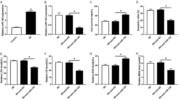
Rosuvastatin protected cardiomyocytes from ischemia-reperfusion -induced inhibition of survival and increase of apoptosis
To establish myocardial ischemia-reperfusion injury model, cardiomyocytes were suffered from the insult of ischemia-reperfusion. Com- pared with control group, ischemia-reperfusion group displayed obvious loss of survival and enhance of apoptosis in cardiomyocytes, sug-gesting the successful establishment of the model (Figure 1A and 1B). Moreover, to explore the effect of rosuvastatin on ischemia-reperfu-sion injury, we used three doses of rosuvas-tatin, 2.5 mg/kg (low), 5 mg/kg (medium) and 10 mg/kg (high) to treat cardiomyocytes re- spectively. The results showed that with the increase of rosuvastatin dose, the cell survival rate was gradually enhanced in ischemia-reper-fusion-treated cardiomyocytes, while the
apop-tosis rate was gradually decreased. More than that, medium and high doses of rosuvastatin significantly weakened the effect of ischemia-reperfusion on cardiomyocytes survival and apoptosis. However, low dose of rosuvastatin had no significant effect on cell survival and apoptosis.
Rosuvastatin abated effect of ischemia-reperfusion on LDH, CK, SOD and MDA level in cardiomyocytes
To further explore the protective role of ro- suvastatin in cardiomyocytes under ischemia-reperfusion, we detected the releases of LDH, CK, MDA and SOD in the media. As shown in
Figure 2A, 2B and 2D, the levels of LDH, CK and MDA were obviously increased in cardio-myocytes after treatment of ischemia-reperfu-sion compared with those in control group. However, ischemia-reperfusion treatment shar- ply lowered the level of SOD in cardiomyocytes (Figure 2C). Interestingly, after different doses of rosuvastatin treatment, the levels of LDH, CK, SOD and MDA were notably reversed in ischemia-reperfusion-treated cardiomyocytes at medium and high doses of rosuvastatin (Figure 2A-D). Low dose of rosuvastatin showed little effect on the levels of LAH, CK, MAD and SOD in cardiomyocytes.
Knockdown of miR-155 decreased ischemia-reperfusion injury in cardiomyocytes
Figure 1. Rosuvastatin attenuated ischemia-reperfusion-induced survival suppression and apoptosis production in cardiomyocytes. cardiomyocytes were treated with ischemia-reperfusion and then incubated with different concentrations of rosuvastatin. Cell survival (A) and apoptosis (B) were detected using MTT assay or
3E, 3F and 3H), while the level of SOD was ele-vated in anti-miR-155-transfected cardiomyo-cytes (Figure 3G).
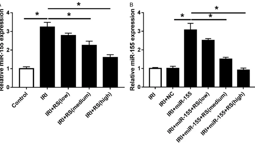
Rosuvastatin decreased miR-155 expression in cardiomyocytes under ischemia-reperfusion
To further explore the relationship between miR-155 and rosuvastatin in ischemia-reperfu-sion-treated cardiomyocytes, we detected the miR-155 expression in cardiomyocytes after treatment of ischemia-reperfusion and rosuv-astatin. Results revealed that with the increase of rosuvastatin dose, miR-155 expression was gradually decreased in ischemia-reperfusion-treated cardiomyocytes (Figure 4A). Furthermo- re, we used different doses of rosuvastatin to
treat cardiomyocytes with high expression of miR-155. The results showed that transfection of miR-155 mimic significantly increased the miR-155 level in ischemia-reperfusion-treated cardiomyocytes, while it was progressively re- duced under the medium and high doses of rosuvastatin (Figure 4B).
Discussion
[image:5.612.93.524.72.463.2]Figure 3. Knockdown of miR-155 promoted cell survival and reduced apoptosis in ischemia-reperfusion-treated cardiomyocytes. (A) The expression of miR-155 was
detected by qRT-PCR in cardiomyocytes with or without treatment of ischemia-reperfusion. Cardiomyocytes were transfected with anti-miR-155 or anti-NC and then
dial tissue damage. However, the underlying mechanism of myocardial ischemia reperfu-sion injury is not completely understood. Many studies have shown that rosuvastatin is effective in curing myocardial ischemia-reper-fusion. For example, rosuvastatin pretreatment was reported to decrease inflammatory and myocardial injury under ischemia-reperfusion condition, which might be mediated through the HMG-CoA reductase pathway [6, 19]. Me- anwhile, rosuvastatin pretreatment also incre- ased SOD activity and decreased LDH, CK, MDA and troponin I/T activities with myocardial ischemia-reperfusion condition [20]. Moreover, postconditioning with rosuvastatin reduced the infarct size and the activity of LDH, CK and MDA, increased SOD activity of myocardial is- chemia-reperfusion injury by regulating the ex- pression of high mobility group box 1 protein [21]. Meanwhile, rosuvastatin improved sys-temic and regional hemodynamics by reduc- ing vascular resistance. And the protective ef- fects of rosuvastatin on vascular and cardio cells was known to promote the production of NO in vascular endothelial cells and alleviate myocardial necrosis after ischemia-reperfusion [22, 23]. In our present study, we found that rosuvastatin obviously attenuated ischemia-re-
perfusion-induced survival suppression, apop-tosis induction and levels of LDH, CK, SOD and MAD in cardiomyocytes, suggesting that rosuv-astatin decreased myocardial ischemia-reper-fusion injury. This also indicated that rosuvas-tatin might serve as a protective agent for th- erapeutic intervention of cardiovascular dise- ases.
[image:7.612.97.514.70.304.2]MiRNAs are associated with cell growth, im- mune response and inflammatory injury in car -diovascular diseases. For instance, miR-125a, miR-139 and miR-314 promoted Urocortin pro-tection to reduce myocardial ischemia-reper- fusion injury [17]. Besides, miR-126 played the protective effects on myocardial infarction by regulating VEGF-A expression [24]. miR-203 was found to be associated with inflammatory injury in myocardial ischemia-reperfusion injury [25]. Of note, miR-155 was another important biomarker for the diagnosis of cardiovascular diseases. Bao et al. suggested that miR-155 and miR-148 reduced cardiac injury of acute viral myocarditis via inhibiting NF-kB pathway [26]. Moreover, miR-155/MMP-16 axis inhibit-ed cell migration in human cardiomyocyte pro-genitor cells [27]. Various studies reported that miR-155 is widely implicated in the inflamma -tory response of myocardial tissues and cells. Figure 4. Rosuvastatin decreased miR-155 expression in ischemia-reperfusion-treated cardiomyocytes. A. The ex-pression of miR-155 was detected by qPR-PCR after treatment of ischemia-reperfusion and rosuvastatin. B. The expression of miR-155 was measured in miR-155-transfected cardiomyocytes via qRT-PCR after treatment of
On the other hand, miR-155 was an important regulatory molecule in ischemia-reperfusion in- jury. The expression of miR-155 was raised in myocardial ischemia-reperfusion injury and im- proved the cytokine expression and ROS ex- pression by down-regulating SOCS-1 [28]. Wu et al. reported that miR-155/FoxO3a/ARC axis resulted in renal pyroptosis with the renal isch-emia-reperfusion injury conditions [29]. In our study, we found that miR-155 expression was enhanced in cardiomyocytes after treatment of ischemia-reperfusion, suggesting that miR- 155 might contribute to myocardial ischemia-reperfusion injury. To verify the biological func-tion of miR-155 in ischemia-reperfusion injury, we obtained lowly-expressed cardiomyocytes for the further investigate. The results showed that knockdown of miR-155 promoted survival rate and inhibited its apoptosis. At the same time, anti-miR-155 transfection obviously de- creased the level of LDH, CK and MAD and increased the level of SOD in myocardial cells under ischemia-reperfusion condition. These findings suggested that down-regulation of miR-155 attenuated ischemia-reperfusion in- jury in cardiomyocytes.
Additionally, we observed that the medium and high dose of rosuvastatin could significantly decrease miR-155 expression in ischemia-re- perfusion-treated cardiomyocytes, which is al- so in agreement with a previous study which presented that rosuvastatin reduced the inci-dence of cardiovascular events by suppressing miR-155/SHIP-1 signaling pathway in patients [16]. According to this, we hypothesized that miR-155 played an important regulatory role in the treatment of rosuvastatin to relieve myocar-dial ischemia-reperfusion injury. In this study, the medium and high doses of rosuvastatin decreased miR-155 expression in cardiomyo-cytes transfected with miR-155 mimic, sugge- sting that rosuvastatin reduces myocardial is- chemia-reperfusion injury by inhibiting miR-155 expression.
This study indicated the cardioprotective role of rosuvastatin in vitro. However, there are some limitations in the present work. Functional miR-NAs were known to regulate the related targets in varying conditions. However, the potential target of miR-155 participating in this mecha-nism was absent in this study. Moreover, this study showed little in vivo data on the effect of
rosuvastatin. Hence, the promising target and signaling pathway as well as animal experi-ments should be explored in further study. In this study, our finding suggested that rosuv -astatin reduced myocardial ischemia-reperfu-sion injury by inhibiting the expresischemia-reperfu-sion of miR-155, providing a new point for the treatment of ischemia-reperfusion injury in cardiovascular diseases.
Acknowledgements
This work was supported by the Youth Science and Technology Research Project of Health and Family Planning Commission in Hebei Province (Grant No. 20170231), the Youth Talent Project in Hebei Province, The Excellent Youth Fund Project of Hebei Education Department (Grant No. YQ2013005) and Funding of Key Discipline Construction Projects in Hebei Province ([2013] No. 4 Pathology and Pathophysiology).
Disclosure of conflict of interest
None.
Address correspondence to: Juan Zhao, Department of Pathophysiology, Chengde Medical University, Anyuan Rd, Shuangqiao District, Chengde 067000,
Hebei, P. R. China. Tel: +86-314-2291234; E-mail:
fbbmmzz@126.com
References
[1] Mustoe T. Understanding chronic wounds: a unifying hypothesis on their pathogenesis and implications for therapy. Am J Surg 2004; 187: S65-S70.
[2] Taylor F, Huffman MD, Macedo AF, Moore THM,
Burke M, Davey Smith G, Ward K and Ebrahim
S. Statins for the primary prevention of cardio-vascular disease. Cochrane Database Syst Rev 2013; 1: CD004816.
[3] Fellström BC, Jardine AG, Schmieder RE,
Hold-aas H, Bannister K, Beutler J, Chae DW, Che -vaile A, Cobbe SM, Grönhagenriska C, De Lima JJ, Lins R, Mayer G, McMahon AW, Parving HH, Remuzzi G, Samuelsson O, Sonkodi S, Sci D, Suleymanlar G, Tsakiris D, Tesar V, Todorov V, Wiecek A, Wuthrich RP, Gottlow M, Johnsson E, Zannad F and Group AS. Rosuvastatin and car-diovascular events in patients undergoing he-modialysis. N Engl J Med 2009; 360: 1395-1407.
Rosu-vastatin reduces interleukin-6-induced expres-sion of C-reactive protein in human hepatocytes in a STAT3- and C/EBP-dependent fashion. Int J Clin Pharmacol Ther 2007; 45: 319-327. [5] Bulhak AA, Gourine AV, Gonon AT, Sjöquist PO,
Valen G and Pernow J. Oral pre-treatment with rosuvastatin protects porcine myocardium from ischaemia/reperfusion injury via a mech-anism related to nitric oxide but not to serum cholesterol level. Acta Physiol Scand 2005; 183: 151-159.
[6] Ke D, Fang J, Fan L, Chen Z and Chen L. Regu -latory T cells contribute to rosuvastatin-in-duced cardioprotection against ischemia-re-perfusion injury. Coron Artery Dis 2013; 24: 334-341.
[7] Naito Y, Katada K, Takagi T, Tsuboi H, Kuroda M, Handa O, Kokura S, Yoshida N, Ichikawa H
and Yoshikawa T. Rosuvastatin reduces rat in-testinal ischemia-reperfusion injury associat-ed with the preservation of endothelial nitric oxide synthase protein. World J Gastroenterol 2006; 12: 2024-2030.
[8] Kelle I, Akkoç H, Uyar E, Erdinç M, Evliyaoğlu O, Sarıbaş S, Tunik S and Özoğul C. The combined
effect of rosuvastatin and ischemic pre- or post-conditioning on myocardial ischemia-re-perfusion injury in rat heart. Eur Rev Med Phar-macol Sci 2015; 19: 2468-2476.
[9] Filipowicz W, Jaskiewicz L, Kolb FA and Pillai
RS. Post-transcriptional gene silencing by siR-NAs and miRsiR-NAs. Curr Opin Struct Biol 2005; 15: 331-341.
[10] Bartel DP. MicroRNAs: genomics, biogenesis, mechanism, and function. Cell 2004; 116: 281-297.
[11] Hsu CY, Hsieh TH, Tsai CF, Tsai HP, Chen HS, Chang Y, Chuang HY, Lee JN, Hsu YL and Tsai EM. miRNA-199a-5p regulates VEGFA in endo-metrial mesenchymal stem cells and contrib-utes to the pathogenesis of endometriosis. J Pathol 2014; 232: 330-343.
[12] Huang X and Lu S. MicroR-545 mediates colorectal cancer cells proliferation through up-regulating epidermal growth factor receptor expression in HOTAIR long non-coding RNA de-pendent. Mol Cell Biochem 2017; 431: 45-54. [13] Zhao J, Fu Y, Wu J, Li J, Huang G and Qin L. The
diverse mechanisms of miRNAs and lncRNAs in the maintenance of liver cancer stem cells. Biomed Res Int 2018; 2018: 8686027. [14] Aronica E, Fluiter K, Iyer A, Zurolo E, Vreijling J,
Van Vliet EA, Baayen JC and Gorter JA.
Expres-sion pattern of miR-146a, an inflammation-as -sociated microRNA, in experimental and hu-man temporal lobe epilepsy. Eur J Neurosci 2010; 31: 1100-1107.
[15] Tili E, Michaille JJ, Wernicke D, Alder H, Costin-ean S, Volinia S and Croce CM. Mutator activity
induced by microRNA-155 (miR-155) links
in-flammation and cancer. Proc Natl Acad Sci U S
A 2011; 108: 4908-4913.
[16] Xie W, LI P, Wang Z, Chen J, Lin Z, Liang X and Mo Y. Rosuvastatin may reduce the incidence of cardiovascular events in patients with acute coronary syndromes receiving percutaneous coronary intervention by suppressing miR-155/SHIP-1 signaling pathway. Cardiovasc Ther 2014; 32: 276-282.
[17] Díaz I, Calderón-Sánchez E, Toro RD, Ávila-Mé-dina J, de Rojas-de Pedro ES, Domínguez- Ro-dríguez A, Rosado JA, Hmadcha A, Ordóñez A and Smani T. 125a, 139 and miR-324 contribute to Urocortin protection against myocardial ischemia-reperfusion injury. Sci Rep 2017; 7: 8898.
[18] Thomas H, Diamond J, Vieco A, Chaudhuri S, Shinnar E, Cromer S, Perel P, Mensah GA, Na-rula J, Johnson CO, Roth GA and Moran AE. Global atlas of cardiovascular disease 2000-2016: the path to prevention and control. Glob Heart 2018; 13: 143-163.
[19] Ma M, Uekawa K, Hasegawa Y, Nakagawa T, Katayama T, Sueta D, Toyama K, Kataoka K, Koibuchi N, Kuratsu J and Kim-Mitsuyama S.
Pretreatment with rosuvastatin protects ag- ainst focal cerebral ischemia/reperfusion in-jury in rats through attenuation of oxidative
stress and inflammation. Brain Res 2013;
1519: 87-94.
[20] Wang L, Lin R, Guo L and Hong M. Rosuvas-tatin relieves myocardial ischemia/reperfusion
injury by upregulating PPAR-γ and UCP2. Mol
Med Rep 2018; 18: 789-798.
[21] Du X, Hu X and Wei J. Postconditioning with ro-suvastatin reduces myocardial ischemia-reper-fusion injury by inhibiting high mobility group box 1 protein expression. Exp Ther Med 2014; 7: 117-120.
[22] Jones SP, Gibson MF, Rimmer DM, Gibson TM, Sharp BR and Lefer DJ. Direct vascular and cardioprotective effects of rosuvastatin, a new HMG-CoA reductase inhibitor. J Am College Cardiol 2002; 40: 1172-1178.
[23] Susic D, Varagic J, Ahn J, Slama M and Frohlich
ED. Beneficial pleiotropic vascular effects of
rosuvastatin in two hypertensive models. J Am College Cardiol 2003; 42: 1091-1097. [24] Fei L, Zhang J, Niu H, Yuan C and Ma X. Effects
of rosuvastatin and MiR-126 on myocardial in-jury induced by acute myocardial infarction in rats: role of vascular endothelial growth factor a (VEGF-A). Med Sci Monit 2016; 22: 2324-2334.
[25] Wang S, Yu W, Chen J, Yao T and Deng F. LncRNA MALAT1 sponges miR-203 to promote
[26] Bao JL and Lin L. MiR-155 and miR-148a
re-duce cardiac injury by inhibiting NF-κB path -way during acute viral myocarditis. Eur Rev Med Pharmacol Sci 2014; 18: 2349-2356. [27] Liu J, van Mil A, Aguor EN, Siddiqi S, Vrijsen K,
Jaksani S, Metz C, Zhao J, Strijkers GJ, Do- evendans PA and Sluijter JP. MiR-155 inhibits cell migration of human cardiomyocyte progen-itor cells (hCMPCs) via targeting of MMP-16. J Cell Mol Med 2012; 16: 2379-2386.
[28] Eisenhardt SU, Weiss JB, Smolka C, Maxeiner
J, Pankratz F, Bemtgen X, Kustermann M,
Thiele JR, Schmidt Y, Bjoern Stark G, Moser M, Bode C and Grundmann S. MicroRNA-155 ag-gravates ischemia-reperfusion injury by
modu-lation of inflammatory cell recruitment and the
respiratory oxidative burst. Basic Res Cardiol 2015; 110: 32.