Original Article
Effects of regulatory dendritic cells on graft-versus-host
disease and graft-versus-leukemia in mice after
allogeneic bone marrow transplantation
Xiao-Tong Yue1,2*, Xin-Xin Wei1*, Wen-Hua Jia1,2, Hui Mao1, Wan-Ru Chen1, Hong-Hong Gu1, Jia-Qing Wu2, De-Peng Li1, Kai-Lin Xu1, Yi-Hong Huang1,2
1Department of Hematology, The Affiliated Hospital of Xuzhou Medical University, 99 Huaihai Road West, Xuzhou
221002, Jiangsu, P.R. China; 2Department of Hematology, The Third Affiliated Hospital of Xuzhou Medical
University, Xuzhou 221003, Jiangsu, P.R. China. *Equal contributors.
Received March 19, 2017; Accepted June 14, 2018; Epub August 15, 2018; Published August 30, 2018
Abstract: We aimed to investigate the effects of regulatory dendritic cells (DCreg) on acute graft-versus-host disease (GVHD) and graft-versus-leukemia (GVL) in leukemia mouse model after allogeneic bone marrow transplantation (al-lo-BMT). DCreg with donor bone marrow cells and splenocytes were subsequently transplanted into myeloablatively irradiated mice. The mice were divided into the TBI (Total body irradiation), leukemia, allo-BMT (bone marrow and spleen cells from donors were injected into recipients after TBI), allo-BMT with immature DC (imDC), and allo-BMT with DCreg (DCreg) groups. DCreg were cultured for longer time than imDC and stimulated by LPS for 1 d. Survival
time in the DCreg and imDC groups was significantly prolonged compared with that of allo-BMT group (P < 0.05).
Moreover, mild histological changes of GVHD or leukemia were observed in mice from DCreg group, with significantly decreased clinical GVHD scores compared with those in allo-BMT and imDC groups. Serum interferon-γ level was decreased in imDC and DCreg groups compared with that in allo-BMT group with even more significantly reduced
in DCreg (P < 0.01). Additionally, Serum interleukin-10 level was gradually increased in imDC and DCreg groups (P < 0.01) but decreased in allo-BMT group. In conclusion, DCreg cell infusion reduced the incidence and ameliorated the severity of GVHD in allo-BMT mouse model, while preserved the effect of GVL.
Keywords: Regulatory dendritic cells, allograft, transplantation, immunotolerance, antileukemia
Introduction
Allogeneic bone marrow transplantation (allo-BMT) is a therapeutic approach for the treat-ment of hematological malignancies, including leukemia, lymphoma, and myelodysplastic syn-drome. However, graft-versus-host disease (GVHD) and leukemia recurrence are critical factors affecting the survival rate and progno-sis after transplantation [1, 2]. Immunosupp- ressive agents and T cell depletion are curr- ently being used in clinic to prevent GVHD. Nevertheless, the efficacy is limited by severe infections and leukemia recurrence. To allevi-ate the complications and improve the thera-peutic outcome for patients, it is urgent to develop new strategies and methods which can effectively prevent GVHD but also preserve or even enhance GVL effects simultaneously [3-5].
tolerance and proliferation potential, and could induce T cell resistance against specific anti -gens, consistent with the findings of Wakkach et al. [10]. Additionally, CD11clowCD45RBhighDC could inhibit mixed lymphocyte reactions (MLR) and, more importantly, this tolerance could be transferred to other individuals.
In mice, adoptive infusions of CD11clowCD45- RBhighDC induced OVA-specific immunological unresponsiveness in T cells and mediated anti-gen-specific Tr1 production [6, 11]. Meanwhile, the survival time of mice receiving allo-BMT was increased significantly and the inflamma -tory response was inhibited after infusion of CD11clowCD45RBhighDC [11, 12]. According to Chorny et al. [13], vasoactive intestinal pep-tides cultured DCreg not only ameliorated GVHD in mice, but also maintained the GVL effects. Consistent with this, our previous find -ing also showed that infusion of immature DC (imDC) and gene modified imDC into mice could effectively prevent acute GVHD development [14]. However, whether the persistent tolerance of DCreg could improve the transplant graft tolerance and prevent GVHD still remains unknown. In this study, we assessed the effects of CD11clowCD45RBhighDC on GVHD and GVL in leukemia mouse model after allo-BMT.
Materials and methods
Materials
RPMI 1640 culture medium, fetal bovine serum, and IMDM culture medium were pur-chased from Gibco (Shanghai, China). Tris, boric acid, osmic acid, L-glutamine, glutaraldehyde, and HEPES were provided by Sigma (St. Louis, MO, USA). mouse GM-CSF, IL-4, IL-10, and TGF-β1 were bought from PeproTech (Rocky Hill, NJ, USA). FITC labeled mouse CD11c antibody and FITC-I-A/I-E antibody, PE labeled CD80, CD86, and CD45RB antibodies, and isotype control antibody was purchased from eBioscience (San Diego, CA, USA). Anti-mouse H-2Kb-FITC and anti-mouse H-2Kd-PE were obtained from BD Biosciences (San Jose, CA, USA). FACS Calibur flow cytometer was offered by BD Biosciences. Animals
C57BL/6 (H-2b) mice (SPF grade) were used as the donors, and BALB/c (H-2d) mice (SPF grade) were used as the recipients. All mice were
between 8-12 weeks old and purchased from the Shanghai SLAC Laboratory Animal Co., Ltd. [SCXK (Hu) 2012-0002]. The mice were kept in the animal center (SPF grade) of Xuzhou Medical University. The cages were sterilized by immersing in 1:10000 of Gaolvjing and were changed every day. Food, water, and padding were all sterilized. Erythromycin (250 mg/L) and gentamicin (320 mg/L) were added into the drinking water a week before transplanta-tion. All procedures performed in the experi-ments were in accordance with animal ethical standards.
SHI-1 cell thawing and passage
A highly invasive monocytic leukemic cell line SHI-1, with specific t (6; 11) (q27; q23) chromo -somal abnormality and MLL/AF6 fusion tran-script [15], was kindly provided by the Institute of Hematology of Jiangsu Province. The frozen SHI-1 cells were taken from liquid nitrogen con-tainers and put into 37°C water bath to thaw the cells within 1 min. The SHI-1 cell suspen-sion was transferred into a sterilized 15 ml cen-trifuge tube and washed with basal IMDM ture medium twice. Then, the cells were cul-tured in culture flasks with 10 ml of 10% IMDM complete culture medium at 37°C with 5% CO2. When the culture medium became yellow and the cell number increased distinctly, the cell suspension was collected and centrifuged at 800 × g, the supernatant was discarded, 2 ml of IMDM complete culture medium was added, and the cells were passaged (1:3). The third cell generation was used for the following experiments.
Separation of bone marrow cells and spleen cells
Acquisition and identification of DC
Bone marrow-derived MNC was obtained from C57BL/6 mice under aseptic conditions and seeded into a 6-well culture plate with RPMI 1640 complete culture medium. Different cyto-kines were added into corresponding groups to culture different DC types. For the imDC induc-tion, we used RPMI 1640 culture medium con-taining rmGM-CSF (20 ng/mL) and rmIL-4 (10 ng/mL) to culture the cells for 5 days. To induce mature DC, we used RPMI 1640 culture medi-um containing rmGM-CSF (20 ng/mL) and rmIL-4 (10 ng/mL) to culture the cells for 6 days, and then stimulated the cells by LPS for 24 h to allow them to mature. To induce DCreg, we used RPMI 1640 culture medium containing rmGM-CSF (20 ng/mL), rmIL-10 (20 ng/mL) and transforming growth factor-β1 (TGF-β1, 20 ng/mL) to culture the cells for 6 days, and then stimulated the cells by LPS for 24 h to allow them to mature. The DC morphology was observed with optical microscope, cell ultra-structure was observed with electronic micro-scope, and cell immunophenotype was mea-sured by flow cytometry.
The ability of DCs in stimulating T lymphocyte proliferation was measured with a CCK-8 meth-od, and in vitro chemotactic cell capabilities were measured with a transwell system. In brief, DCs were collected from different groups followed by addition of mitomycin (25 μg/ml) and incubated for 45 min at 37°C, 5% CO2. After washing two times, DCs (1 × 10⁵/well) were co-cultured with T lymphocytes in 1640 medium in 96-well plate with triplicates for each group for 120 h at 37°C, 5% CO2. At the last 16 h, 10 ml CCK-8 reagent was added into each well and incubated for 4 h followed by measuring the absorbance at 450 nm using a microplate reader. Only T lymphocytes were added for the control group. Proliferation rate was calculated as the OD value of experimental group divided by the OD value from the control group × 100%.
Meanwhile, DC cells migration was measured by Transwell system. Briefly, cultured DCs were collected from each group and added into the upper layer of the transwell (150 μl at 5 × 10⁵/ ml). At the meantime, 450 μl 10% FBS 1640 medium was added into the lower layer. Then the transwell system was placed at an incuba-tor (37°C, 5% CO2) for 72 h followed by
collect-ing the cells from lower layer and counted the number of cells. The migration rate was calcu-lated as number of the cells migrated into the lower layer divided by the original number of cells × 100%.
Establishing the mouse leukemia model and grouping
Total body irradiation of 60Co γ-ray (total dose: 7.5 Gy, dose rate: 0.66 Gy/min, irradiation time: 682 s) was performed for the BABL/c recipient mice on the day of transplantation for 4 hours, and then bone marrow transplantation was performed. The mice were randomly divided into 5 groups (TBI, leukemia, allo-BMT, imDC, and DCreg) using a random number table with 15 mice in each group (not including the mice that were sacrificed for chimera analysis or cytokine measurement). For the mice in the TBI group, 0.3 ml of sterilized normal saline was injected through the caudal vein; for the mice in the leukemia group, syngeneic bone marrow cells (5 × 10⁶), spleen cells (5 × 10⁵), and SHI-1 cells (1.2 × 10⁷) were injected through the cau-dal vein into each mouse after TBI; for the mice in the allo-BMT group, bone marrow cells (5 × 10⁶) and spleen cells (5 × 10⁵) from the donors were injected through the caudal vein into each recipient after TBI; for the mice in the imDC group, bone marrow cells (5 × 10⁶) and spleen cells (5 × 10⁵) from the donors, SHI-1 cells (1.2 × 10⁷), and imDC (5 × 10⁶) were injected through the caudal vein into each recipient after TBI; and for the mice in the DCreg group, bone marrow cells (5 × 10⁶) and spleen cells (5 × 10⁵) from the donors, SHI-1 cells (1.2 × 10⁷), and DCreg (5 × 10⁶) were injected through the caudal vein into each recipient after TBI.
Observation index after transplantation
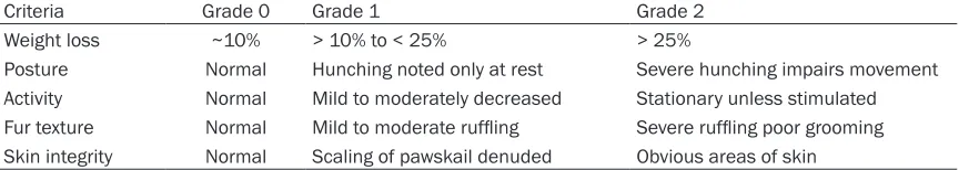
poor appetite, poor mobility, reduced body weight, roachback, ruffled fur, and diarrhea. GVHD severity was evaluated by a scoring sys-tem (Table 1) reported by Cooke et al. [16], and scores were evaluated from 5 aspects includ-ing weight loss, posture, activity, fur texture, and skin integrity with the highest score of 10. 4) Development of leukemia: WBC > 20 × 109/L, hepatomegaly, splenomegaly, and a large amount of leukemia cells in peripheral blood; and histopathology showed a large num-ber of acute monocytic leukemia cell infiltration in liver, spleen, lung, and other tissues. 5) Transplantation-related death: deaths caused by bleeding or infection induced by hematopoi-etic inhibition within 2 weeks after transplanta-tion (except deaths caused by leukemia or GVHD) were defined as transplantation-related death.
Pathological examination
The liver, small intestine, lung, and spleen were collected from the moribund recipients with GVHD or recipients sacrificed on +30 d in each group. The tissues were then sliced, fixed with 100 g/L of formaldehyde, paraffin-embedded, HE stained, and observed with optical micro-scope to detect pathological GVHD changes and leukemia cell infiltration. A pathological scoring system developed by Blazar and Kaplan
et al. [17, 18] was used to evaluate the patho-logical changes of the target organs in mice with acute GVHD.
Detection of bone marrow cell chimeras
Bone marrow cells of 1 × 10⁶ were randomly selected from recipients at +18 d or from the ones that survived more than 30 d. After cells were treated with FITC-anti-H-2Kb and PE-anti-H-2Kd, flow cytometer was used to evaluate the percentage of lymphocytes that expressed donor-derived H-2Kb.
Detection MLL-AF6 fusion gene with nested RT-PCR
Total RNA was extracted from liver and spleen tissues from the mice using TRIzol one-step method, then reverse RNA transcription was performed using M-MLV (2 μg) reverse tran -scriptase to obtain cDNA. PCR was performed according to the methods reported in previous studies [19]. Agarose gel electrophoresis (1.5%) was undertaken to analyze the PCR products. A gel imaging system was used to observe and photograph the images under ultraviolet.
ELISA detection of the peripheral blood cyto
-kine
Peripheral blood from the recipient mice (1.5 mL) was collected via the fossa orbitalis vein on days 0, 5, 10, 15, 20, and 30 post-BMT and centrifuged to obtain serum, which was pre-served at -80°C until use. The levels of IFN-γ and IL-10 in peripheral blood serum were detected with ELISA kits according to the instructions by the manufacturer.
Statistical analysis
SPSS 16.0 software (SPSS Inc, Chicago, IL, USA) was used for statistical analysis. Data were described as means ± standard divisions (SD), and comparison of the differences be- tween different groups was assessed by one-way analysis of variance (one-one-way ANOVA).
q-test was used for the pairwise comparison, and the Kaplan-Meier test was applied for sur-vival rate comparison. P < 0.05 was considered statistically significant.
Results
Mature DC and DCreg enhanced proliferation of allogeneic lymphocytes
DC morphology and immunophenotype after in
[image:4.612.91.523.87.164.2]vitro induction were published in our previous
Table 1. Analysis of GVHD severity by a scoring system
Criteria Grade 0 Grade 1 Grade 2
Weight loss ~10% > 10% to < 25% > 25%
Posture Normal Hunching noted only at rest Severe hunching impairs movement Activity Normal Mild to moderately decreased Stationary unless stimulated Fur texture Normal Mild to moderate ruffling Severe ruffling poor grooming
study [12]. The regulatory DC subgroup was featured as CD11clowCD45RBhigh DCreg. MLR results showed that imDC only mildly stimulat-ed T lymphocyte proliferation (cell proliferation rate: 270.76 ± 12.25%), while mature DC effec -tively stimulated T lymphocyte proliferation (cell proliferation rate: 348.94 ± 14.45%, P < 0.01). Conversely, the T lymphocyte proliferation was significantly lower after stimulation with DCreg (cell proliferation rate: 237.05 ± 7.60%) com -pared with imDC (P < 0.01) and mature DC stimulation (P < 0.01).
The mature DC, imDC and DCreg possessed strong capability of migration
To understand the DC capability of migration, we investigated the migration of three DC groups by chemotactic assay. The migration rate of mature DC, imDC, and DCreg were 8.13 ± 0.97%, 15.94 ± 0.87% and 26.81 ± 0.43%, respectively, suggesting that the migration capability of DCreg was the strongest in the three DC group.
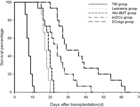
The imDC and DCreg potently prolonged mice survival after transplantation
In order to investigate the effect of different DC on mice survival, mice survival time was com-pared. In the TBI group the first death was found on +6 d, and then the mice gradually
on +19 d, with 46.7% mice alive on +30 d, and 13.3% alive on +60 d, with an AST of 36.00 ± 13.76 d. Taken together, these results indicat-ed that DCreg significantly prolongindicat-ed the sur -vival time in allo-BMT model mice (P < 0.05) (Figure 1).
DCreg infusion significantly prevented the de
-velopment of GVHD
To identify the effect of Dcreg on GVHD, we evaluated clinical acute GVHD phenotypes in- cluding tiredness, poor mobility, reduced body weight, roachback, ruffled fur, hair loss, diar -rhea, and even bloody stool in allo-BMT, imDC, and DCreg groups. The most severe symptoms were found in allo-BMT group, while the least severe symptoms were found in DCreg group. The GVHD clinical score was 7.60 ± 0.99, 4.93 ± 0.96, and 3.8 ± 0.68 in the allo-BMT, imDC, and DCreg groups, respectively. This result showed that DCreg infusion significantly pre -vented the development of GVHD compared with other two groups (P < 0.05).
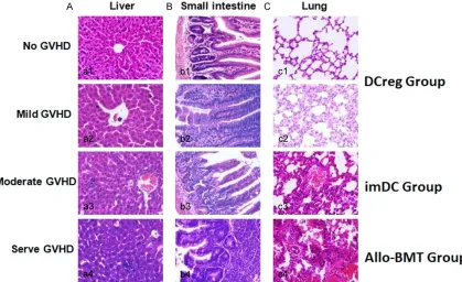
The imDC and DCreg reduced the pathological GVHD score compared with allo-BMT
[image:5.612.92.371.73.286.2]In order to assess the effect of imDC, DCreg, and allo-BMT on GVHD score, the pathological changes of liver, small intestine, and lung were investigated in accordance with the clinical Figure 1. Post-BMT survival curves of mice in different. transplantation
groups. imDC group versus allo-BMT group, P < 0.05; DCreg group versus imDC and allo-BMT group, P < 0.05.
phenotypes in each group after transplanta-tion. In allo-BMT and imDC groups, there were obvious derangement of liver cells, focal necro-sis, lymphocyte infiltration in the periportal area, destruction and collapse of the small bile duct in liver tissues, intestinal epithelial cells necrosis and partial defect excalation with sig-nificant inflammatory cells infiltration in the underlayer, intestinal villous atrophy degenera-tion, and even severe structural damage to the small intestine. Furthermore, inflammatory cells infiltration and lung structure damages were observed in 25% pulmonary vessels. Meanwhile, inflammatory cells were infiltrated into 2-3 parenchymal cell layers. However, in
The incidence of leukemia in allo-BMT, imDC and DCreg groups was similar
[image:6.612.97.516.74.330.2]To study the effect of allo-BMT, imDC and DCreg on the occurrence of leukemia, mice in each group were observed. Leukemia cells infiltra -tion in eyes was found on +18 d in mice from leukemia group, and all mice in this group died of leukemia within 24 d. Significantly enlarged liver, spleen and leukemia cell infiltrated dif -fused lesions were detected (Figure 3A). In his-topathological examinations, higher numbers of leukemia cells were infiltrated in the liver and spleen (Figure 3B). The WBC count in the peripheral blood was 19~34 × 109/L, and 28%-Figure 2. Histopathological changes of liver, small intestine, and lung in mice (HE, × 400). A: Liver; B: Small intes-tine; C: Lung; a1, b1, c1: Liver, small intestine, and lung in mice which showed no evidence of GVHD; a2, b2, c2: Liver, small intestine, and lung in mice with mild GVHD; a3, b3, c3: Liver, small intestine, and lung in mice with moderate GVHD; a4, b4, c4: Liver, small intestine, and lung in mice with severe GVHD. A pathological scoring system developed by Blazar and Kaplan et al. [17, 18] was used as the criteria for mild, moderate and severe GVHD.
Table 2. GVHD pathologic scoring of liver, small intes-tine, and lung in mice after transplantation (n = 3, mean ± SD)
Groups Liver Small intestine Lung Allo-BMT 6.33 ± 0.58 7.00 ± 1.00 6.67 ± 0.58 imDC 4.67 ± 0.58* 4.67 ± 0.58* 2.33 ± 1.16*
DCreg 3.00 ± 1.00*,# 2.67 ± 0.58*,# 2.00 ± 1.00*,#
*P < 0.05 vs. allo-BMT group; #P < 0.05 vs. imDC group.
[image:6.612.89.323.449.502.2]43% white blood cells were leukemia cells. Leukemia incidence was 13.3%, 20%, and
Complete bone marrow chimeras pointed to successful allo-BMT
Bone marrow cells were harvested from mice that lived more than 30 d in imDC and DCreg groups, and flow cytometer was used to detect bone marrow cell chimeras. The results showed that 95-100% of the cells were positive for H-2Kb, which suggested complete cell chime-rism and demonstrated that the allo-BMT per-formed in the present study was successful (Figure 5). In addition, significantly increased numbers of white blood cells were observed on d 14 (1.54 ± 0.50 × 10⁹/L) in mice than those on d 7 (0.34 ± 0.27 × 10⁹/L) after allo-BMT (P < 0.05), suggesting hematopoiesis recovery after transplantation.
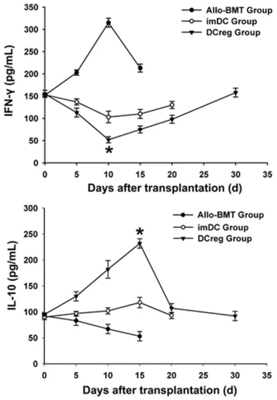
Serum interleukin-10 level was gradually increased in imDC and DCreg groups but de
-creased in allo-BMT group
[image:7.612.90.374.68.314.2]The cytokine changes in mice after transplan- tation were shown in Figure 6. Plasma IFN-γ level was increased gradually in allo-BMT group and peaked on +10 d, followed by a decrease gradually. In imDC and DCreg groups, the plas-ma IFN-γ level was decreased gradually and reached a lowest level on +10 d, followed by an increase gradually. The decreased plasma IFN- γ level was more pronounced in DCreg group than that in imDC group (P < 0.01). The IL-10 level was decreased gradually in allo-BMT group but increased gradually in imDC and Figure 3. Analysis of leukemia
cell infiltration into Liver. In mice
with injection of SHI-1 cells, liver was isolated for analysis
of leukemia cells infiltration (A)
[image:7.612.91.290.346.412.2]and liver pathology (× 400) (B).
Table 3. Incidence of Leukemia
Groups Leukemia incidence (%)
Leukemia 100
Allo-BMT 13.3 (2/15)
imDC 20 (3/15)
DCreg 13.3 (2/15)
DCreg group vs. imDC and allo-BMT group, P > 0.05.
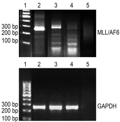
Figure 4. RT-PCR analysis of MLL/AF6 fusion gene. 1: 100 bp Marker; 2: Mouse liver; 3: Mouse spleen; 4: Healthy mouse liver; 5: ddH2O, blank control.
13.3% in allo-BMT, imDC, and DCreg groups, respectively, with no statistically significant among groups (P > 0.05) (Table 3).
MLL/AF6 expression was observed in liver and spleen tissues of leukemia group
[image:7.612.89.288.445.647.2]DCreg groups, reaching a peak on +15 d, and then decreased gradually. The increased IL-10 level was more pronounced in DCreg group than that in imDC group (P < 0.01).
GVL’s effects. Recent immunological findings show that acquiring graft-specific immune tol -erance could be an optimal approach to resolve this problem, and inducing immune tolerance with immune competent cells has become a research hotspot [22-24].
[image:8.612.93.377.74.208.2]DC is known to be the most powerful APC in the body and the only one that could activate naive T lymphocytes. It could not only initiate immune tolerance but also regulate immune response. As DCreg plays an important role in initiating and maintaining immune tolerance, it has been gained increasing attention in the immune tolerance field [25-27]. During hematopoietic stem cell differentiation into DC, different cyto-kines could substantially influence the differen -tiation. In our previous studies [12], mice bone marrow cells were stimulated with cytokines including GM-CSF, IL-10, and TGF-β1, followed by differentiation into DCreg. The high CD45RB and low CD11c, CD80, CD86, and MHC-II (IA/ IE) are expressed in DCreg cells and are defined as immature DC cells. This novel DC subgroup, namely CD11clowCD45RBhighDC, has high ability on chemotaxis migration, low ability on stimu-lating allogeneic T lymphocyte proliferation, and shows immuno-hypo-responsiveness in mixed lymphocyte reactions, offering a new method for inducing graft tolerance. In the present study, leukemia allo-BMT mouse model was induced to investigate the effects of infusing CD11clowCD45RBhighDC on GVHD and GVL’s effects in mice after bone marrow transplantation.
Figure 5. Chimeric rate of recipient mice who survived 30 days after trans-plantation in imDC and DCreg group. Flow cytometric analysis of bone mar-row cells from mice surviving for > 30 d revealed that 96.8% of cells were H-2 d positive.Bone marrow cells were dualfluorescence stained with FITC-labelled anti-H-2 Kb monoclonal antibody and PE-FITC-labelled anti-H-2 Kd mono-clonal antibody. FITC, fluorescein isothiocyanate; PE, phycoerythrin; Mice without BMT was served as a negative control.
Figure 6. Post-BMT plasma interferon-γ and IL-10 levels in different transplantation groups. Peripheral blood from the recipient mice was collected at indi-cated time points post-BMT and centrifuged to
ob-tain serum for analysis of the levels of IFN-γ and IL-10
by ELISA. *P < 0.01 vs. group allo-BMT and imDC.
Discussion
[image:8.612.92.286.309.589.2]In our previous studies, SHI-1, a human mono-cytic leukemia cell line with high infiltration and invasion, was used to induce humanized leuke-mia mouse model [15, 28, 29]. In this study, more SHI-1 cells were infused and leukemia incidence increased accordingly. All mice in leu-kemia group developed leuleu-kemia after infusion of SHI-1 cells in the present study. Extensive leukemia cell infiltration was found in the liver, spleen, and eyes of the leukemic mice with all mice died of monocytic leukemia. Spleen tis-sues from moribund mice were collected to detect the MLL/AF6 fusion gene by nested RT-PCR, and the first transcripts were identified on day 20 after transplantation, in accordance with previous findings [30, 31]. We compared the anti-leukemia effects among imDC, DCreg, and allo-BMT groups, and found that only a small proportion of mice that received alloge-neic transplantation developed leukemia. The survival rate and AST were significantly higher in imDC and DCreg groups than those in allo-BMT group, which were more pronounced in DCreg group than those in imDC group. Also, no leukemia-related complications were found in recipients that lived for a long time. These find -ings showed that infusion of imDC or DCreg into allo-BMT mice could preserve GVL’s effects to a certain degree as demonstrated by signifi -cantly higher GVL effects in DCreg group than imDC group. With regard to GVHD development, GVHD severity was significantly ameliorated in imDC group than allo-BMT group, while the severity was even lower in the DCreg group, in which the survival time was significantly longer. These findings suggested that infusion of imDC could reduce GVHD severity, but these effects were limited by several factors. After infusion, antigen stimulation increased the potential of mature imDC. Costimulatory molecule expres-sions were up-regulated with increased capa-bility of presenting antigens and this reduced immune tolerance gradually decreased the GVHD-alleviating effects. However, DCreg could preserve imDC features and effectively de- creased GVHD severity and increased the sur-vival rate.
GVHD is an immune response induced by the activated donor T cells recognizing the recipi-ents’ incompatible antigens. Recent studies suggested that GVHD is caused by a “cytokine storm” [32, 33]. Most recent studies suggest that Th1 cells could release pro-inflammatory cytokines including IL-2, IFN-γ, and TNF, which
could aggravate GVHD. Th2 cells could release Th2-type cytokines including IL-4, IL-5, and IL- 10 to antagonize the pro-inflammatory effects of Th1 cells and prevent GVHD [34, 35]. In the present study, we found that both imDC and DCreg reduced IFN-γ levels and increased IL-10 release, further confirming that imDC and DCreg could induce the donor CD4+ T cells
which are activated by the recipients’ antigens to develop into immune tolerance cells. DCreg could also produce IL-10, promote the CD4+ T
cells to be differentiated into Treg cells (which could release IL-10), and induce the naïve T cells to develop into CD4+CD25+Foxp3+ Treg
cells [36, 37] and thus increase mice survival time after transplantation. However, DCreg’s GVHD-alleviating effects are also limited, and most mice died of GVHD. We speculated that the followings might be involved: 1) after infu-sion, the in vitro-induced DCreg could be stimu-lated by exogenous antigens and inflammations and mature gradually, thus finally losing the ability to induce immune tolerance; and 2) APC in the recipients could also play a role in GVHD development. The APC from donors was selec-tively intervened, while APC activation in the recipients could not be sufficiently inhibited. Therefore, in future studies, we plan to optimize the experimental conditions, perform multiple infusions at different time points as well as in- crease the infusion dose gradually. Additionally, genetically modified DCreg could also be used to induce stable immune tolerance.
In summary, donor-derived DCreg infusion could effectively reduce GVHD incidence and severity in leukemia allo-BMT mouse model, while the GVL effects could be preserved sig-nificantly. These findings provide an effective alternative approach for applying DCreg during allo-HSCT.
Acknowledgements
The present study was supported by the Medi- cal Scientific Research Foundation of Jiangsu Province (grant no. H201427), the Xuzhou Sci- ence and Technology Plan Program (grant no. KC16SH016) and The Ordinary College Post- graduates Practice and Innovation Projects of Jiangsu Province in 2015-2017 (grant nos. SJLX15_0722 and SJZZ16_0288).
Disclosure of conflict of interest
Address correspondence to: Yi-Hong Huang and Xiao-Tong Yue, Department of Hematology, The
Affiliated Hospital of Xuzhou Medical University, Jiangsu, P.R. China. Tel: +86-13905218950; Fax: +86-051683638101; E-mail: hxr1583@sina.com; huangyihong_l@163.com (YHH); 1272344199@
qq.com (XTY)
References
[1] Mielcarek M, Storer B, Martin PJ, Forman SJ, Negrin RS, Flowers ME, Inamoto Y, Chauncey TR, Storb R, Appelbaum FR and Bensinger WI. Long-term outcomes after transplantation of HLA-identical related G-CSF-mobilized periph-eral blood mononuclear cells versus bone mar-row. Blood 2012; 119: 2675-2678.
[2] Morecki S, Yacovlev E, Gelfand Y, Shabat Y and Slavin S. Induction of graft-versus-leukemia (GVL) effect without graft-versus-host disease (GVHD) by pretransplant donor treatment with immunomodulators. Biol Blood Marrow Transplant 2009; 15: 406-415.
[3] Li N, Chen Y, He W, Yi T, Zhao D, Zhang C, Lin CL, Todorov I, Kandeel F, Forman S and Zeng D. Anti-CD3 preconditioning separates GVL from GVHD via modulating host dendritic cell and donor T-cell migration in recipients conditioned with TBI. Blood 2009; 113: 953-962.
[4] Zhang P, Chen BJ and Chao NJ. Prevention of GVHD without losing GVL effect: windows of opportunity. Immunol Res 2011; 49: 49-55. [5] Cao J, Chen C, Zeng L, Li L, Li Z and Xu K. Engi-
neered regulatory T cells prevent host disease while sparing the graft-versus-leukemia effect after bone marrow transplan-tation. Leuk Res 2010; 34: 1374-1382. [6] Sato K, Yamashita N, Baba M and Matsuyama
T. Modified myeloid dendritic cells act as regu -latory dendritic cells to induce anergic and regulatory T cells. Blood 2003; 101: 3581-3589.
[7] Zhang B, Liu R, Shi D, Liu X, Chen Y, Dou X, Zhu X, Lu C, Liang W, Liao L, Zenke M and Zhao RC. Mesenchymal stem cells induce mature den-dritic cells into a novel Jagged-2-dependent regulatory dendritic cell population. Blood 2009; 113: 46-57.
[8] Liu QY, Yao YM, Zhang SW, Yan YH and Wu X. Naturally existing CD11c(low)CD45RB(high) dendritic cells protect mice from acute severe
inflammatory response induced by thermal in -jury. Immunobiology 2011; 216: 47-53. [9] Yan YH, Shang PZ, Lu QJ and Wu X. Triptolide
regulates T cell-mediated immunity via induc-tion of CD11c(low) dendritic cell differentia-tion. Food Chem Toxicol 2012; 50: 2560-2564.
[10] Wakkach A, Fournier N, Brun V, Breittmayer JP, Cottrez F and Groux H. Characterization of den-dritic cells that induce tolerance and T regula-tory 1 cell differentiation in vivo. Immunity 2003; 18: 605-617.
[11] Fujita S, Seino K, Sato K, Sato Y, Eizumi K, Yamashita N, Taniguchi M and Sato K. Regu- latory dendritic cells act as regulators of acute
lethal systemic inflammatory response. Blood
2006; 107: 3656-3664.
[12] Fujita S, Sato Y, Sato K, Eizumi K, Fukaya T, Kubo M, Yamashita N and Sato K. Regulatory dendritic cells protect against cutaneous chronic graft-versus-host disease mediated
through CD4+CD25+Foxp3+ regulatory T cells.
Blood 2007; 110: 3793-3803.
[13] Chorny A, Gonzalez-Rey E, Fernandez-Martin A, Ganea D and Delgado M. Vasoactive intestinal peptide induces regulatory dendritic cells that prevent acute graft-versus-host disease while maintaining the graft-versus-tumor response. Blood 2006; 107: 3787-3794.
[14] Huang Y, Feng S, Xu Y, Chen W, Wang S, Li D, Li Z, Lu Q, Pan X and Xu K. Suppression of graft-versus-host disease and retention of graft-ver-sus-tumour reaction by murine genetically en-gineered dendritic cells following bone marrow transplantation. Mol Med Rep 2015; 11: 3820-3827.
[15] Chen S, Xue Y, Zhang X, Wu Y, Pan J, Wang Y and Ceng J. A new human acute monocytic leu-kemia cell line SHI-1 with t(6;11)(q27;q23), p53 gene alterations and high tumorigenicity in nude mice. Haematologica 2005; 90: 766-775.
[16] Cooke KR, Kobzik L, Martin TR, Brewer J, Delmonte J Jr, Crawford JM and Ferrara JL. An experimental model of idiopathic pneumonia syndrome after bone marrow transplantation: I. The roles of minor H antigens and endotoxin. Blood 1996; 88: 3230-3239.
[17] Blazar BR, Taylor PA, McElmurry R, Tian L, Panoskaltsis-Mortari A, Lam S, Lees C, Wald- schmidt T and Vallera DA. Engraftment of
se-vere combined immune deficient mice receiv -ing allogeneic bone marrow via In utero or postnatal transfer. Blood 1998; 92: 3949-3959.
[18] Kaplan DH, Anderson BE, McNiff JM, Jain D, Shlomchik MJ and Shlomchik WD. Target anti-gens determine graft-versus-host disease phe-notype. J Immunol 2004; 173: 5467-5475. [19] Mitterbauer G, Zimmer C, Pirc-Danoewinata H,
[20] Warren EH and Deeg HJ. Dissecting graft-ver-sus-leukemia from graft-versus-host-disease using novel strategies. Tissue antigens 2013; 81: 183-193.
[21] Sinkovics JG. Antileukemia and antitumor ef-fects of the graft-versus-host disease: a new immunovirological approach. Acta Microbiol Immunol Hung 2010; 57: 253-347.
[22] Alpdogan O and van den Brink MR. Immune tolerance and transplantation. Semin Oncol 2012; 39: 629-642.
[23] Cools N, Ponsaerts P, Van Tendeloo VF and Berneman ZN. Balancing between immunity and tolerance: an interplay between dendritic cells, regulatory T cells, and effector T cells. J Leukoc Biol 2007; 82: 1365-1374.
[24] Huang Y, Feng S, Tang R, Du B, Xu K and Pan X.
Efficacy of pretreatment of allografts with me -thoxypolyethylene glycol-succinimidyl-propion-ic acid ester in combination with an anti-OX40L monoclonal antibody in relieving graft-versus-host disease in mice. Int J Hematol 2010; 92: 609-616.
[25] Raich-Regue D, Glancy M and Thomson AW. Regulatory dendritic cell therapy: from rodents to clinical application. Immunol Lett 2014; 161: 216-221.
[26] Li YP, Latger-Canard V, Marchal L, Li N, Ou-Yang JP and Stoltz JF. The regulatory role of dendritic cells in the immune tolerance. Biomed Mater Eng 2006; 16: S163-S170.
[27] Min WP, Zhou D, Ichim TE, Strejan GH, Xia X, Yang J, Huang X, Garcia B, White D, Dutartre P, Jevnikar AM and Zhong R. Inhibitory feedback loop between tolerogenic dendritic cells and regulatory T cells in transplant tolerance. J Immunol 2003; 170: 1304-1312.
[28] Wang C, Chen Z, Li Z and Cen J. The essential roles of matrix metalloproteinase-2, mem-brane type 1 metalloproteinase and tissue in-hibitor of metalloproteinase-2 in the invasive capacity of acute monocytic leukemia SHI-1 cells. Leuk Res 2010; 34: 1083-1090.
[29] Feng S, Huang Y and Chen Z. Does VEGF se-creted by leukemic cells increase the permea-bility of blood-brain barrier by disrupting tight-junction proteins in central nervous system leukemia? Med Hypotheses 2011; 76: 618-621.
[30] Reikvam H, Hatfield KJ, Oyan AM, Kalland KH,
Kittang AO and Bruserud O. Primary human acute myelogenous leukemia cells release ma-trix metalloproteases and their inhibitors:
re-lease profile and pharmacological modulation.
Eur J Haematol 2010; 84: 239-51.
[31] Cavallo F, Forni M, Riccardi C, Soleti A, Di Pierro F and Forni G. Growth and spread of human malignant T lymphoblasts in immunosup-pressed nude mice: a model for meningeal leu-kemia. Blood 1992; 80: 1279-1283.
[32] Ball LM and Egeler RM; EBMT Paediatric Working Party. Acute GvHD: pathogenesis and
classification. Bone Marrow Transplant 2008;
41: S58-S64.
[33] Sun Y, Tawara I, Toubai T and Reddy P. Pathophysiology of acute graft-versus-host dis-ease: recent advances. Transl Res 2007; 150: 197-214.
[34] Coghill JM, Sarantopoulos S, Moran TP, Murphy
WJ, Blazar BR and Serody JS. Effector CD4+ T
cells, the cytokines they generate, and GVHD: something old and something new. Blood 2011; 117: 3268-3276.
[35] Sun K, Li M, Sayers TJ, Welniak LA and Murphy WJ. Differential effects of donor T-cell cyto-kines on outcome with continuous bortezomib administration after allogeneic bone marrow transplantation. Blood 2008; 112: 1522-1529.
[36] Luo X, Tarbell KV, Yang H, Pothoven K, Bailey SL, Ding R, Steinman RM and Suthanthiran M. Dendritic cells with TGF-beta1 differentiate
na-ive CD4+CD25- T cells into islet-protectna-ive Foxp3+ regulatory T cells. Proc Natl Acad Sci U
S A 2007; 104: 2821-2826.
[37] Yamazaki S, Patel M, Harper A, Bonito A, Fukuyama H, Pack M, Tarbell KV, Talmor M, Ravetch JV, Inaba K and Steinman RM. Effe-
ctive expansion of alloantigen-specific Foxp3+ CD25+ CD4+ regulatory T cells by dendritic