Original Article
Downregulation of miR-27a-3p may
increase hematoma volume in rats
with intracerebral hemorrhage
Xiaoxia Fang1, Wei Hu2, Lan Zhou2, Yueliang Zhang2, Yanpeng Sun2, Zuneng Lu1
1Department of Neurology, Renming Hospital of Wuhan University, Wuhan, Hubei, China; 2Department of
Neurol-ogy, Taihe Hospital Affiliated to Hubei Medical College, Shiyan, Hubei, China
Received April 23, 2018; Accepted June 27, 2018; Epub October 15, 2018; Published October 30, 2018
Abstract: Objective: The aim of this study was to investigate expression and mechanisms of action of miR-27a-3p
in hematoma tissue of rats with intracerebral hemorrhage (ICH). Methods: Intracerebral injections of 2 μl of bacte -rial collagen were applied to establish rat models of ICH in control and experimental groups. In the experimental group, miR-27a-3p inhibitors were injected into the right ventricle of rats before initiation of the model. RT-PCR was conducted to measure relative expression of miR-27a-3p in the hematoma of ICH rats. Results: Relative expression
of miR-27a-3p in brain hematoma tissue in the experimental group was significantly lower than the control group
on days 1 and 3 (t = 21.140, P < 0.001 on day 1; t = 12.480, P < 0.001 on day 3). The volume of hematoma in rats
of the experimental group was significantly greater than the control group on day 1 (t = 13.110, P < 0.001). miR-27a-3p inhibitors significantly downregulated miR-miR-27a-3p (t = 5.061, P < 0.001). In rats with ICH, downregulation of miR-27a-3p increased hematoma volume (t = 7.453, P < 0.001) and aggravated neurological deficits (t = 7.992, P
< 0.001). Conclusion: miR-27a-3p may be involved in the formation and development of cerebral hematoma after ICH. Therefore, miR-27a-3p could become a biological therapeutic target for ICH.
Keywords: miR-27a-3p, cerebral hemorrhage, volume of hematoma, neurological deficit
Introduction
Intracerebral hemorrhage (ICH) is an acute cerebrovascular disease with a high incidence rate, accounting for 30% of cases of acute cerebrovascular diseases in middle-aged and elderly populations. It also has a rather high mortality rate. Even most survivors have resid-ual disabilities [1]. With changes in lifestyle and dietary patterns, the population with ICH has become younger and younger, resulting in an increased number of cases [2]. After ICH, he- matomas rapidly form and last for a long time. They do not subside easily. Hematomas expand peripherally, gradually inducing space-occupy-ing effects and causspace-occupy-ing intracranial hyperten-sion, cerebral palsy, and secondary cerebral edema. They also lead to changes in coagula-tion, fibrinolysis, and the microcirculatory sys -tem of surrounding tissues, eventually bring- ing about death [3, 4]. Some studies have shown that when the volume of hematomas
has space-occupying effects, these gradually expanding hematomas are closely associated with poor prognosis of patients. Hematoma vol-ume is one of the main factors affecting prog-nosis of patients with ICH [5, 6]. Therefore, the search for biological indicators closely associ-ated with volume in patients with ICH is im- portant for improving early diagnosis, treat-ment of ICH, and prognosis of patients.
cells to participate in a series of processes such as regulation of ontogeny, cell prolifera-tion, apoptosis, and differentiation. miRNAs are closely related to the formation and progres-sion of various malignant tumors [8, 9]. Many studies have revealed abnormal expression lev-els of some specific miRNAs in ICH. miRNAs have been tightly linked to occurrence and development of ICH. miR-27a-3p, widely distrib-uted in human digestive, nervous, and other organ systems, is closely related to Alzheimer’s disease and malignant tumors [10].
In recent years, research on miR-27a-3p has been mainly focused on mechanisms of malig-nant tumors. Toles of miR-27a-3p in ICH and the relation between the volume of ICH hema-tomas and miR-27a-3p, however, have not been elucidated. This present study established a rat model of ICH to examine expression of miR-27a-3p and to analyze its involvement and mechanisms of action in ICH.
Materials and methods
Animals
Experimental animals included 87 purebred and inbred Sprague-Dawley (SD) rats, aged 3 to 7 weeks, with body weights of 250-300 g. SD rats were purchased from Bioray Labo- ratories Inc. and maintained in a clean stable environment, with an indoor temperature of 22-26°C and humidity of 52% to 58%. This ani-mal experiment was approved by the Hospital Ethics Committee of Renming Hospital of Wuhan University. Experimental procedures were performed in compliance with the “Guide for the Care and Use of Laboratory Animals by the National Institutes of Health” [11]. Pento- barbital sodium was applied as anesthesia dur-ing the surgical procedure to minimize the suf-fering of experimental animals.
Creation of the rat ICH model
According to the principle of similar body weight among groups, rats were subdivided into an experimental group and control group, with 35 rats in the experimental group and 30 in the control group. As reported by Lv et al. [12], the rat ICH model was established in the following manner: SD rats were anesthetized with pento-barbital sodium (40 mg/kg) and fixed on a loca -tor with the head fully exposed. The scalp was
isolated through a midline incision and a small hole ~1.5 mm in diameter was drilled in the skull. A microneedle was introduced into the right basal ganglia area under the guidance of the locator. Rats in the experimental group were injected with 2 μl of bacterial collagen into the basal ganglia area, while rats in the control group were injected with 2 μl of normal saline. Injections lasted 3 minutes. After 10 minutes, the microneedle was removed, skull incision of rats was closed, and scalp incision was sutured. Next, 0.3 mL of gentamicin was injected to pre-vent infection. On days 1, 3, and 7 after initia-tion of the model, the rats were euthanized. Intact brain tissue was collected and hemato-ma tissue was isolated and preserved at -80°C for later use.
Intraventricular injection of mimic inhibitors
According to a study by Liang et al. [13], miR-27a-3p inhibitors were prepared and corre-sponding NCs (Beijing Bioneeds Biotechnology Co., Ltd.) were injected, respectively, into the right ventricle of the remaining 22 rats (11 rats for miR-27a-3p and 11 rats for NC). The stereo-taxic technique was chosen, using a Harvard pump (11 plus microinjection pump) to inject RNA into right ventricles of anesthetized rats, with the bregma as the base point, 1 mm back-wards, 2 mm on the right side, and 4.5 mm in depth. The rat model was established within 50 minutes after injection.
Hematoma volume measurement in rats
After washing, hematoma tissue was fixed in a 4% paraformaldehyde solution to measure the width and length of the largest section of the hemorrhage area of the rats. Coronal equidis-tant sections were made and hematoxylin and eosin (HE) staining was performed. XSP-8C/ 8CA trinocular biological microscope (Shang- hai Dianying Optical Instrument Co., Ltd.) was applied to examine and take pictures of tis-sues. Image analysis software was used to measure hematoma volume.
Neurological deficit scores of rats
non-neurolog-Table 2. General information on the three groups of rats [n (%)] (_x ± s) Category Research group (n = 33) Control group (n = 30) t/χ2 P
Gender 1.395 0.496
Male (%) 18 (54.55) 7 (70.00)
Female (%) 15 (45.45) 3 (30.00)
Weekly age 0.831 0.660
≤ 8 weeks 11 (33.33) 4 (40.00)
> 8 weeks 22 (66.67) 6 (60.00)
Body mass 2.578 0.282
≤ 180 g 8 (24.24) 5 (50.00)
> 180 g 25 (75.76) 5 (50.00)
Room temperature (°C) 24.26 ± 1.19 23.76 ± 1.01 1.514 0.226 Indoor humidity (%) 54.19 ± 1.47 54.87 ± 1.28 1.317 0.099
ical deficit scored as 0, paralysis with asymme -try in the activity of the limbs scored as 1, pa- ralysis with a failure to climb or turn around scored as 2, paralysis with one paralyzed side scored as 3, and no activity and loss of con-sciousness scored as 4. Rats were scored in an awakened state after model establishment. A score of 1-2 implied a successful model, while 0 or 4 represented a failure of modeling.
RT-PCR analysis
For total RNA extraction, this study processed myocardial tissue cryopreserved at -80°C by means of TRIzol Reagent (Thermo Fisher Sci- entific [China] Co., Ltd.) with vortexing at room temperature for 30 minutes for thorough lysis. The extraction process strictly followed manu-facturer instructions. The absorption value of RNA was measured on an Ultrospec III ultravio-let spectrophotometer (Takara, Japan) and the purity of total RNA was evaluated by agarose gel electrophoresis. An RT-PCR assay was per-formed with the miR-27a-3p fluorescence quan -titative PCR kit (Invitrogen, USA). Reverse tran-scription (RT) reaction (reaction volume 18.6 μl) was prepared as follows: added RNA (10 μl) and oligo-dT (1.3 μl) into thin-walled tubes, mixed them well, heated at 65°C for 30 min-utes, and added 7.3 μl of the MIX solution to the mixture. Components were reacted at 37°C
Software supplied by the manufacturer was used for analysis of amplification data and
GAPDH served as the internal reference gene. Results were processed by the 2-ΔCt method. Primers for miR-27a-3p and GAPDH gene sequences are shown in Table 1.
Statistical analysis
SPSS 18.0 software (Yijun [Shanghai] Infor- mation Technology Co., Ltd.) was used for sta-tistical analysis. Measurement data are ex- pressed as mean ± standard deviation and comparison of measurement data between groups was performed by t-test. Chi-squared test was carried out to compare data between groups, while one-way analysis of variance was conducted to compare multiple groups. P < 0.05 implies statistical significance.
Results
General condition of rats
[image:3.612.90.399.83.126.2]After model establishment, two rats in the experimental group showed failed modeling. Thus, the success rate was 94.29% (33/35). Both the control group and inhibition group were set up successfully. Gender, age, body weight, indoor temperature, and indoor humid-ity of rats in the three groups had no influence on this experiment (P > 0.05) (Table 2).
Table 1. miR-27a-3p and GAPDH gene sequence primers
Gene Upstream primer sequences Downstream primer sequences miR-27a-3p 5’-GGGTTCACAGTGGCTAAG-3’ 5’-CAGTGcGTGTCGTGGA-3’ GAPDH 5’-GGTGAAGGTCGGTGTGAACG-3’ 5’-CTCGCTCCTGGAAGATGGTG-3’
for 2.5 hours and heated at 65°C for 30 minutes; Di- luted synthesized cDNA (20 to 100 μl) with deion -ized water and stored the solutions at -20°C for later use. PCR used
[image:3.612.90.397.164.334.2]Changes in miR-27a-3p expression in cerebral
hematoma on days 1, 3, and 7 after initiation of the ICH model
miR-27a-3p levels in the cerebral hematoma of the experimental group were 1.018 ± 0.346, 2.536 ± 0.431, and 4.935 ± 0.539, respective-ly, on days 1, 3, and 7. Levels in the control group were 5.287 ± 0.564, 5.459 ± 0.633, and 5.137 ± 0.497, respectively. Relative expres-sion of miR-27a-3p in the brain hematoma in the experimental group was significantly lower than the control group (t = 21.140, P < 0.001) on the first day. On the third day, it was signifi -cantly lower than the control group (t = 12.480, P < 0.001) and significantly increased com -pared with the first day (t = 9.109, P < 0.001). On the seventh day, no significant differences
were observed between the experimental gr- oup and control group (t = 0.913, P = 0.371), and levels were significantly higher than on the third day (t = 11.530, P < 0.001) (Figure 1).
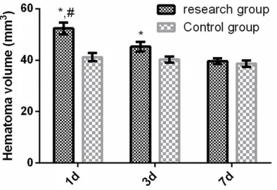
Changes in brain hematoma volume in the rat
model of ICH on days 1, 3, and 7
Volumes of the hematoma in brains of rats in the experimental group were 52.38 ± 2.26, 45.26 ± 1.81, and 39.61 ± 1.16 mm3 on days 1, 3, and 7, respectively, while those in the con-trol group were 41.22 ± 1.53, 40.26 ± 1.19, and 38.67 ± 1.24 mm3. Volumes of the hema-toma in rats of the experimental group were significantly larger than the control group on day 1 (t = 13.110, P < 0.001). On day 3, they were significantly greater than the control gr-oup (t = 7.394, P < 0.001), significantly smal-ler volumes on day 1 (t = 8.156, P < 0.001). On day 7, no significant differences were observed between the experimental group and control group (t = 1.795, P = 0.088) and levels were significantly greater than on the third day (t = 8.717, P < 0.001) (Figure 2).
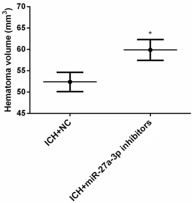
Effects of miR-27a-3p on hematoma volume
and neurological function of rats with ICH
To analyze the effects of miR-27a-3p on hema-toma volume in rats with ICH, miR-27a-3p mimic inhibitors or corresponding NCs were injected into the right ventricle of rats to assess change of hematoma volume and neurological function of rats with ICH on day 1. Results showed that miR-27a-3p inhibitors significantly downregulated miR-27a-3p (t = 5.061, P < 0.001). Downregulation of miR-27a-3p appear- ed to increase hematoma volume after ICH (t = 7.453, P < 0.001). Downregulation of miR-27a-3p aggravated neurological deficits of rats after initiation of ICH (t = 7.992, P < 0.001) (Figures 3-5).
Discussion
[image:4.612.92.287.75.219.2]ICH is a very destructive disease. Treatment methods for ICH, however, remain limited. The 30-day mortality rate of spontaneous ICH has reached 50%, with approximately 80% of survi-vors suffering neurological function disorders. Only 20% of surviving patients have been restored to living independently [15, 16]. In most patients with spontaneous ICH, hemor-rhages are rapidly stopped in the brain. Re- Figure 1. Changes of miR-27a-3p expression in
ce-rebral hematomas of the ICH Rat Model. Expression levels of miR-27a-3p in cerebral hematomas of the ICH rat model on days 1, 3, and 7 were determined by RT-PCR. *P < 0.01 compared with day 7; #P < 0.01
compared with day 1.
Figure 2. Changes of brain hematoma volume in the rat ICH model on days 1, 3, and 7. *P < 0.01
[image:4.612.92.289.312.447.2]limit expansion to the periphery, and to elimi-nate hematomas altogether [17, 18]. The vol-ume of ICH hematomas reflect the severity of the disease, while control of hematoma volu- me often determines the prognosis of patients [19]. Therefore, finding biological markers clo-sely associated with volume of hematomas in patients with ICH may be a key to successful treatment of ICH.
[image:5.612.324.522.72.280.2]miRNAs are noncoding RNA molecules that par-ticipate in biological processes such as cell pro-liferation, apoptosis, differentiation, metabo-lism, and death by regulating gene expression through transcription or after transcription [20]. miRNAs perform a key function in the develop-ment of various cell types and are closely link- ed to cell differentiation, morphogenesis, and tumorigenesis [21]. Studies have revealed that miRNAs may play cancer-promoting or tumor-suppressing roles in the development of vari-ous human tumors and can serve as an effec-tive molecular biological indicators for early diagnosis, treatment, and prognostic assess-ment of tumors [22]. One study by Xu et al. [23] indicated that when a neuroglioma is under the conditions of hypoxia, miR-27a-3p regulates the adaptability of breast cancer cells to the hypoxic state through hypoxia-inducible factor (HIF). Results of this present study showed that miR-27a-3p in brain hematomas of rats in the experimental group was significantly lower than Figure 3. Changes of relative expression of
miR-27a-3p in the inhibition group after intraventricular injec-tion of a miR-27a-3p inhibitor. *P < 0.01 compared
[image:5.612.93.286.74.288.2]with the ICH+NC group.
Figure 4. Effects of downregulation of miR-27a-3p on hematoma volume in ICH rats. *P < 0.01 compared
with the ICH+NC group.
[image:5.612.90.286.358.566.2]bleeding exacerbates the deterioration of neu-rological function. Intracranial hemorrhages occur within 24 hours in 30% of patients with spontaneous ICH [15]. The mechanism of brain injury caused by ICH is mainly related to mechanical occupancy effects of hematoma. Researchers are currently trying to determine how to reduce or reverse the occupancy effects,
Figure 5. Influence of downregulation of miR-27a-3p
on neurological function in ICH rats. *P < 0.01
that in the control group and peaked. On day 3, it was significantly lower than that of the con-trol group and significantly increased compared with the control group on day 1. On day 7, no significant differences from the control group was observed and levels were significantly higher than on day 3. Volume of hematomas in rats with ICH in the experimental group was sig-nificantly greater than the control group on day 1. Volume was significantly greater on day 3 than that in the control group, while significant -ly decreasing on day 3. No significant differ -ences were observed on day 7 compared with the control group and levels significantly in-creased relative to day 3. Accordingly, it was speculated that miR-27a-3p may be involved in the occurrence and development of cerebral hematomas after ICH. With downregulation of miR-27a-3p, the volume of cerebral hemato-mas in rats gradually increased. Therefore, miR-27a-3p may participate in the regulation of volume of ICH hematomas.
miRNAs can bind to the 3’ untranslated region (UTR) of a target gene to form a correspond- ing RNA-induced silencing complex, inhibiting translation of mRNA and downregulating ex- pression of the target gene. A study by Zhao et al. [24] revealed that miR-27a-3p had multi-ple target genes, including BCLAF and BBC3, closely associated with apoptosis-related gene
Bcl-2, according to software analysis of target genes. A study by Yuan et al. [25] showed that relative expression of Bcl-2 in hematomas of ICH rats was higher compared with that of the control group, suggesting that hematomas of ICH may cause brain damage by increasing expression of apoptosis-related gene Bcl-2. By injecting miR-27a-3p inhibitors into the lat-eral ventricle of rats, this study found that miR-27a-3p inhibitors may significantly downregu -late miR-27a-3p. This downregulation may in- crease hematoma volume of rats after ICH and aggravate neurological deficits of rats after ICH. These data suggest that downregulation of miR-27a-3p may aggravate brain injuries caused by ICH. Thus, miR-27a-3p may regulate hematoma volume in rats and it is possible to relieve ICH brain damage through regulation of miR-27a-3p expression.
This present study considered the reproducibil-ity and reliabilreproducibil-ity of animal experiments. All
pur-chased rats were rigorously screened. Gender, age (weeks), body weight, indoor temperature, and indoor humidity had no effect on the results, thereby ensuring the scientific rigor of this study. In this study, the mechanisms of action of miR-27a-3p in a ICH rat model was preliminarily discussed. More detailed verifica -tion of mechanisms was not provided. The- refore, this present study had certain limita-tions. Due to the complexity of an in vivo envi-ronment, whether observed miR-27a-3p mech-anisms exist in the human body and whether they are affected by peripherally related genes requires further clinical study.
Moreover, miR-27a-3p may be involved in the occurrence and development of cerebral hema-tomas after ICH. With downregulation of miR-27a-3p, the volume of cerebral hematomas in rats gradually increased. Thus, miR-27a-3p may play a part in the regulation of the volume of hematomas. It may be possible to alleviate brain injuries due to ICH by regulating expres-sion of miR-27a-3p. This miRNA could become a biological therapeutic target for brain injuries caused by ICH.
Disclosure of conflict of interest
None.
Address correspondence to: Zuneng Lu, Depart- ment of Neurology, Renming Hospital of Wuhan University, Jiefang Road 238, Wuchang District, Wuhan 430060, Hubei, China. Tel: +86-13593760- 874; E-mail: luznlll@163.com
References
[1] Qureshi A, Palesch Y, Barsan W, Hanley D, Hsu C, Martin R, Moy C, Silbergleit R, Steiner T and Suarez J. Intensive blood-pressure lowering in patients with acute cerebral hemorrhage. J Neurosurg Anesthesiol 2017; 29: 176-177. [2] Lattanzi S, Cagnetti C, Provinciali L and
Silves-trini M. How should we lower blood pressure after cerebral hemorrhage? A systematic re-view and meta-analysis. Cerebrovasc Dis 2017; 43: 207-213.
[4] Wang Y, Chen Q, Tan Q, Feng Z, He Z, Tang J, Feng H, Zhu G and Chen Z. Simvastatin accel-erates hematoma resolution after
intracere-bral hemorrhage in a PPARγ-dependent man -ner. Neuropharmacology 2018; 128: 244-254. [5] Cao S, Zheng M, Hua Y, Chen G, Keep RF and
Xi G. Hematoma changes during clot resolu-tion after experimental intracerebral hemor-rhage. Stroke 2016; 47: 1626-1631.
[6] Rodriguez-Luna D, Coscojuela P, Rubiera M, Hill MD, Dowlatshahi D, Aviv RI, Silva Y, Dzi-alowski I, Lum C and Czlonkowska A. Ultraearly hematoma growth in active intracerebral hem-orrhage. Neurology 2016; 87: 357-364. [7] Rupaimoole R and Slack FJ. MicroRNA
thera-peutics: towards a new era for the manage-ment of cancer and other diseases. Nat Rev Drug Discov 2017; 16: 203-222.
[8] Naga Prasad SV, Duan ZH, Gupta MK, Suram-pudi VS, Volinia S, Calin GA, Liu CG, Kotwal A, Moravec CS, Starling RC, Perez DM, Sen S, Wu Q, Plow EF, Croce CM, Karnik S. Unique
microR-NA profile in end-stage heart failure indicates alterations in specific cardiovascular signaling
networks. J Biol Chem 2009; 284: 27487-27499.
[9] Liu N, Zhang L, Wang Z, Cheng Y, Zhang P, Wang X, Wen W, Yang H, Liu H and Jin W. Mi-croRNA-101 inhibits proliferation, migration and invasion of human glioblastoma by target-ing SOX9. Oncotarget 2017; 8: 19244. [10] Uemura M, Nakata W, Kawashima A, Ujike T,
Nagahara A, Fujita K and Nonomura N. Overex-pression of miR-27a-3p is an independent prognostic factor for recurrence in clear cell renal cell carcinoma. European Urology Sup-plements 2017; 16: e1704-e1706.
[11] Toussaint I. Alternative methods for killing lab-oratory animals: summary of a published advi-sory report by the netherlands national com-mittee for the protection of animals used for
scientific purposes and for education (NCad).
Lab Anim 2017; 51: 222-223.
[12] Lv LJ, Li J, Qiao HB, Nie BJ, Lu P, Xue F and Zhang ZM. Overexpression of GRP75 inhibits
inflammation in a rat model of intracerebral
hemorrhage. Mol Med Rep 2017; 15: 1368-1372.
[13] Liang J, Tang J, Shi H, Li H, Zhen T, Duan J, Kang L, Zhang F, Dong Y and Han A.
miR-27a-3p targeting RXRα promotes colorectal cancer progression by activating Wnt/β-catenin path -way. Oncotarget 2017; 8: 82991.
[14] Muengtaweepongsa S, Prapa-Anantachai P and Dharmasaroja PA. Not only the sugar, early infarct sign, hyperdense middle cerebral
ar-tery, age, neurologic deficit score but also
atrial fibrillation is predictive for symptomatic
intracranial hemorrhage after intravenous re-combinant tissue plasminogen activator. J Neurosci Rural Pract 2017; 8: 49-54.
[15] Marini S, Morotti A, Ayres AM, Crawford K, Kourkoulis CE, Lena UK, Gurol EM, Viswana-than A, Goldstein JN and Greenberg SM. Sex differences in intracerebral hemorrhage ex-pansion and mortality. J Neurol Sci 2017; 379: 112-116.
[16] Giede-Jeppe A, Bobinger T, Gerner ST, Sembill JA, Sprügel MI, Beuscher VD, Lücking H, Hoel-ter P, Kuramatsu JB and Huttner HB. Neutro-phil-to-lymphocyte ratio is an independent pre-dictor for in-hospital mortality in spontaneous intracerebral hemorrhage. Cerebrovasc Dis 2017; 44: 26-34.
[17] Orito K, Hirohata M, Nakamura Y, Takeshige N, Aoki T, Hattori G, Sakata K, Abe T, Uchiyama Y and Sakamoto T. Leakage sign for primary in-tracerebral hemorrhage: a novel predictor of hematoma growth. Stroke 2016; 47: 958-963. [18] Carcel C, Sato S, Zheng D, Heeley E, Arima H,
Yang J, Wu G, Chen G, Zhang S, Delcourt C, La-vados P, Robinson T, Lindley RI, Wang X, Chalmers J, Anderson CS; Intensive Blood Pressure Reduction in Acute Cerebral
Hemor-rhage Trial 2 Investigators. Prognostic signifi -cance of hyponatremia in acute intracerebral hemorrhage: pooled analysis of the intensive blood pressure reduction in acute cerebral hemorrhage trial studies. Crit Care Med 2016; 44: 1388-1394.
[19] Fiorella D, Arthur A, Bain M and Mocco J. Mini-mally invasive surgery for intracerebral and in-traventricular hemorrhage: rationale, review of existing data and emerging technologies. Stroke 2016; 47: 1399-1406.
[20] Lennox KA, Vakulskas CA and Behlke MA.
Non-nucleotide modification of anti-miRNA
oli-gonucleotides. In: editors. Drug target miRNA. Springer 2017; 51-69.
[21] Xie F, Yuan Y, Xie L, Ran P, Xiang X, Huang Q, Qi G, Guo X, Xiao C and Zheng S. miRNA-320a in-hibits tumor proliferation and invasion by tar-geting c-Myc in human hepatocellular carcino-ma. Onco Targets Thery 2017; 10: 885-894. [22] Pichler M, Stiegelbauer V,
Vychytilova-Faltejs-kova P, Ivan C, Ling H, Winter E, Zhang X, Goblirsch M, Wulf-Goldenberg A and Ohtsuka
M. Genome-wide miRNA analysis identifies
miR-188-3p as a novel prognostic marker and molecular factor involved in colorectal carcino-genesis. Clin Cancer Res 2017; 23: 1323-1333.
pro-mote cell proliferation in glioma cells via coop-erative regulation of MXI1. Int J Oncol 2013; 42: 757-766.
[24] Zhao N, Sun H, Sun B, Zhu D, Zhao X, Wang Y, Gu Q, Dong X, Liu F and Zhang Y. miR-27a-3p suppresses tumor metastasis and VM by down-regulating VE-cadherin expression and inhibiting EMT: an essential role for Twist-1 in HCC. Sci Rep 2016; 6: 23091.
![Table 2. General information on the three groups of rats [n (%)] (_x ± s)](https://thumb-us.123doks.com/thumbv2/123dok_us/1374730.671078/3.612.90.399.83.126/table-general-information-groups-rats-n-x-s.webp)