Original Article
Correlation analysis between bone metabolism
factors and the stability of dental implant in the
postoperative recovery of dental implanted patients
Puyu Ma1, Haoyang Wu2
1School of Stomatology, Zhengzhou University, Zhengzhou, China; 2Department of Implantation, Oral Center, First Affiliated Hospital of Zhengzhou University, Zhengzhou, China
Received May 9, 2017; Accepted March 8, 2018; Epub April 15, 2018; Published April 30, 2018
Abstract: The aim of this research was to investigate the effect of osteoprotegerin and RANKL on the change of
implant stability quotient (ISQ). Seventy-eight patients were implanted Straumann soft tissue level implants (Switzerland) through a non-submerged method. Survival rate of dental implants and postoperative complications was examined. Modified plaque index (mPLI) and modified sulcus bleeding index (mSBI) were evaluated. In the 1st,
2nd, 3rd, 4th, 6th, 8th and 12th week after operation, perio-implant crevicular fluid (PICF), gingival crevicular fluid (GCF)
and ISQ were measured, respectively. Osteoprotegerin and RANKL levels in GCF and PICF samples were detected by ELISA. There was no mechanical complication. And there was no obvious marginal bone loss. Osteoprotegerin in GCF and PICF increased in the 2nd week, which was much higher than that in other time points (P<0.05). RANKL in
GCF was the highest in the 12th week (P<0.05), and RANKL in PICF was the highest in the 3rd week (P<0.05). And
there was no difference between osteoprotegerin and RANKL in GCF and PICF (P>0.05). Osteoprotegerin/RANKL ra-tio in PICF was higher than that in GCF in the 1st week (P=0.034<0.05). Moreover, ISQ was the lowest in the 4th week
than in the 1st, 2nd, 6th, 8th and 12th week (P<0.05). ISQ decreased when osteoprotegerin rose, and the trends of ISQ
and RANKL were similar. When osteoprotegerin/RANKL ratio increased, ISQ decreased. There were respectively opposite and same trend between ISQ and osteoprotegerin and RANKL. On the basis of the relationship between ISQ and osteoprotegerin and RANKL, there might be a method to improve the stability by regulating osteoprotegerin and RANKL in GCF and PICF.
Keywords: Implant, implant stability quotient, osteoprotegerin, gingival crevicular fluid, perio-implant crevicular fluid
Introduction
Tooth loss is a common symptom of clinical diseases, which could reflect the condition of patients’ dental diseases [1, 2]. In most coun-tries, tooth loss is often considered as an effec-tive indication of good or bad oral health. Thus, it is necessary to constantly monitor and take care of oral health, and dental diseases need relevant lifelong treatment [3]. For patients with tooth loss, the depth and width of the tooth have an effect on the success rate of dental implant, and it is also the key factor for a successful implantation. Bad bone quality in implantation site would lead to a low success rate of dental implant [4]. The closure of bone and soft tissue, the masticatory pressure and
functional recovery of dental implant are impor-tant evaluation indexes for a successful implan-tation. And good long-term results of these indexes are significant in improving patients’ quality of life.
easy and noninvasive, and it can perform a quantitative assessment on dental implant sta-bility [7].
The condition of soft tissue surrounding the implantation site has a certain effect on a suc-cessful implantation. Gingival crevicular fluid (GCF) in periodontium and perio-implant cre-vicular fluid (PICF) have attracted more and more attention. GCF is the physiological solu-tion exuding from plexus vasculosus in gingival dermis, and it can also participate in the inflam-matory response as the inflaminflam-matory exudates [8]. GCF was first found in the early 19th century [9, 10]. Afterwards, Waerhaug described GCF as periodontal disease related liquid compound in his classic study [11, 12]. GCF has been found for many years, but its generation and function are still unknown to people. And the function of PICF also draws more and more attention from researchers gradually, but it is still unclear whether PICF has the same func-tion as GCF and what is the funcfunc-tion of PICF in the early stage of dental implant. However, some research indicated that PICF contained higher content and activity of type II collagen than GCF [13].
In addition, the process of dental implant involves bone remodeling which relates to the dynamic equilibrium of bone resorption and bone formation, and the process is closely related to the function of osteoclast and osteo-blast. Both GCF and PICF contain osteoprote-gerin (OPG), receptor activator of kappa B (RANK) and receptor activator of nuclear factor kappa B ligand (RANKL). These factors could induce the interaction between osteoblast and
and argumentation on the effect of RANKL/ RANK/OPG in implant osseointegration. In this research, we observed and analyzed the chang-es of detection indexchang-es in GCF and PICF after dental implantation, and compared and ana-lyzed the effect of OPG and RANKL on the change of implant stability quotient (ISQ). Materials and methods
Data of patients
The data of patients in the department of sto-matology of our hospital from May 2011 to December 2016 were retrospectively analyzed. Inclusion criteria: 1) patients were healthy with-out obviously serious illness and within 20< age <65; 2) patients suffered from tooth loss for at least three months in planning implanta-tion region; 3) no bone grafting was needed; 4) there is no medication history of antibiotics within three months before the research; 5) for patients suffering from periodontitis, they needed basic periodontal inflammatory treat-ment including oral hygiene firstly; after the treatment periodontal probing depth ≤5 mm and gingival bleeding index ≤2 within the oral cavity; 6) patients were not addicted to tobacco or were non smokers; or patients were with less than 10 cigarettes per day. In addition, diabetic patients, heavy smokers, gravidas, alcoholics and drug abusers were excluded. All selected patients were given regular follow-up.
[image:2.612.90.368.73.225.2]Seventy-eight patients were selected in this research: 26 males and 52 females with an average age of 41.6. This research had obtained the ethics committee approval from our hospital. All patients in this research signed informed consent forms.
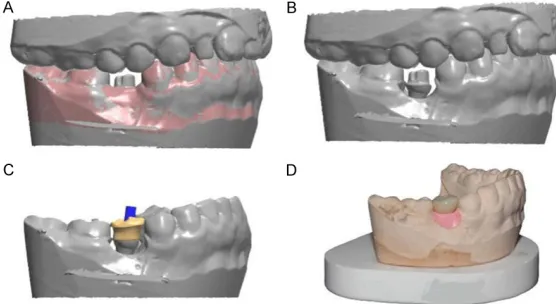
Figure 1. The model of implant-abutment.
Dental implantation
According to the standard operating procedure, all patients were implanted (Straumann soft tis-sue level implants, Switzerland) by a non-sub-merged method. Patients rinsed their mouths with 0.1% of chlorhexidine gargle for 1 min. Perioral disinfection was performed by povi-done iodine. Local infiltration anesthesia was implemented. H-shape incision was carried out in tooth loss region. Opened mucoperiosteal flaps. Fixed points by round bur, punched a hole layer by layer, and cooled the hole. The depth of the hole was measured by sounding scale. Then the implant was implanted, and abutment was placed (Figure 1). Mucoperiosteal flap was reposited and sutured. Before and after the end of the operation took and saved photos.
Observation indexes
Survival rate of dental implants: According to the standard issued by Buser [18], the sur-vival rate of dental implants was examined. The specific standards were as follows: ① Clinical examination showed that the implants were stable and no mobility; ② Patients did not have any subjective feeling, such as pain or numb-ness; ③ There was no repeated outbreak of inflammatory response around the dental implant; ④ Imageological diagnosis results showed that there was no continuous cast shadow around the dental implant.
Postoperative complications: The main com- plication related to implant abutment was
Biological complications: Biological complica-tions were evaluated by the health status of peri-implant soft tissue. We recorded the condition of peri-implant soft tissue of patients during the follow-up. The main evaluation index-es were modified plaque index (mPLI) and mod-ified sulcus bleeding index (mSBI) [19]. MPLI could implement an objective and effective assessment on peri-implant plaque. The higher the mPLI value was, the worse the oral hygiene became. No dental plaque was a score of 0. As the smooth neck of implant was slightly scratched by the top of the probe, observed dental plaque was a score of 1. A macroscopic plaque was a score of 2. Mass plaque accumu-lation was a score of 3. mSBI could effectively reflect the health status, mainly the bleeding condition, of peri-implant mucosa. No bleeding detected along implant gingival margin by the probe was a score of 0. Scattered hemorrhagic spot was a score of 1. Hemorrhagic spot inside gingival sulcus in a linear pattern was a score of 2. Severe or spontaneous bleeding was a score of 3. Specific scoring standards of mPLI and mSBI were shown in Tables 1 and 2.
Detection indexes
[image:3.612.90.357.84.165.2]Before operation, GCF was absorbed by absor-bent paper from the loci locating in the medial and distal surface of implant and its two adja-cent natural teeth. The weight of absorbent paper before and after the absorption of GCF was measured by AE240 electronic balance (METTLER, Sweden), then the weight of GCF Table 1. Scoring standards of mPLI
mPLI Content
0 No dental plaque
1 Dental plaque was observed as the smooth neck of implant was slightly scratched by the top of probe
2 Macroscopic plaque 3 Mass plaque accumulation
Note: mPLI: modified plaque index.
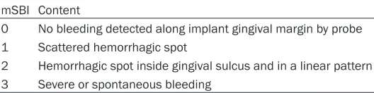
Table 2. Scoring standards of mSBI
mSBI Content
0 No bleeding detected along implant gingival margin by probe 1 Scattered hemorrhagic spot
2 Hemorrhagic spot inside gingival sulcus and in a linear pattern 3 Severe or spontaneous bleeding
Note: mSBI: modified sulcus bleeding index.
[image:3.612.92.356.207.273.2]was calculated. GCF was cryopreserved at -70°C. The same method was used to measure the weight of PICF. On further consultation in the 1st, 2nd, 3rd, 4th, 6th, 8th and 12th week after operation, patients were measured PICF and GCF, respectively. Osstell Mentor RFA (Integration Diagnostics, Savedalen, Sweden) was used to detect implant stability quotient (ISQ) on each further consultation because ISQ is very important to evaluate the integra-tion of implant and bone. The detecintegra-tion was taken once by the probe on the medial surface of implant, buccal side and the lingual side, respectively. The mean value of ISQ in the three sites was ISQ value of implant. Data obtained were entered in a database. Then using ISQ-related monitoring data, each implant was completed by inserting a final crowns.
Implant periapical film was taken immediately after the operation and on further consultation, and the shadow and bone resorption around the implant were detected. The levels of OPG and RANKL in GCF and PICF samples were detected by ELISA, and the changes of OPG and RANKL in the early period of postoperation were observed.
Statistical analysis
SPSS21.0 statistical software was used to ana-lyze the levels of OPG and RANKL in GCF and PICF at different points in time. The levels were shown as mean ± standard deviation. The changes of ISQ, OPG and RANKL in GCF and PICF were analyzed by ANOVA for repeated measurement. The differences between OPG
dibular teeth missing (17 males and 25 females). And 78 implants (Straumann, Wand- enburg, Switzerland) were inserted. All patients accepted further consultation after the 1st, 2nd, 3rd, 4th, 6th, 8th and 12th week. The retention rate, ISQ, OPG and RANKL in GCF and PICF were detected.
Retention rate of implants and postoperative complications
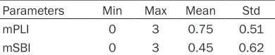
Mechanical complications and biological com-plications for all patients post operation were observed. There were no broken, loose, fall and obvious periodontal inflammations character-ized by gum red and swell. Postoperative mPLI and mSBI were 0.75±0.51 (0-3) and 0.45±0.62 (0-3), respectively. The imaging examination results showed that there was no obvious mar-ginal bone loss. Details about soft tissue condi-tion were shown in Table 4.
On the basis of Buser’s standard of implant retention rate, the implants of all patients were stable. And there was no recurrent inflamma-tion around the implants. The results of ima-geological examination demonstrated that there was no continuous cast shadow sur-rounding dental implants. As a conclusion, the implant retention rate of our patients was 100%.
The change of OPG and RANKL in GCF and PICF
[image:4.612.90.312.83.150.2]During the 12-week follow-up visit, all patients were reexamined OPG and RANKL in GCF and PICF in the 1st, 2nd, 3rd, 4th, 6th, 8th and 12th week Table 3. Basic information of patients
Terms posterior teethMaxillary Mandibular teeth Number Gender Male 18 17 35
Female 18 25 43 Age (years, mean) 41.6±14.2
Table 4. The results of soft tissue examina-tion
Parameters Min Max Mean Std mPLI 0 3 0.75 0.51 mSBI 0 3 0.45 0.62
Note: mPLI: modified plaque index; mSBI: modified sulcus bleeding index.
and RANKL in GCF and PICF at same points in time were detected by t test. Relations of ISQ to OPG and RANKL at different points in time were analyzed by logistic regression, excluding the effect of age and gender. When
p value was less than 0.05, the differences were significant.
Results
Basic information
[image:4.612.91.286.196.235.2]man-after operation. The result showed OPG in GCF and PICF increased in the 2nd week which was much higher than those at other time (P<0.05).
repeated measures analysis of variance. In the 1st week, there was a slight rise of ISQ and then decreased. The lowest ISQ was appeared in the 4th week, and then an increase followed. Statistical analysis showed that ISQ in the 1st, 2nd, 6th, 8th and 12th week were significantly higher than that in the 4th week (P<0.05) (Table 8). This related to the resorption- immersion remodeling process which occurred in the 2-4 weeks after operation.
Relationships between ISQ and OPG/RANKL
[image:5.612.93.315.86.204.2]Compared with the trend of ISQ, OPG and RANKL, OPG and RANKL in GCF were more closely related to ISQ. ISQ decreased when OPG rose, and the trends of ISQ and RANKL were similar. And OPG/RANKL ratio in GCF and PICF also had a relationship with the change of ISQ. When the ratio increased, ISQ decreased. Table 5. OPG in GCF and PICF
Time/week OPG t P
GCF PICF
1st 418.7±18.7a 431.2±19.6a -1.012 0.295
2nd 434.4±20.8 486.4±28.8 -0.839 0.414
3rd 415.2±17.1a 443.5±32.6a -0.679 0.500
4th 391.4±16.8a 419.6±26.3a -1.121 0.305
6th 390.5±17.9a 411.2±24.3a -0.434 0.657
8th 384.1±21.2a 402.7±22.9a -0.613 0.590
12th 381.1±18.9a 397.6±23.6a -0.815 0.375 Note: Repeated measures analysis of variance, P<0.05. a: means there is a significant difference when compared with the OPG of 2nd week, P<0.05. OPG: osteoprotegerin; GCF: gingival
[image:5.612.93.315.280.400.2]crevicular fluid; PICF: perio-implant crevicular fluid.
Table 6. RANKL in GCF and PICF
Time/week RANKL t P
GCF PICF
1st 251.4±12.7 232.1±20.6c 0.791 0.471
2nd 241.3±16.8 256.3±26.7 0.314 0.817
3rd 237.7±18.3b 267.2±29.7 -0.579 0.691
4th 238.5±20.6b 251.8±26.1c 0.509 0.613
6th 241.2±19.6b 243.6±20.1c 0.497 0.561
8th 247.6±18.4 238.7±19.6c 0.348 0.590
12th 256.8±19.7 237.8±20.1c 0.051 0.914 Note: Repeated measures analysis of variance, P<0.05. b: means there is a significant difference when compared with the RANKL of 12th week, P<0.05. c: means there is a significant
difference when compared with the RANKL of 3rd week, P<0.05.
RANKL: receptor activator of nuclear factor kappa B ligand; GCF: gingival crevicular fluid; PICF: perio-implant crevicular fluid.
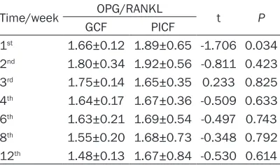
Table 7. OPG/RANKL Ratio
Time/week OPG/RANKL t P
GCF PICF
1st 1.66±0.12 1.89±0.65 -1.706 0.034
2nd 1.80±0.34 1.92±0.56 -0.811 0.423
3rd 1.75±0.14 1.65±0.35 0.233 0.825
4th 1.64±0.17 1.67±0.36 -0.509 0.633
6th 1.63±0.21 1.69±0.54 -0.497 0.743
8th 1.55±0.20 1.68±0.73 -0.348 0.792
12th 1.48±0.13 1.67±0.84 -0.530 0.614
Note: ANOVA, P<0.05. The ratio OPG/RANKL of PICF was
remarkably higher than GCF at 1st week. There was no
dif-ference at other time. OPG: osteoprotegerin; RANKL: recep-tor activarecep-tor of nuclear facrecep-tor kappa B ligand; GCF: gingival crevicular fluid; PICF: perio-implant crevicular fluid.
However, there was no difference between OPG in PICF and GCF (P>0.05) (Table 5). This demonstrated that 1-2 weeks after opera-tion the proliferaopera-tion and differentiaopera-tion of osteoblast have been initiated. And in the 2nd week, the level of OPG in GCF also increased which indicated there were plenty of active osteoblasts and the periodontium of adja-cent teeth may also participate in the bone remodeling.
Moreover, analysis on RANKL in GCF and RANKL showed that RANKL in GCF was the highest in the 12th week (P<0.05), and RANKL in PICF was the highest in the 3rd week (P<0.05). And there was no difference between RANKL in GCF and PICF (P>0.05) (Table 6). This might be due to OPG high affinity combined with RANKL and reduced the differentiation of osteoclast by inhibiting function of RANKL.
On the basis of OPG and RANKL, we calcu-lated OPG/RANKL in GCF and PICF at each time point. In the 1st week, OPG/RANKL in PICF was much higher than that in GCF (P=0.034<0.05). This also demonstrated the initiation of proliferation and differentiation of osteoblast. There was no difference at other time points (Table 7).
ISQ detection
[image:5.612.93.293.498.616.2]The changes of OPG, RANKL and OPG/RANKL appeared before the change of ISQ.
Discussion
Damage and missing of implants could have a great influence on life quality. Recently, with the development of science and technology, dental implants which have accepted a well effect in the reconstruction of teeth, were more and more widely used and gradually become the common treatment method of dental missing [20, 21]. And it is also one of the most success-ful recovery techniques in clinical treatment [22]. There will be variable problems during the operation and postoperation, which have a great effect on the success of implantation. Implant stability index could effectively evalu-ate the implant stability, which has a remark-able clinical value in estimating the recovery of implants and could decide whether it could load immediately or not [23]. Moreover, doctors could estimate the implant osseointegration and mechanical characteristics through con-tinuous monitoring of implant stability in order to play guidance in future treatment.
However, the sclerotin around the implants, adjacent teeth, inserting ways, material of implants and the recovery condition post oper-ation could affect the stability of implant-abut-ment [24]. Otherwise, smoking, diseases such as diabetes also have an impact on it [24]. RANKL/RANK/OPG system could influence tooth resorption by the regulation of differentia-tion and activadifferentia-tion of osteoclast. And it also involves in the reconstruction of periodontal tissue and the regulation of tooth eruption, tooth germ formation and tooth absorption [25-27]. All of these were reflected in our research. Otherwise, the balance between bone resorp-tion and formaresorp-tion is essential for the recon-struction of bone, in which osteoclast and osteoblast participated. RANK expresses in the surface of osteoclast precursors, and RANKL expresses in the surface of osteoblasts/matrix cells. The combination of RANK and RANKL
could activate osteoclasts, which is dose-dependent to extend the survival time of osteo-clasts, and to improve the ability of osteoclast movement and the formation of bone resorp-tion pits [28, 29]. And some research shows that RANKL not only regulates the activation of osteoclast, also plays an effect on the absorption function of osteoclast [30]. For the function of RANKL/RANK/OPG system in the regulation of differentiation and activation of osteoclast, the expression of OPG and RANKL could influence the formation and reconstruc-tion of new bone [31]. RANKL could promote the differentiation and maturation of osteo-clasts, inducing bone resorption. However, OPG belongs to tumor necrosis factor superfamily and is a natural inhibitor of RANKL. It could competitively inhibit the combination of RANKL and RANK and to inhibit bone resorption [32]. So the changes of OPG and RANKL are differ-ent which is demonstrated by this research. In this research, OPG in GCF and PICF increas- ed in the second week post operation, which was higher than the other time, especially OPG in PICF (P<0.05). There was no difference between OPG in GCF and PICF. However, there was different variation trend of RANKL in GCF and PICF post operation. The trend of RANKL in GCF was decreased first and then rose. The level of RANKL in GCF in the third, fourth and fifth week after operation was remarkably lower than that in the 12th week. However, the level of RANKL in PICF in the 3rd week was higher than those in the 1st, 4th, 6th, 8th and 12th week (P<0.05). There was no difference between RANKL in GCF and in PICF. The differ-ent trends of OPG and RANKL were mainly caused by the competitive inhibition between OPG and RANKL.
In this research, there were relationships between ISQ and OPG or RANKL. The higher the OPG was, the lower of ISQ would be. However, trends of RANKL and ISQ were same. This was related to the competitive inhibition between OPG and RANKL, which both could combine with RANK to regulate osteoclast. So Table 8. ISQ of different time
Time/week 1st 2nd 3rd 4th 6th 8th 12th
ISQ 66.5±1.1a 64.8±1.2a 62.9±0.8 62.2±0.9 69.8±1.0a 72.6±1.1a 74.9±1.0a
Note: Repeated measures analysis of variance, P<0.05. a: means there is a significant difference when compared with the ISQ
in future treatment, we could improve the implant osseointegration and primary stability by regulating the level of OPG and RANKL.
Disclosure of conflict of interest
None.
Address correspondence to: Haoyang Wu, Depart-
ment of Implantation, Oral Center, The First Affiliated Hospital of Zhengzhou University, 1 East Jianshe Road, Zhengzhou 450000, Henan, China. Tel: +86-371-55966339; Fax: +86-371-6707-7303; E-mail: [email protected]
References
[1] Petersen PE, Bourgeois D, Ogawa H, Estupinan-Day S and Ndiaye C. The global burden of oral diseases and risks to oral health. Bull World Health Organ 2005; 83: 661-9.
[2] Baelum V, van Palenstein Helderman W, Hugoson A, Yee R and Fejerskov O. A global perspective on changes in the burden of caries and periodontitis: implications for dentistry. J Oral Rehabil 2007; 34: 872-906; discussion 940.
[3] Kassebaum NJ, Bernabé E, Dahiya M, Bhandari B, Murray CJ and Marcenes W. Global burden of severe tooth loss: a systematic review and meta-analysis. J Dent Res 2014; 93: 20S-28S. [4] Atieh MA, Alsabeeha NH, Payne AG, Duncan W,
Faggion CM and Esposito M. Interventions for replacing missing teeth: alveolar ridge preser-vation techniques for dental implant site devel-opment. Cochrane Database Syst Rev 2015; CD010176.
[5] Ersanli S, Karabuda C, Beck F and Leblebicioglu B. Resonance frequency analysis of one-stage dental implant stability during the osseointe-gration period. J Periodontol 2005; 76: 1066-71.
[6] Glauser R, Sennerby L, Meredith N, Rée A, Lundgren A, Gottlow J and Hämmerle CH. Resonance frequency analysis of implants subjected to immediate or early functional oc-clusal loading. Successful vs. failing implants. Clin Oral Implants Res 2004; 15: 428-34. [7] Meredith N, Alleyne D and Cawley P. Quan-
titative determination of the stability of the im-plant-tissue interface using resonance fre-quency analysis. Clin Oral Implants Res 1996; 7: 261-7.
[8] Barros SP, Williams R, Offenbacher S and Morelli T. Gingival crevicular fluid as a source of biomarkers for periodontitis. Periodontol 2000 2016; 70: 53-64.
[9] BG V, Special Dental Pathology. Chicago: Medico-Dental Publishing Co. 1915; 17.
[10] SER A. Essai sur l’anatomie et la physiologie des dents: ou, Nouvelle théorie de la dentition. Méquignon-Marvis 1817.
[11] Waerhaug J. The source of mineral salts in sub-gingival calculus. J Dent Res 1955; 34: 563-8. [12] Waerhaug J and Steen E. The presence or ab-sence of bacteria in gingival pockets and the reaction in healthy pockets to certain pure cul-tures; a bacteriological and histological investi-gation. Odontol Tidskr 1952; 60: 1-24. [13] Xu L, Yu Z, Lee HM, Wolff MS, Golub LM, Sorsa
T and Kuula H. Characteristics of collage-nase-2 from gingival crevicular fluid and implant sulcular fluid in periodontitis and peri-implantitis patients: pilot study. Acta Odontol Scand 2008; 66: 219-24.
[14] Rakic M, Lekovic V, Nikolic-Jakoba N, Vojvodic D, Petkovic-Curcin A and Sanz M. Bone loss biomarkers associated with peri-implantitis. A cross-sectional study. Clin Oral Implants Res 2013; 24: 1110-6.
[15] Pérez-Sayáns M, Somoza-Martín JM, Barros-Angueira F, Rey JM and García-García A. RANK/ RANKL/OPG role in distraction osteogenesis. Oral Surg Oral Med Oral Pathol Oral Radiol Endod 2010; 109: 679-86.
[16] Boyce BF and Xing L. Functions of RANKL/ RANK/OPG in bone modeling and remodeling. Arch Biochem Biophys 2008; 473: 139-46. [17] Zhu WQ, Wang X, Wang XX and Wang ZY.
[Molecular mechanism of bone remodelling during mandibular distraction osteogenesis in rats]. Zhonghua Kou Qiang Yi Xue Za Zhi 2007; 42: 729-32.
[18] Buser D, Mericske-Stern R, Bernard JP, Behneke A, Behneke N, Hirt HP, Belser UC and Lang NP. Long-term evaluation of non-sub-merged ITI implants. Part 1: 8-year life table analysis of a prospective multi-center study with 2359 implants. Clin Oral Implants Res 1997; 8: 161-72.
[19] Mombelli A, van Oosten MA, Schurch E Jr and Land NP. The microbiota associated with suc-cessful or failing osseointegrated titanium im-plants. Oral Microbiol Immunol 1987; 2: 145-51.
[20] Weber HP and Sukotjo C. Does the type of im-plant prosthesis affect outcomes in the par-tially edentulous patient? Int J Oral Maxillofac Implants 2007; 22 Suppl: 140-72.
[21] Lekholm U, Gunne J, Henry P, Higuchi K, Lindén U, Bergström C and van Steenberghe D. Survival of the Branemark implant in partially edentulous jaws: a 10-year prospective multi-center study. Int J Oral Maxillofac Implants 1999; 14: 639-45.
and bacterial penetration on different implant-abutment connection systems. Braz Dent J 2010; 21: 123-9.
[23] Yang H and He HY. Analysis on the factors of dental implant osseointegration. Journal of Oral Science Research 2008; 24: 708-711. [24] Chuang SK, Wei LJ, Douglass CW and Dodson
TB. Risk factors for dental implant failure: a strategy for the analysis of clustered failure-time observations. J Dent Res 2002; 81: 572-7.
[25] Neves JS, Salmon CR, Omar NF, Narvaes EA, Gomes JR and Novaes PD. Immunolocalization of CSF-1, RANKL and OPG in the enamel-relat-ed periodontium of the rat incisor and their implications for alveolar bone remodeling. Arch Oral Biol 2009; 54: 651-7.
[26] Nakao K, Goto T, Gunjigake KK, Konoo T, Kobayashi S and Yamaguchi K. Intermittent force induces high RANKL expression in hu-man periodontal ligament cells. J Dent Res 2007; 86: 623-8.
[27] Kim T, Handa A, Iida J and Yoshida S. RANKL expression in rat periodontal ligament subject-ed to a continuous orthodontic force. Arch Oral Biol 2007; 52: 244-50.
[28] Yang S, Chen W, Stashenko P and Li YP. Specificity of RGS10A as a key component in the RANKL signaling mechanism for osteoclast differentiation. J Cell Sci 2007; 120: 3362-71.
[29] Sobacchi C, Frattini A, Guerrini MM, Abinun M, Pangrazio A, Susani L, Bredius R, Mancini G, Cant A, Bishop N, Grabowski P, Del Fattore A, Messina C, Errigo G, Coxon FP, Scott DI, Teti A, Rogers MJ, Vezzoni P, Villa A and Helfrich MH. Osteoclast-poor human osteopetrosis due to mutations in the gene encoding RANKL. Nat Genet 2007; 39: 960-2.
[30] Wada T, Nakashima T, Hiroshi N and Penninger JM. RANKL-RANK signaling in osteoclastogen-esis and bone disease. Trends Mol Med 2006; 12: 17-25.
[31] Zhang DG. The expression of PDGF, OPG and RANKL in mandibular fracture healing process. Third Military Medical University 2005; 5. [32] Yasuda H, Shima N, Nakagawa N, Yamaguchi
K, Kinosaki M, Mochizuki S, Tomoyasu A, Yano K, Goto M, Murakami A, Tsuda E, Morinaga T, Higashio K, Udagawa N, Takahashi N and Suda T. Osteoclast differentiation factor is a ligand for osteoprotegerin/osteoclastogenesis-inhibi-tory factor and is identical to TRANCE/RANKL.