CROWDED CHARGES IN ION CHANNELS
BOB EISENBERG1,2
1Department of Molecular Biophysics and Physiology, Rush University,
Chicago, IL 60305, USA
2Mathematics and Computer Science Division, Argonne National Laboratory,
9700 South Cass Avenue, Argonne, IL 60439, USA
CONTENTS
I. Physical Chemistry and Life A. Ion Channels
II. Physical Chemistry and Biological Problems
A. Complexity in Structure and Physics Produces Simplicity in Biological Function B. Ion Channels
C. Channels Open and Close
D. Sigworth’s Equation: Single Channel Currents E. Opening Time Course of Channels
F. Open Probability G. Relation to Nerve Function
III. Action Potential is a Cooperative Phenomenon A. Allosteric Mechanisms
B. Conformation Changes
C. Cooperative Behavior Produced by Current Flow IV. Computation of the Action Potential
A. Changes in Ion Concentration B. Gating Processes
C. Selectivity and Permeation
D. Physiological Models of Permeation and Selectivity E. Rate Models have their Place
1. Energy Profiles are Well Defined 2. Existence of a Large Barrier 3. Assumption of a Single Path 4. Motion Along the Reaction Path 5. Rate Equations Predict too Little Current
Advances in Chemical Physics, Volume 148, First Edition. Edited by Stuart A. Rice and Aaron R. Dinner.
© 2012 John Wiley & Sons, Inc. Published 2012 by John Wiley & Sons, Inc.
F. Structural Models of Permeation and Selectivity
G. Reduced Models are the Essence of Biology and Engineering H. The Scientific Method and Channels
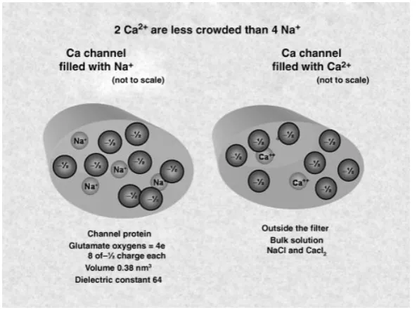
V. Reduced Models of Calcium and Sodium Channels A. Different Ions Carry Different Signals B. Selectivity of the Calcium Channel C. Reduced Model of the Calcium Channel
D. The Ionic Environment of the Channel is Remarkably Crowded E. History of Reduced Models of Ca2+Channels
F. Reduced Model Properties
VI. Crowded Ions: Properties of the Model of Calcium Channels A. Limitations in Analysis
B. Crowded Charge as a Biological Adaptation C. Necessity of Calibration
VII. Balanced Forces and Structures in Crowded Systems A. The Structure of Forces in Proteins
B. Importance of Structures
C. Some Structural Properties are So Important that they Cannot be Observed D. Self-Organized Structures
E. Limitations of Crystallography VIII. Inverse Methods and Selectivity Models
A. Building Calcium Channels B. Mutations of Channels C. Mutations of Models
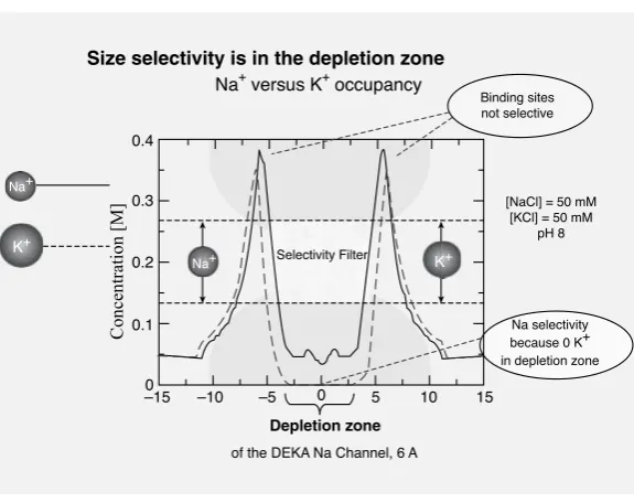
D. Dielectric Miracle: Na+Versus K+Selectivity E. Na+Versus Ca2+Selectivity in the Calcium Channel F. Na+Versus K+Selectivity in Sodium Channels G. Control Parameters
IX. Reduced Model of Transport Through a Channel
X. Reduced Model of a “Transport” Channel the Ryanodine Receptor XI. Conclusions and Implications of the Crowded Charge Reduced Model
A. What are the Source of Problems with High-Resolution Models? B. Biological Control by Trace Concentrations of Ions
C. All the Scales Need to be Dealt with At Once D. Reduced Models Deal with the Range of Scales E. Interacting Ions in Solutions and Channels F. Ionic Solutions as Complex Fluids XII. Variational Approach
A. Scaling inEnVarA
B. Scaling in Space inEnVarA
C. Scaling in Time inEnVarA
D. Scaling of Parameters inEnVarA
E. Scaling of the Protein
XIII. Outlooks: Unsolved Problems in Physical Chemistry A. Unsolved Problems in Physical Chemistry of Solutions B. Unsolved Problems in Applied Mathematics
C. Unsolved Problems in Molecular Biology and Biophysics D. Unsolved Problems in Channels
XIV. Conclusion A. A Full Circle
Appendix A: Models of Chemical Kinetics and the Law of Mass Action Rate Constants Vary
Rate Constants are not Constant in Crowded Conditions Mathematics must Deal with Interactions
Acknowledgments References
I. PHYSICAL CHEMISTRY AND LIFE
Ions in water are the liquid of life. Life occurs almost entirely in “salt water.” Life began in salty oceans. Animals kept that salt water within them when they moved out of the ocean to drier surroundings. The plasma and blood that surrounds all cells are electrolytes more or less resembling seawater. The plasma inside cells is an electrolyte solution that more or less resembles the seawater in which life began. Water itself (without ions) is lethal to animal cells and damaging for most proteins. Water must contain the right ions in the right amounts if it is to sustain life.
Physical chemistry is the language of electrolyte solutions and so physical chemistry, and biology, particularly physiology, have been intertwined since phys-ical chemistry was developed some 150 years ago. Physiology, of course, was studied by the Greeks some millennia earlier, but the biological role of electrolyte solutions could not be understood until ions were discovered by chemists some 2000 years later.
Physical chemists and biologists come from different traditions that separated for several decades as biologists identified and described the molecules of life. Communication is not easy between a fundamentally descriptive tradition and a fundamentally analytical one. Biologists have now learned to study their well-defined systems with physical techniques, of considerable interest to physical chemists. Physical chemists are increasingly interested in spatially inhomoge-neous systems with structures on the atomic scale so common in biology. Physical chemists will find it productive to work on well-defined systems built by evolution to be reasonably robust, with input–output relations insensitive to environmen-tal insults. The overlap in science is clear. The human overlap is harder because the fields have grown independently for some time, and the knowledge base, as-sumptions, and jargon of the fields do not coincide. Indeed, they sometimes seem disjoint, without overlap.
developed by Liu [1–5]—not too widely used among physical chemists. My goal is to provide the knowledge base, and identify the assumptions, that biologists use in studying ion channels, avoiding jargon. Although we do not know enough to write atomic, detailed physical models of the process by which ions move through channels, rather simple models of selectivity and permeation work quite well in important cases. Those physical models and cases are the main focus of this chapter because they demonstrate the strong essential link between the traditional treat-ments of ions in chemical physics and the biological function of ion channels.
At first, ion channels may seem to be an extreme system. They are as small as they can be, given the particulate nature of matter. Ion channels are atomic valves that allow a handful of atoms to control macroscopic flows of current, and thus macroscopic properties of cells, tissues, animals, and life. They do this by working at the extremes of forces as well as sizes. They have enormous densities of ions crowded into tiny spaces with huge electric and chemical fields and forces of excluded volume. These enormous densities are as far as one can imagine from the vanishing densities of simple or ideal fluids. So ion channels may seem a special case not of general interest to physical chemists.
I hope to show that the special case of ion channels gives general insight. “If you look closely enough at a keyhole, you can look through it, and sometimes even glimpse an horizon or even stars” (John Edsall, personal communication). Whenever a physical system is controlled by a small space, whenever a chemi-cal engineer uses a tiny valve, whenever a boundary layer near an electrode is a determinant of electrochemical function, one can expect crowded charges in tiny spaces. In those physical systems, crowded charges are likely to involve the same physics as crowded charges in biological channels. A general theme can be viewed through the biological channel.
The general theme that emerges is that everything interacts with everything else in any crowded environment, including ions in channels. I will argue that crowded conditions require a mathematics that deals naturally with interactions. I will argue that the law of mass action (with constant constants) does not apply in crowded cases. I will argue that crowded systems are complex, not simple fluids. Interactions in complex fluids have been analyzed with Chun Liu’s variational methodEnVarA
that naturally deals with interactions [2–5] and we are now applying that approach to ionic solutions [6, 7].
A number of topics are discussed several times from different perspectives in this chapter more in the tradition of an essay than a scientific paper. The motivation is to provide physical, chemical, and biological views of the key topics. I hope those already familiar with these ideas will have patience with this approach.
A. Ion Channels
we shall see. Ion channels have been studied in astonishing detail [8, 9] despite their staggering diversity [10–13]. Ion channels have enormous biological and medical importance [8]. Thousands of diseases are produced by genetic defects in channels, including many diseases of profound importance, like cystic fibrosis, epilepsy, atrial and ventricular fibrillation, and so on, as documented in many papers [14–98] among thousands of others. Many of these diseases are caused by problems in the construction of channels, or the insertion of channels in the wrong places in the wrong cells, or in the regulation and control of channels. This chapter is not focused on such biological problems, because we do not know enough yet to write physical models of the problematic biological systems.
This chapter is written to show the interactions of physical chemistry and molec-ular biology in channels, in theory, simulations, experiments, and mathematics, as well as in the text itself. Channels are defined, along with enough discussion to show how they are used in biology, without (I hope) overwhelming the reader with complexity. The selectivity properties of channels are discussed at length because in some cases these can be understood quite completely with simple ideas from the primitive (implicit solvent) model of electrolyte solutions of classical physical chemistry. Selectivity implies interactions. In the world of ideal point particles K+ and Na+are identical! The need to analyze finite size particles that interact because of their size and electric field is a recurrent theme. The need to analyze flows is also a recurrent theme, although most of that analysis is yet to be done. Flow must be analyzed because ion channels, like most devices, work far from equilibrium.
Equilibrium is death to biology. A variational methodEnVarAis introduced that allows automatic extension of equilibrium analysis to nonequilibrium.EnVarAwas developed to deal with complex fluids, with flowing interacting subelements far more complex than hard sphere ions. I argue that electrolytes can be viewed more realistically as a complex fluid than as a simple fluid of classical theory. The variational method is then applied to a few cases of interest. The article tries to go full circle: describing ion channels, using classical physical chemistry to deal with an important biological property of channels, introducing the new variational approach to deal with flows through channels, and finally arguing that this varia-tional approach provides a new perspective on ions in solution as well as channels. I propose that ions in solutions are complex fluids in which interactions dominate: “everything interacts with everything else.”
II. PHYSICAL CHEMISTRY AND BIOLOGICAL PROBLEMS
Many scientists want to apply physical chemistry to biological problems. The question is, what problems? The challenge is how to do it. The answers to these questions are hard for human reasons, I believe.
Biologists want to understand how machines, systems, and devices work well enough to make life better, in health and disease.
Biologists have little interest in how living systems work under “nonphysio-logical conditions.” Only anatomists study “fixed” (dead) material and they do so because fixed material is easier to view than moving, living systems. Structure is important, but it is important mostly because it can move and do something.
Biologists are much like engineers. Chemical engineers are as much physical chemists as they are engineers. Physical chemistry is linked to important parts of biology much as it is linked to chemical engineering. Physiologists and physical chemists dealt with the same issues until molecular biology came along and focused physiological attention on proteins, rather than the ions that surround them.
Engineers have a particular approach to problems shared by biologists. Engi-neers study devices as they function in a particular case. EngiEngi-neers want to study an amplifier as it amplifies. (I use electrical examples because of my limited knowl-edge of chemical engineering.) Engineers are not eager to study amplifiers when they cannot amplify, when they are “dead.”
Little work is done on amplifiers at equilibrium, with power leads soldered together and held at ground (zero) potential. Engineers (like biologists) usually study systems in a limited set of conditions in which the systems actually work. Few systems actually work at equilibrium. Most systems require specific “power supplies.” Most systems are tolerant to some changes in conditions, but fail to work at all outside a certain range.
Biological systems only function when gradients of ions are in a certain lim-ited range. Gradients of chemical and electric potential are the power supplies of biological systems. Gradients of ionic solutions drive signaling in the nervous system, the control of muscle contraction, the secretion of hormones, enzymes, and urine. Biologists are interested in ions because they power so much biology. For them, this is a universe. For a physical chemist, it is not even a solar system in the universe of all ionic phenomena.
Physical chemists have a broader view than engineers and biologists. They are interested in everything that ionic solutions can do in any temperature or pressure, in solutions made of many types of ions. They study the general properties of ionic solutions. The special properties of ions important in biology—Na+, K+, Ca2+, and Cl−in∼150 mM aqueous solutions around 300 K—strike them as a particular, perhaps boring, special case, while biologists (and physicians) call that case life!
Machines are robust in some ways and delicate in other. Rather small changes of ingredients or structure will stop the machine, and may in fact “gum it up” so it will not ever work. Think of kerosene in the gas tank of a car.
Electrochemical devices, like electrical devices in general, require particular power supplies. With the wrong voltages, they cannot work and can in fact be damaged irreversibly. Similarly, ions in channel proteins perform useful functions only under special conditions. They need certain concentrations and gradients of electrical and chemical potential to function. They need certain concentrations of control ions that regulate channels much as the accelerator of a car regulates the speed of the car. The wrong ions or wrong concentrations of ions can irreversibly denature proteins just as the wrong voltages applied to an amplifier will irreversibly “denature” it.
Biological cells and molecules only function under restricted conditions. Ani-mals are the same. We are all too familiar with the fragility of life. Outside a nar-row range of temperature, we are uncomfortable. Outside a slightly wider range of temperature we die. The properties of biological cells and proteins have the same sensitivity. Indeed, one of the roles of a biological organism is to maintain the special chemical and physical conditions that its cells, tissues, and proteins need to function. Homeostasis and “fitness of the environment” are main themes in the classical physiology of the nineteenth and twentieth century. The organism buffers the cell and its molecules from the outside world much as our houses and clothes buffer humans from what other animals experience.
Biologists and engineers know that their machines require power and special-ized conditions to perform their function. Biologists and engineers know the im-portance of structure. They know that the essence of their machines are the special structures that use power to convert inputs into outputs of general use. Biologists and engineers are trained to study systems that are alive and performing their “de-sign” function. Biologists and engineers only study systems in the range that they actually function. It seems obvious to them that an amplifier should not be stud-ied without its power supply, or with its power inputs soldered together. It seems obvious that function of living systems cannot be reproduced in death.
It seems obvious to biologists and engineers that no general analysis is possible. It seems obvious to them that the desire of physical chemists for a general approach cannot be satisfied. Biologists and engineers think a truly general approach is impossible.
Machines are designed to use complex structures and specific power supplies to execute simple functions. An amplifier can often be described by a single number, its gain. Many transistors arranged in a complex circuit are needed to produce the simple behavior of an amplifier. Each transistor is described by complex field equations—coupled partial differential equations in fact not so different from the equations of ions in solutions and channels [101–104]. The transistors are con-nected in a circuit of some complexity. The physical layout of the transistors is a structure much more complex than the circuit diagram. All of that complexity is needed to produce a simple property, in this case the gain of an amplifier.
The general behavior of the machine—for example, the gain of the amplifier— can be simple and powerfully described by simple equations, often much simpler than the general equations needed to describe the underlying physics or structure of the machine. But that simplification is possible only because of the complexity of structures involved, and the restricted set of conditions under which the machine operates. A very complicated circuit is needed to make a linear amplifier, and that circuit only works when given the right power supply. But the resulting linear function can be described (for most purposes) by a single number, the gain.
A. Complexity in Structure and Physics Produces Simplicity in Biological Function
Molecular biology illustrates these facts very well. Molecular biology shows how complex structures and physics are used to make simple function. The revolution of molecular biology is so important because it has revealed some universals of life. These universals are complex structures that use physics to write the code of life’s molecules. All life is inherited but almost the only thing inherited is a blueprint to make proteins. The only things the blueprint (DNA) describes are proteins. Attention is thus focused in biology on a very narrow world of proteins and nucleic acids. Proteins and nucleic acids exist in a narrow range of temperatures and pressures. These are the only conditions compatible with life. The study of life is not general. It is the study of these systems of complex structure at those temperatures and pressures.
Ions set the necessary milieu for proteins and nucleic acids that shields protein permanent charge, so electric fields and potentials are small. Without the shielding of charge, electric fields in proteins would be very large, likely to damage the very structure that allows them to function. Ion channels illustrate this generality: without proper concentrations and gradients of ions, most channel proteins simply do not function.
Ions have another role. Ions act as controllers for most intracellular proteins. Many intracellular proteins are controlled by the concentration of ions, much as a gas pedal controls a car, or a dimmer (or rheostat) controls a light. Quite often proteins are controlled by the concentration of Ca2+. The controlling concentra-tions are very small making the physical chemistry of trace soluconcentra-tions of particular biological interest. Ca2+concentrations of 10−7M are typical; many controllers of proteins work in lower concentrations. Some “hormones” function at concen-trations of 10−11M.
The role of ions in life is too much to review in general. But the specific role of ions in channel proteins is nearly manageable and so I write about them.
B. Ion Channels
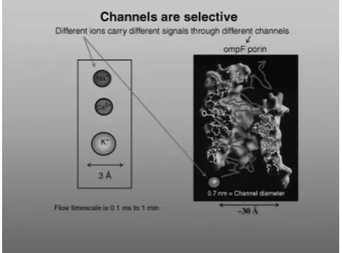
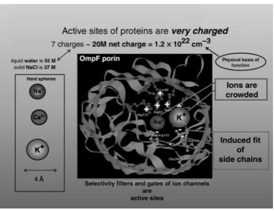
[image:9.432.95.336.337.515.2]Ion channels are proteins with a hole down their middle that control the flow of ions through otherwise impermeable membranes (Figs. 1 and 2). Ion channels are the
Figure 2. A “cross section” of a channel structure drawn to emphasize the crowding of ions in a channel. The structure of the channel OmpF is known from crystallography [105–111]. Figure adapted from Schirmer [112]. (See the color version of this figure in Color Plates section.)
nano (nearly pico) valves of life with as general a role as transistors in integrated circuits. Ion channels form a useful path into biology for physical chemists, one that carries familiar ions over potential barriers. Ion channels can help carry physical chemists over the social and intellectual barriers formed by exhaustive descriptive biology. The study of ion channels involves physical chemists in a biological problem of great generality and importance, one that nonetheless can be attacked (in large measure) by physics (and chemistry) as usual, without invoking vital new principles of organization or complexity. We present a brief description of ion channels now so the gate to physical understanding is open and not organically blocked by unknown structures.
the focus on a specific, well defined, reasonably robust system that uses a definite set of forces and structures to maintain a definite set of properties, namely its input–output relation. The physical chemist finds it much easier to study ionic solutions around 200 mM ionic strength made of alkali metal ions and alkali earth ions than to study all solutions of all elements at all concentrations. The physical chemist (or at least the chemical engineer) can find the problem of determining the transfer function of an electrochemical cell more approachable than the problem of determining all properties of the solutions inside the cell. Biology provides “the existence theorem” that guarantees that a reduced model can describe an ion channel quite well.
The selectivity of ion channels is an easy subject to study in the physical tradi-tion. The role of structural change (in the channel protein) is minimal and stereo-typed, so elaborate descriptions of different types of conformational change are not needed. The physics of selectivity is also quite stereotyped and so only a few mechanisms are likely to be involved, although the balance between the energies of different ions (and processes) is likely to be quite different in different channels. Physical scientists often dislike descriptive detail. The selectivity of ion chan-nels requires less descriptive detail than many other properties of chanchan-nels (or proteins), making ion channels a (relatively) easy biological object to study for physical scientists.
Biologists on the other hand relish descriptive detail and many find selectivity boring for that reason. Selectivity requires description in numbers, not names and biologists are often glad to leave that to their physically oriented colleagues. The common themes of all ion channels seem too common—and boring—to some biologists. I hope they do not seem too challenging for physical chemists.
C. Channels Open and Close
Not all properties of ion channels are as simple as selectivity. Channels open and close and open again and that process involves conformational changes of the protein and of the electrical and chemical potential fields within the channel. When channels are open, ions flow through a single structure that does not change on the biological timescale (slower than say 100s). Ions flow through a “hole in the wall” following the laws of electrodiffusion, at a fixed temperature, with a simple contribution from hydrodynamics. Ion channels also share themes with each other and with enzymes in general [113]. A general physical analysis of some functions of channels is possible. Ion channels are much less complex objects to analyze physically than amplifiers, particularly once the channel is open.
The open channel so far falls into two types, one in which side chains of the protein mix with ions and water in the pore of the protein (calcium and sodium channels) and one in which the side chains face away from the pore and the pro-tein forms a (relatively) strong “smooth” surface (as far as we can tell) (potassium channels).
Ion channelsare nanovalves that control most biological systems by control-ling the flow of ions, water, and electric current across otherwise impermeable biological membranes. The membranes are lipid bilayers that define cells and structures within cells, like the cell nucleus or mitochondria as well as the cell itself. Evolution—like an engineer—isolates subsystems from their neighbors by insulators. Engineers use silicon dioxide. Evolution uses lipid bilayers. Subsystems are connected through “channels” that cross the insulation.
Ion channels exist so ions can cross otherwise impermeable membranes in disjoint physically separate pathways (“parallel resistors” if one likes engineering images). Ion channels have internal structure that allows ions to move and external structure that allows them to exist in lipid membranes.
Physical chemists need to be reminded of the importance of structures. Com-plex structures are found in nearly all devices and biological systems. ComCom-plex structures must be understood as much as physics must be understood if we are to control and build devices and biological systems. The complex structures need description. No engineer would consider studying a machine without its parts list. A great deal of biology has been devoted to constructing that parts list, for centuries at the macroscopic level, and now at the molecular level. It takes a great deal of work to separate and identify the parts of a complex system, if you do not have a blueprint. Molecular biology learned part of the blueprint when it learned of the existence of genes and their biochemical basis, DNA and RNA. The blueprint provides (most of) the information needed to make proteins. It determines the se-quence of amino acids joined into a linear chain to make the protein. That is the only function of the blueprint. But the linear chain is like a chain of beads that folds to make the devices and machines of life. We do not know the rules that fold the chain into devices and indeed sometimes additional information is needed in the form of structural templates. That is why so much effort has been spent on the protein folding problem. Much progress has been made but the truth remains that we must catalogue the structures of proteins as we catalogue their sequence of amino acids if we are to have a reasonably complete parts list. Our parts list must include three-dimensional structures as well as sequences. Biology thus necessar-ily involves a great deal of description. That description is needed before the role of structure can be understood in biological function.
systems is an obvious strategy for building complexity from simpler building blocks. Valves are the physical building blocks that control flow from system to system. Ion channels are the nano (nearly pico) valves of life. Vacuum tubes (“valves” to twentieth century speakers of UK English), and field effect transistors (FETs) are a great deal easier to use than bipolar transistors because they are much more independent. The input characteristics of a FET are far more independent of output characteristics (of itself or its connecting devices) than bipolar transistors and this makes design much easier and more robust.
A large fraction of the proteins in an organism are channel proteins that use ions to carry a current of particles, just as a large fraction of the devices in a computer are FETs that use their own channels as nanovalves to carry the current of (quasi)-particles, holes, and “electrons.” The biologist and computer designers need to know about channels and transistors because they are such a large fraction of their systems. The physical chemists and physicists need to know about channels and transistors because they are so interesting, providing mechanisms by which tiny structures and powers can control large movements and flows. Channels and transistors are valves. And valves use interesting physics.
The analogy between channels and FETs is useful and productive [102–115] because they both follow similar mathematics even though the charged objects are quite different. The charged objects of channels are ions, atoms with permanent charge that do not change during the great majority of biological processes.
The electrons of semiconductors are more ephemeral. They are mathemat-ical constructs with quite distinct properties from the electrons in a vacuum tube. Surprisingly few scientists are aware that the negative quasiparticles of silicon/germanium have little in common with the isolated electrons of physics textbooks and cathode ray tubes. I wish the “electrons” of silicon/germanium were named (something like) “semielectrons” (for semi[conductor]electrons) so they are not confused with isolated electrons that flow in a vacuum. A mathe-matical construct and a physical electron are really quite different things. It is a naughty convenience to use the same word “electron” for both, at least in my view [102].
D. Sigworth’s Equation: Single Channel Currents
little. I mention enough of the properties of ensembles so the reader will know the main additional features that occur when channels work together. It would not be good for the reader to be entirely divorced from the biological reality of cells and tissues [116–131].
Consider an ensemble ofNindependent channels of the same type. Ion channels control current flow in a simple way, summarized by Sigworth’s equation [132]. The equation describes the current flowIthroughNsingle channels measured in a voltage clamp experiment in which all single channels have the same (open chan-nel) conductance, voltage and concentration across them and all single channels open with probabilitypto an identical mean current level< i >. (Historical note: I choose the name for this equation for personal and historical reasons. Most chan-nologists learned of the equation from Sigworth [132]. I certainly did, in a seminar he gave us early in his career, while still a graduate student, if I remember correctly. I do not know if there was a significant previous history.)
I=N < i > p= N(s) Number of channels available
· < i (s)>
Amplitude of single channel
· p(ms)
Probability of opening of one channel
(1)
Much of the lore of studying channels (“channology” if you will) is the applica-tion of Sigworth’s equaapplica-tion to the particular case where there is just one channel protein being recorded at a time. I try to make explicit what experimentalists learn implicitly “in the lab.” This knowledge is needed by physical scientists, if their work is to deal with real data. Theory and simulations are most useful when they produce results indistinguishable in form and format from experiments.
Sigworth’s equation is used in the lab for the case where only one channel protein of one type can open. It is used when there is negligible probability of more than one channel being open. Experiments with single channels are often designed to show openings of only one type of channel to only one level. Indeed, one of the most important features of the single channel revolution of Neher and Sakmann [133–136] is often unremarked. If an experiment records a single channel molecule, it records a single type of molecule. One molecule can only be of one type. Currents measured from that one molecule do not have to be “deconvolved” into the currents of a multiplicity of channels.
that is, they almost all involved more than one channel type, and often unknown channel types as well.
Single channel experiments are designed to study one channel protein of one type. That is appropriate when the goal is to understand that channel protein. That is the goal of much of molecular biophysics.
But channel proteins function in physiological systems made of many types of proteins. In those systems many types of channels are present that interact to make the system function. Physiology as a profession tried to link these scales for several centuries, culminating in the successful analysis of the neuron from molecule to membrane to cell, even to cells meters long.
The physiological tradition is not taught to most molecular biologists, however, in my experience. Most molecular biologists need to be taught what steps are needed to connect channels and cell function. They need to be taught the enormous importance of intermediate scale models. No engineer would think of making an all atom model of an amplifier. Indeed, most engineers would not make a model of the physical layout of an amplifier. Many would avoid the complete circuit of the amplifier. They would rather use the simplest equivalent circuit that illustrated the properties they needed to know. Classical physiologists used the same approach to the nerve cell. Thus, the linkage of atoms (e.g., ions), molecules (e.g., channel proteins), cell structures (e.g., membranes), cells (e.g., nerve fibers) into a single system is very well understood in classical neurobiology. Neurobiology has learned to use a multiscale approach just as an engineer would, with different resolution descriptions of ions, channels, membranes, axons, and nerve cells. A traditional [149] and a modern textbook [150] illustrate this material very well, much better than I can here.
More complex cases of more than one channel, or channel type, or of many superposed channel openings—repeated openings of the same channel molecule, or overlapping openings of several different channel molecules—can be handled in theory by multiple convolutions. This is straightforward when individual openings are independent and uncorrelated [136, 151] Openings of different channel proteins are likely to be uncorrelated if the proteins are widely separated. On the other hand, multiple openings of the same channel molecule are likely to be correlated. Nearby channel proteins that interact “directly” will have correlated openings, by definition. In these cases, theoretical methods must include every way a channel can open, for example, by using enough nested convolutions to describe all interactions that can open a channel.
Equation (1) has an explicit separation of scales that is important in its use. If the timescales are not widely separated, the distinctions I am about to make do not hold, and a more elaborate analysis is needed, and it is likely to be different for each type of channel and each situation.
than seconds. Regulatory and metabolic processes can change the number on the timescale of minutes, and (mechanistically obscure) processes labeled “slow in-activation” can change the number of available channels on the timescale of (say) seconds. The longer timescales are thought to reflect the construction of the channel and the regulation of its biochemical state by covalent bond changes like phospho-rylation [152]. Those timescales are supposed to be captured by theNvariable in Eq. (1) although that description is obviously an oversimplification. The inactiva-tion, regulatory, and metabolic processes might change. Description by a constant
Nwould be misleading in that case. The properties ofN need to be checked by experiment in each case.
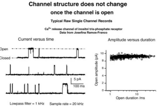
In Eq. (1) each channel protein conducts a current ofi(s) amps, where the (s) indicates that this current reaches steady state in less than a microsecond [153, 154]. The current through a single channel in Eq. (1) follows a rectangular time course. The current (in the mean, averaging out noise, by averaging over an ensemble of measurements) is zero for a stochastic time after a voltage change (or other perturbation) is applied. The current then suddenly switches to a new level, the open channel current. The new level is maintained without drift or internal correlations (once noise is averaged out [139, 140, 144, 155–159]) for a stochastic duration, and then the channel suddenly closes, usually to the same zero level it opened from. The channel may reopen. If it does, the channel will suddenly open to the same level, within measurement error. Figures 3 and 4 show some classical sudden openings of single channels and illustrate the difference between gating and permeation.
E. Opening Time Course of Channels
Everyone wants to know how fast a channel opens. The time course of a single channel opening has been studied by Miodownik-Aisenberg [153] and indepen-dently by Tang et al. [154]. Neither group chose to publish a full length paper (personal communication) because their results were so complex. On a timescale in which a single channel opening could be resolved (say 100 ns), an enormous range of behaviors was seen, that could not be easily summarized by either group. Some openings were sudden, some were very gradual, some were gradual and then sudden. It was not even possible to define a clear 10–90% risetime. What is possible is to say that after a few microseconds, this complex opening behavior seems to have no further effect on the current through an open single channel. We do not know what questions to ask of the opening of the channel. We do know what to ask of the open channel. So we study the open channel from now on, and ignore the opening process. It is important for scientists not to ask questions they know they cannot answer, particularly if they do not even know what question to ask.
Figure 3. Typical “raw” recording of current through a single channel. The left-hand panel shows a plot of current versus time recorded by Dr. Ramos-Franco from a single channel calcium release channel, of the insoitol tris-phosphate receptor with a sampling rate of 20 k samples per second, through a 1 kHz low pass filter. I am grateful for her permission to show these results. Under the conditions shown, the channel is closed most of the time, opening suddenly from a level of nearly zero, to a level of approximately 9 pA. The openings occur at stochastic intervals, and have stochastic durations. Successive records are not identical, but are reproducibly distributed around a mean value. The right-hand panel shows that the amplitude of the open (single) channel current is independent of duration. This is a general property of single channel recordings and is nearly their “operational definition.” The text argues that the amplitude can be independent of duration only if the “structure” of the channel does not change significantly: if the structure changed by even 0.1 ˚A, the current would change because the charges of the protein are so close to the ions in the channel. Indeed, the probably are mixed with them in an ionic and “electric stew” (references in text). The word “structure” means the average location of atoms, averaged over the duration of a few sampling intervals, here say 2×50 = 100s.
experimental error. Channels open and close stochastically, all to the same level, if no perturbation is applied. The duration of opening is stochastic, usually expo-nentially distributed, often distributed as the sum of a few exponentials [161–163]. The fraction of time the channel is open is a reflection of the gating process that opens the channel, widely assumed to be a change in conformation of the protein, although the relevant conformation in general is that of the (electrochemical) po-tential profile along the channel and not the anatomical shape of the channel protein [103, 104, 114, 164, 165].
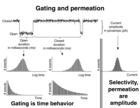
Figure 4. Gating, permeation, and selectivity. The figure shows a typical single channel record (upper left, amplitudes are typically 10 pA, durations typically 10 ms). The histograms show the distribution of amplitudes (on the right-hand side) and durations (on the left-hand side) in log and linear plots. Selectivity properties of channels measure the current flow or binding in the open channel. They are properties of the amplitude of the open single channel current. Gating properties are produced by different mechanisms, with different structures, pharmacology, voltage, and time dependence. Most of this review is about selectivity. Not enough is yet known to make physical models of gating.
dimensions (on the 10s and slower timescale, see Figs. 3 and 4). Flexibility seen in simulations is on a much faster timescale [171–174].
Selectivity is produced by physical forces that depend on the ion charge and size and chemical interactions with parts of the channel protein that form what is called “the selectivity filter” [175–189]. Those chemical interactions can arise from physical interactions of ions with side chains of the protein, physical interactions with the permanent electrical charges in the protein, physical interactions with the induced polarization (i.e., dielectric) charge on the protein, chemical interactions involving changes in the distribution of charge within the molecular and atomic orbitals of the protein. (If the changes in distribution of charge in the orbitals are proportional to the local electric field, the change in charge is identified as the induced polarization charge of a dielectric. If they are not proportional, I call them “chemical.”) In any case, those interactions do not vary significantly on the timescale of 10s or longer if they did, the single channel current level would not be “constant” independent of duration of opening, or time after an opening.
the protein (whether partial charges in a carbonyl or full charges in a carboxylate) are within an angstrom of permeating ions. The change in the electric field from Coulomb’s law is large if these groups move. The electric force has an exponential effect on current flow. Thus, tiny changes of structure (<0.5 ˚A) would have an easily measured effect on open channel current, if the structural change lasts long enough, longer than (say) 10s.
Sigworth’s equation is applied to cases in which the electrical potential across all the channels is the same, as in voltage clamp experiments. Sigworth’s equation is not applied to nerve or muscle cells when they are carrying action potential signals because the voltage is then changing. The potential in voltage clamp experiments (but not in real cells) is usually controlled in steps that change from one value to another at one time or another (in the tradition of Hodgkin et al. [166,168]). The chemical potential (e.g., the concentration of ions) on each side of the channel is assumed not to change significantly with current flow or time in the classical tradi-tion, although significant exceptions to this rule are common and indeed inevitable in important cases like calcium channels, where the concentration of one of the permeating ions is very low on one side of the channel or the other. Experiments and theory should always be wary of the assumption of “constant” concentration (independent of time and current amplitude) because it is an approximation that cannot be generally true [190].
the flow of current in transmission lines, submarine cables, and long cylindrical cells, called cable theory [149, 150, 193, 194].
We return from history to the modern study of single channels. In the idealized case, the individual channel behaves very much like a pore with a gate. The pore conducts current carried by ions, with a diameter about half that of the pore. The flow is controlled by a gating process that turns the current through the pore on and off but does not otherwise affect that current. At least this is the classical view, and a most useful initial approximation.
F. Open Probability
The probability that a single channel opens varies on a millisecond timescale and so is written asp(ms) in our version of Sigworth’s equation (1). Thisp(ms) is the probability of any one of theNchannels opening. It is not the probability of a particular channel opening. If one wishes to write the probability of any channel opening as a functional of the properties of one channel opening, one must deal with the variable latency and duration of individual (i.e., one channel) openings. One must perform multiple nested convolutions, being sure to include the possibility of multiple openings, of the same or of different channels, and durations that depend on the number of openings, as well as the duration of previous openings, and so on. The convolutions must sum all the possible openings one would see if one made a very large number of measurements of the response of a single channel protein to repeated steps of voltage in a voltage/patch clamp experiment. One must be sure to include all theNchannels that can conduct current. Usually those channels are assumed to open independently (if the voltage is maintained constant), but such is not expected to be the case if the concentration change produced by channel openings correlates their properties, or if the channels interact through their internal charge movements (i.e., through electric fields within the protein and membrane), or by mechanical interaction, or by mysterious allosteric forces of unspecified physical origin.
G. Relation to Nerve Function
We turn now to the properties of ensembles of currents in one important case, the action potential of nerve fibers, the natural activity of nerve axons, which are the “wires” that conduct signals long distances in the nervous system. This detour from physical chemistry seems necessary to lend biological and medical reality to our discussion.
I include the following description of textbook material [149, 150] meant mostly for physical scientists new to channel research.
Channel proteins are typically some 10 nm in diameter, while the pores through which ions move are typically 0.6–1 nm in diameter. Channel proteins are typically (but not always: important exceptions include the node of Ranvier and parts of the endoplasmic and sarcoplasmic reticulum) separated by substantial amounts of lipid membrane. Channel proteins are thought to be built to operate independently so it is natural to assume no interactions between channel proteins if they are all held subject to the same electrical and chemical potential gradients in a voltage clamp experiment. Many channel proteins are modulated by nearby proteins however, that themselves are not channels, for example, the neurologically important GABA receptor complex [195].
Channels arenotindependent when functioning in a natural unconstrained way. Most channels are independent in a voltage clamp experimental apparatus but not in nature in their natural setting in cell and organelle membranes. When experiments are done without voltage clamp, the responses to stimuli and drugs are complex, obviously not just the sums of components, in many cases. When experiments are done with a voltage (and “space”) clamp system, responses to stimuli are much simpler, often just the sums of components [167, 168].
Channels in nerve cells interact with each other strongly through the electric field generated in part by the current through the channel. Channels control the electric field by regulating the current flow through the membrane. The current flow through the membrane in turn changes the potential. In the classical case of the voltage clamped squid axon,channels interact only through the electric field cre-ated by their current because the currents flow only for a few milliseconds. Even in squid axons, however, current flow of potassium over somewhat longer timescales than initially studied [160, 166] produces significant concentration changes that produce interactions of channels [196]. The accumulation of potassium after a series of action potentials in fact may be of crucial importance in a number of biological functions (memory) and medical disorders (epilepsy) [197, 198].
Confusion can be easy here if the experimental situation is not precisely de-scribed. The movement of current through a single channel rarely if ever is large enough to change the concentration of ions significantly, unless the concentration on one side of the channel is very very small (as with Ca2+ions [199]). But the movement of current through a macroscopic number of the same channel may change the concentration quite significantly. (One must never forget that the con-centration of messenger molecules like Ca2+is in fact very very small, e.g., 10−7 M. Thus, calculations of Ca2+flows must not assume that the intracellular con-centration of Ca2+ is constant, or independent of current flow or experimental conditions. The same is the case for calculations of other messenger molecules.)
surrounding solutions that seem (traditionally) to be uninteresting. Thus, they work hard to remove the effects of gradients of electrical potential outside the channel in the baths (by “series resistance compensation,” starting with [160] and then developing into a substantial literature [200, 201]) and to remove the effects of gradients of chemical potential (i.e., concentration) in the baths, by judicious restriction of timescales, choice of ionic solutions, and even selection of experi-mental records. “Concentration polarization”—as such effects were once quaintly called—are particularly difficult to deal with because they necessarily involve water flow and convection (at least in general and probably nearly always). The rotating disk electrode method of electrochemistry [202–205] was developed to control such effects by establishing a known and controllable convection field that dominates the spatial distribution of concentration. Biologists seem not yet to have used rotating disks or other methods to establish convection gradients and con-trol concentrations outside channels, membranes, or cells. Such methods might be particularly helpful in studying water and solution flow, which are plagued by controversy, perhaps arising from their sensitivity to bath conditions. It is interest-ing to note that controversy decreased dramatically in the fields of polarography and electrodics (i.e., electrochemistry involving chemical reactions at electrodes) once the rotating disk electrode was introduced.
Time dependent concentrations involving water flow are also particularly hard to deal with in theories or simulations. Simulations on atomic scales ignore such flows typically because they occur on timescales so remote from the timescales of the simulations. (Water flows take milliseconds to second; atomic motions take femtoseconds.) Theories have difficulty knowing how to mix different types of flow (i.e., macroscopic pressure volume flows, diffusion, and electrical migration) and how to deal with interactions of different solutes and ions.
Recently, variational analysis has been applied to this problem [6] and progress seems possible. Energies and dissipations of different components are combined in the energy variational approach and Euler–Lagrange equations are then derived, as we show later in this paper. These partial differential equations are the unique con-sequence of the contributions of individual components. The form and parameters of the partial differential equations are determined by algebra without additional physical content or assumptions. The partial differential equations of mixtures automatically combine physical properties of individual (unmixed) components without arbitrary parameters. It will be interesting to see how far this approach is able to simplify the formidably complex descriptions [206–208] of water flow in complex ionic solutions with many components.
III. ACTION POTENTIAL IS A COOPERATIVE PHENOMENON
of sodium channel opening, channel current, and potassium channel opening at one location with the same openings at another location produces the action potential that carries information long distances in nerve fibers [170, 209–211]. Hodgkin proved [210, 212] (when he was an undergraduate) that the cooperative behavior that produces propagation of the action potential is entirely electrical and does not involve any “propagating wave” of chemical activity in the nerve membrane itself. Hodgkin’s papers are beautiful examples, easy to read today, of what can be done with minimal apparatus and maximal thought.
Hodgkin once told me that he had quite purposely written the papers to show they could have been performed with apparatus available around 1900. Modern readers of Hodgkin simply need to know that “electrotonus” is an old way of stating, more or less, the change in membrane potential as a function of time and distance. (Readers of the “There will always be an England” features—found in the New Yorker magazine for many decades—need to know that Hodgkin did in fact use an oscilloscope and not an electromechanical apparatus to record potential: being aggressively archaic does not mean being foolish.)
Later work showed that the action potential itself, as well as its propagation, is a passive process that does not involve the chemical interaction of different parts of the membrane (i.e., that does not involve chemical interaction of different channel proteins, in modern language). Channels cooperate to make an action potential
onlythrough the electric field that the channel currents create (in the classical case [170]). In the less classical case [213], concentration changes produced by these currents also produce interactions. In no case are covalent bond changes or ATP hydrolysis involved in the action potential.
Allosteric interactions between different channel proteins are not involved in the main signaling phenomena of the nervous system, although it is certainly true that the effect of membrane potential on the opening of a single channel might be called allosteric. Binding proteins near ion channels can importantly modify the properties of ion channels [195] but they are not involved in the voltage or agonist responses of the classical sodium and acetylcholine channels of neurobiology.
A. Allosteric Mechanisms
The physics that produces allosteric interactions is not widely discussed, perhaps because the inventors of the phrase “allosteric” thought physical mechanisms un-interesting or unapproachable on the atomic scale.
Indeed, some scientists have avoided the atomic scale altogether. Some sug-gested that an important allosteric interaction is produced by rigid rods [214] that reach across an intracellular space to join two membranes. Such models seem to me more appropriate for the macroscopic deterministic scale of laboratories than the atomic Brownian scale of atoms.
enormous number of times in the shortest biological timescale (say 10s) as it moves more or less at the speed of sound [217] in a system with almost no empty space. Indeed, friction limits all motion [215, 216] to the overdamped case [218] in condensed phases like ionic solutions. (It is important to remember that ice floats on water, even salt water, thus showing that the density of liquid ionic solutions is more than that of the relevant solid. Thus, the empty space in liquid ionic solutions islessthan the empty space in solid solvent. Gas-phase models of biochemical reactions are more or less worthless, in this view, because the density of ideal gases is nearly zero.)
My view [103, 104, 164, 165] is that local (i.e., incompletely shielded) changes in electric field within the membrane, within a Debye length of proteins, are likely to be involved in these long-range allosteric effects as well as conformation changes. These local electric fields are likely to be focused by regions of low dielectric coefficient such as found on the inside of the KcsA channel [219]. The focusing of electric fields by dielectrics can create an enormous range of machines in the hands of engineers [220]. It would be surprising if evolution did not do some of what engineers do so much.
B. Conformation Changes
A macroscopic description of the forces driving conformation changes seems nec-essary in both physical and biological models. The gap between atomic scales and macroscopic scales seems too large to deal with in the foreseeable future, as we discuss later in this paper.
Conformation changes and allosteric forces arise from the random motions of atoms computed on a 10−16s timescale in molecular dynamics but the resulting conformation changes of channels are so slow (from 10−5to 10 s) that comput-ing them directly is likely to be difficult. It will be even more difficult to do the computations, given the size of the system (involving 109memory locations so positions can be specified in three dimensions to 0.1% of a 1000 ˚A computational box) and the ratio of timescales (1011–1017) and number densities involved. (The number density of water is some 108×larger than the number density of intra-cellular Ca2+.) Doing the calculations in a reliable way with known numerical and mathematical error bounds requires calibration against known macroscopic properties of physical systems [221, 222].
as I know, no calibrations of biological (i.e., mixed) solutions have been made successfully. Attempts to calibrate NaCl solutions are beginning [116–131, 223, 224] and are reasonably successful when dealing with properties of the neutral combination NaCl, but are not so successful when dealing with cations and anions separately. Cations go through channels and act in enzymes separately and so cal-culations involving them must calibrate ions separately. Indeed, sodium channels are called sodium channels and not sodium chloride channels precisely because sodium—not sodium chloride—goes through them. Calibrations of solutions with divalents have not been completed as far as I know. Mixtures of ions have not been attempted, even though mixtures are invariably present and important in biologi-cal solutions. Thus, atomic sbiologi-cale simulations of conformation changes in realistic ionic conditions with calibrated results are not on the immediate horizon.
In my view, uncalibrated analysis is dangerous, not really part of a proper scien-tific process. Uncalibrated analysis can go on and on, sucking resources from other kinds of science that might lead to reproducible results and answers to questions. In my view, multiscale analysis is needed in which parts of the problem are handled on atomic scale and parts on macroscopic scale. The coupling of scales is part of the model in any multiscale analysis. Multiscale analysis requires treatment of reduced models, and the interactions of reduced models. What is important is that the reduced models can be checked against experiments in a range of condi-tions relevant to their use. In my view, physical chemistry can be of enormous help in dealing with biological problems if it addresses multiscale problems explic-itly and insists that structural biologists and simulators deal with the macroscopic reality of ionic solutions. Studying individual trajectories, whether imagined or calculated, does not deal with the macroscopic reality of trajectories and ionic interactions. Trajectories and ionic interactions are incredibly complex, involv-ing a more than astronomical number of terms if all ions are coupled by the electric field.
The models of structural biologists and molecular dynamics must produce re-sults comparable with experiments if they are to be compared to experiments. Graphs must have the same variables whether they come from experiment or the-ory. To replace graphs with verbal discussions, or individual trajectories, is to replace science with metaphor. However beautiful, metaphors are not helpful in actually building machines and devices. They are hardly likely to be more helpful in understanding and building biological machines and devices. There is no engi-neering without numbers; there should be little molecular biology or biophysics without numbers, for the same reasons.
One of the most important targets of multiscale analysis should be the con-formation changes of proteins. These occur on timescales from femtoseconds to seconds, and length scales from atoms to whole proteins.
them. Naming them as allosteric might in fact be a step away from understanding if newcomers or structural biologists felt that the adjective “allosteric” had a specific physical meaning. The arrows of allosteric models might seem to be disguises for the law of mass action. Indeed, the arrows of allosteric models do become specific statements involving the law of mass action once the rate constants of that law are identified with physical processes. But that identification is not one of the main topics of discussion in the usual treatments of allostery. Physical chemistry is needed to replace the metaphor of allostery with the reality of numbers.
Later we will discuss the law of mass action and the physical origin of its “constants.” Briefly stated, we will show that the law of mass action is usually used in a misleading way. The law is most helpful if reactants are ideal. It is most helpful if reactants are uncharged solutes, and rate constants are identified as a property of unidirectional fluxes into absorbing boundaries. But reactants are often not ideal. Indeed, reactants are present in enormous concentrations in channels and close to active sites of enzymes where they are important. Reactants in those locations more resemble an ionic liquid [225–229] (e.g., melted NaCl) than a solution of infinitely dilute uncharged solutes. Indeed, ions in solution always come “in pairs” (more precisely in groups of anions and cations that are exactly balanced in charge so the systems are neutral taken as a whole). Ions come in pairs because anions and cations interact so strongly. Anions and cations cannot be independent solutes and also form solutions that are exactly electrically neutral. Ionic solutions are electrically neutral because their ions interact. If the reactants in the law of mass action are not ideal, the rate constants must be variables, and rather strongly varying variables at that. In fact, the variables often change exponentially with potential [230–235]. The law of mass action with constant rate constants seems a poor initial guess at a transport law for ions in multimolar concentrations near active sites or in channels.
The forces that drive conformation changes (and determine the rate constants of models of conformation change) are not known, but are likely to be electrical and mechanical arising from excluded volume of ions and side chains [176]. The forces driving conformation changes are closely related to the mechanical forces in classical Donnan systems [184, 236] that produce osmotic pressure.
C. Cooperative Behavior Produced by Current Flow
The ionic current that flows through channels in an action potential flows down the length of the fiber a long distance (think mm in myelinated nerve fibers of ver-tebrates and cm in squid nerves) to a neighboring region. This longitudinal current (that happens to be conducted by different ions from those that cross the membrane to make the action potential at the original location) changes the potential in that neighboring region. That change in potential changes the probability of a channel being open quite dramatically, which in turn produces an action potential in this new region, and the process continues down the nerve fiber, creating a propagating action potential. The process is rather like that in an underwater cable with repeater stations some distance apart that restore a digital signal to its original specification.
IV. COMPUTATION OF THE ACTION POTENTIAL
A complete mathematical theory of the propagating action potential has been avail-able for a long time [9, 237–239]. Measurements of channel currents in voltage clamped axons allow computation of the propagating action potentials [170] with-out the introduction of any new physics. Thus, a complete mathematical theory of a major biological function is possible and was essentially complete (although without molecular explanation) in 1952. Molecular explanation followed in the work of many channologists [9, 237–239], culminating in the single channel mea-surements [136] and Nobel Prizes to Sakmann and Neher with structural insight provided later by Mackinnon [240–242]. A complete mathematical theory of a major biological function starting from atoms and finishing with nerves of the length of a giraffe leg has been available for several decades. Sadly, we do not have room here (or time in our lives) to write a full description of this remarkable accomplishment of physiologists and biophysicists. Good accounts can be found in Refs [8, 9, 149, 150, 209, 243, 244].
spatial and temporal dependence of the natural propagating action potential. The spatial dependence is removed (“short circuited by a space clamp,” in lab jargon) by inserting a wire down a cylindrical nerve fiber. The wire keeps the potential at all locations (hopefully) the same at all times [246–249]. The spatial depen-dence can be removed by studying spherical cells that do not allow propagation [250, 251].
The temporal variation of electrical potential is prevented by electronic circuitry and the experimental setup, the voltage clamp apparatus, see Ref. [252] for history, and Ref. [136] for recent implementation in “whole cell clamp.” A variation called the patch clamp is used if the system includes only one or two channels [136, 138–140, 142–144, 149, 156, 253].
A. Changes in Ion Concentration
The voltage clamp experimental apparatus is designed to isolate single types of channels or individual channel molecules so they can be studied independently, as we have just discussed. In nature, channels interact by passing current that changes the electrical potential across the membrane of the cell and the thus across neighboring channels. The current through the channels is carried by one type of ion depending on the selectivity of the channels. The current inside the cell is carried by a different type of ion depending on the types of ions inside the cell. The potential inside the cell changes the potential across the cell membrane and is usually nearly equal to the membrane potential (see Refs [191, 250, 251, 254] for an experimentally important exception). The experimental apparatus is designed to prevent this natural interaction so the underlying mechanisms can be isolated and studied. The apparatus does this by controlling electrical potential with an electronic feedback apparatus (and a system of four electrodes to link amplifiers with ion currents) that prevents the natural interactions between channels.
In nature, channels may also interact by changing the concentration of ions near channels (called accumulation or depletion, or “concentration polarization” in the older literature). Such effects are particularly important in calcium channels, because the calcium concentrations inside cells (on the intracellular side of the channel protein) are small [Ca2+]in∼=1×10⁻⁷M compared to the intracel-lular concentrations of potassium [K+]in∼=3×10⁻¹M or sodium [Na+]in∼= 3×10⁻²M. The amount of charge actually carried through a channel is of-ten set functionally by the capacitance of the nearby cell membrane (typically 0.8F cm−2), producing a capacitance of some 63 pF for the 7.85×10⁻⁵cm2of membrane in spherical or cylindrical cell of diameter 5×10⁻³cm with typical electrical properties.
when current flows through the channel. Electrical forces are much stronger than chemical forces (see the first paragraphs of Feynman et al. [255]) so, in some vague sense, we expect the charge carried by current to be more important than the mass, that is, change in concentration, produced by the ions that carry the current.
The importance of charge versus concentration depends on the concentration of ions present before the charge moves. If the electrical charge and current are carried by sodium or potassium ions through the membrane, the concentration of ions is changed by a much smaller fraction than if the current is carried by calcium, because there is so much less calcium present to begin with inside cells. Calcium concentration inside cells can be easily changed by calcium current through chan-nels while potassium and sodium concentration cannot because there is so little calcium inside cells (some 10−7M Ca2+versus 10−2M Na+or K+). In chemical and biological language, calcium is hardly buffered by the “background” con-centration inside cells but sodium and potassium ions are buffered, because the concentration of sodium and potassium inside cells is so much higher than cal-cium [190, 196, 256]. (The reader should be warned not to think of this buffering process as simply as this. Elaborate subcellular machinery is present to control these concentrations and that machinery involves transporters that use chemical energy to control concentrations.) A given amount of current (i.e., charge) passing through a channel changes the concentration of any ion (with the same valence) carrying the charge by the same amount. The relative importance of this change in concentration depends on the background concentration (before the current flow). If the concentration is high, like K+inside a cell, the relative effect of the inward charge movement is small. If the concentration is tiny, like Ca2+inside a cell, the relative effect of the inward charge movement is large.
Changes in calcium concentration inside cells are often used by biological sys-tems as signals (“messengers”) of channel activity for this reason. In a similar, but less dramatic way, changes in potassium concentration outside cells can be used by biological systems as signals of channel activity. The background resting potassium concentration outside cells is typically [K+]out∼=2×10⁻³M compa-rable to the calcium concentration outside cells, but much less than the sodium concentration there. Of course, the concentrations of ions inside and outside cells are also regulated by diffusion and convection and “active” properties of nearby biological structures designed to control concentration.
B. Gating Processes
This chapter does not deal further with the gating process that opens channels because simulations of gating are not quite in our grasp and the physical basis of gating has not yet been described by reduced models. (Historical note: despite their numbers, models based on arrows [9, 257, 258] instead of physics have not proven useful and make little connection to the physical properties of channel properties or ions. A British physiologist, who in fact had written a number of arrow models, once told me “You can tell how much is known by how few papers are written on a subject. When it is understood, little more needs to be said.” He was referring to his early work on the (mesoscopic scale) sliding filament “hypothesis” [259–261] not to his own later arrow models on the atomic scale.)
Some of the many states postulated by arrow-based gating models are likely to exist in a well-defined sense, even if most of them cannot be unambiguously identified or measured, but the problem is that the traditional arrow models of biochemistry, biophysics, and channology (compare [9, 257, 262] and [209, 245]) do not let you know which states are significant and which are not. It seems unlikely that enough data could possibly be measured to determine the arrangement of the arrows in the models containing tens or even hundreds of reactions. Indeed, the amount of information needed is much larger than the number of rate constants themselves, because rate constants vary with conditions and cannot be constants. It seems likely that deviations from expected behavior would always be explained [9, 263] not by questioning the models themselves, but by invoking still more states and rate constants that could themselves be measured only with great difficulty. This process seems unlikely to be an efficient way to understand how channels work, particularly given that the underlying models themselves are not appropriate, at least in my view [103, 104, 164, 165, 264–266].
I believe one needs a physical model that shows specifically how proteins move from state to state, under what forces, and with what dependence on electrical and chemical potentials, physical properties of the channel protein, and so on, if one is to make progress. One needs a physical model that shows how the conformation changes of the channel protein reveal themselves as an ionic conductance or as the nonlinear capacitive current called gating current [267–280]. Physical analysis of gating in this spirit is beginning [278,281–283] but it is not mature enough for me to review.