ARTICLE
Convergence of Genes and Cellular Pathways
Dysregulated in Autism Spectrum Disorders
Dalila Pinto,
1,2,3,4,5,6,85Elsa Delaby,
7,8,9Daniele Merico,
10Mafalda Barbosa,
1,2,3,4Alison Merikangas,
11Lambertus Klei,
12Bhooma Thiruvahindrapuram,
10Xiao Xu,
2,4,5Robert Ziman,
10Zhuozhi Wang,
10Jacob A.S. Vorstman,
13Ann Thompson,
14Regina Regan,
15,16Marion Pilorge,
7,8,9Giovanna Pellecchia,
10Alistair T. Pagnamenta,
17Ba´rbara Oliveira,
18,19Christian R. Marshall,
10,20Tiago R. Magalhaes,
15,16Jennifer K. Lowe,
21Jennifer L. Howe,
10Anthony J. Griswold,
22John Gilbert,
22Eftichia Duketis,
23Beth A. Dombroski,
24Maretha V. De Jonge,
13Michael Cuccaro,
22Emily L. Crawford,
25Catarina T. Correia,
18,19Judith Conroy,
16,26Ineˆs C. Conceic¸a˜o,
18,19Andreas G. Chiocchetti,
23Jillian P. Casey,
15,16Guiqing Cai,
1,2,3Christelle Cabrol,
7,8,9Nadia Bolshakova,
11Elena Bacchelli,
27Richard Anney,
11Steven Gallinger,
28Michelle Cotterchio,
29Graham Casey,
30Lonnie Zwaigenbaum,
31Kerstin Wittemeyer,
32Kirsty Wing,
17Simon Wallace,
33Herman van Engeland,
13Ana Tryfon,
1,2,80Susanne Thomson,
25Latha Soorya,
1,2,81Bernadette Roge´,
34Wendy Roberts,
35Fritz Poustka,
23Susana Mouga,
36,37Nancy Minshew,
12L. Alison McInnes,
1,2,82Susan G. McGrew,
38Catherine Lord,
39Marion Leboyer,
40,41,42,43Ann S. Le Couteur,
44Alexander Kolevzon,
1,2,6Patricia Jime´nez Gonza´lez,
45Suma Jacob,
46,47Richard Holt,
17Stephen Guter,
46Jonathan Green,
48,49Andrew Green,
16,50Christopher Gillberg,
51Bridget A. Fernandez,
52Frederico Duque,
36,37Richard Delorme,
40,53,54,55Geraldine Dawson,
56Pauline Chaste,
12,40Ca´tia Cafe´,
36Sean Brennan,
11Thomas Bourgeron,
40,53,54,57Patrick F. Bolton,
58,59Sven Bo¨lte,
60,83Raphael Bernier,
61Gillian Baird,
62Anthony J. Bailey,
33,84Evdokia Anagnostou,
63Joana Almeida,
36Ellen M. Wijsman,
64,65Veronica J. Vieland,
66Astrid M. Vicente,
18,19Gerard D. Schellenberg,
24Margaret Pericak-Vance,
22Andrew D. Paterson,
10,67Jeremy R. Parr,
68Guiomar Oliveira,
36,37John I. Nurnberger,
69,70Anthony P. Monaco,
17,71Elena Maestrini,
27Sabine M. Klauck,
72Hakon Hakonarson,
73,74Jonathan L. Haines,
25Daniel H. Geschwind,
21Christine M. Freitag,
23Susan E. Folstein,
75Sean Ennis,
16,50Hilary Coon,
76Agatino Battaglia,
77Peter Szatmari,
14James S. Sutcliffe,
25Joachim Hallmayer,
78Michael Gill,
11Edwin H. Cook,
46Joseph D. Buxbaum,
1,2,3,4,6,79Bernie Devlin,
12Louise Gallagher,
11Catalina Betancur,
7,8,9,85,*
and Stephen W. Scherer
10,20,85,*
Rare copy-number variation (CNV) is an important source of risk for autism spectrum disorders (ASDs). We analyzed 2,446 ASD-affected families and confirmed an excess of genic deletions and duplications in affected versus control groups (1.41-fold, p¼1.03105) and an increase in affected subjects carrying exonic pathogenic CNVs overlapping known loci associated with dominant or X-linked ASD and intellectual disability (odds ratio¼12.62, p¼2.731015, ~3% of ASD subjects). Pathogenic CNVs, often showing variable expressivity, included rare de novo and inherited events at 36 loci, implicating ASD-associated genes (CHD2,HDAC4, andGDI1) previously linked to other neurodevelopmental disorders, as well as other genes such asSETD5,MIR137, andHDAC9. Consistent with hypothesized gender-specific modulators, females with ASD were more likely to have highly penetrant CNVs (p¼0.017) and were also overrepresented among subjects with fragile X syndrome protein targets (p¼0.02). Genes affected by de novo CNVs and/or loss-of-function single-nucleotide variants converged on networks related to neuronal signaling and development, synapse function, and chromatin regulation.
1
Introduction
Autism spectrum disorders (ASDs) affect ~1% of the
popu-lation and are characterized by impairments in social
interaction and communication, as well as by repetitive
and restricted behaviors. ASDs include mild to severe levels
of impairment—cognitive function ranges from above
average to intellectual disability (ID)—and are often
accompanied by seizures and other medical problems.
There is a ~4:1 male-to-female gender ratio in ASD.
ASDs are highly heritable,
1and genomic studies have
revealed that a substantial proportion of ASD risk resides
in high-impact rare variation, ranging from chromosome
abnormalities and copy-number variation (CNV)
2–6to
single-nucleotide variation (SNV).
7–11These studies have
highlighted a striking degree of genetic heterogeneity,
implicating both de novo germline mutation and rare
in-herited ASD variation distributed across numerous genes.
De novo CNVs are observed in 5%–10% of screened
ASD-affected individuals, and after further follow-up studies,
some of them have proven to alter high-risk genes (e.g.,
NRXN1
12[MIM 600565]). De novo or transmitted CNVs,
such as 15q11.2–q13 duplications of the affected region
in Prader-Willi syndrome (PWS [MIM 176270]) and
Angel-man syndrome (AS [MIM 105830]), 16p11.2 deletion
(MIM 611913), 16p11.2 duplication (MIM 614671), and
X-linked deletions including the
PTCHD1
-
PTCHD1AS
locus (MIM 300828), have also been found to contribute
to risk.
6,13,14Exome and whole-genome sequencing
studies have estimated at least another ~6% contribution
to ASD
7–10,15and an additional 5% conferred by rare
inherited recessive or X-linked loss-of-function (LoF)
SNVs.
11,16A genetic overlap between ASD and other
neuropsychiatric conditions has also been increasingly
recognized.
Interestingly, CNV testing and exome sequencing have
so far yielded mostly nonoverlapping genes, which might
reflect different mutational mechanisms, although they
27Department of Pharmacy and Biotechnology, University of Bologna, 40126 Bologna, Italy;28Samuel Lunenfeld Research Institute, Mount Sinai Hospital, Toronto, ON M5G 1X5, Canada;29Cancer Care Ontario, Toronto, ON M5G 2L7, Canada;30Department of Preventive Medicine, Keck School of Medicine, University of Southern California, Los Angeles, CA 90089, USA;31Department of Pediatrics, University of Alberta, Edmonton, AB T6B 2H3, Canada; 32School of Education, University of Birmingham, Birmingham B15 2TT, UK;33Department of Psychiatry, University of Oxford and Warneford Hospital, Oxford OX3 7JX, UK;34Unite´ de Recherche Interdisciplinaire Octogone, Centre d’Etudes et de Recherches en Psychopathologie, Toulouse 2 University, 31058 Toulouse, France;35Autism Research Unit, The Hospital for Sick Children, Toronto, ON M5G 1X8, Canada;36Unidade de Neurodesenvolvimento e Autismo do Servic¸o do Centro de Desenvolvimento da Crianc¸a and Centro de Investigac¸a˜o e Formac¸a˜o Clinica, Pediatric Hospital, Centro Hospitalar e Universita´rio de Coimbra, 3000-602 Coimbra, Portugal;37University Clinic of Pediatrics and Institute for Biomedical Imaging and Life Science, Faculty of Medicine, University of Coimbra, 3000-354 Coimbra, Portugal;38Department of Pediatrics, Vanderbilt University, Nashville, TN 37232, USA; 39NewYork-Presbyterian/Weill Cornell Medical Center, New York, NY 10065, USA;40FondaMental Foundation, 94010 Cre´teil, France;41Institut National de la Sante´ et de la Recherche U955, Psychiatrie Ge´ne´tique, 94010 Cre´teil, France;42Faculte´ de Me´decine, Universite´ Paris Est, 94010 Cre´teil, France; 43Department of Psychiatry, Henri Mondor-Albert Chenevier Hospital, Assistance Publique – Hoˆpitaux de Paris, 94010 Cre´teil, France;44Institute of Health and Society, Newcastle University, Newcastle upon Tyne NE1 4LP, UK;45Child Developmental and Behavioral Unit, Hospital Nacional de Nin˜os Dr. Sa´enz Herrera, Caja Costarricense de Seguro Social, San Jose´, Costa Rica;46Institute for Juvenile Research, Department of Psychiatry, University of Illinois at Chi-cago, ChiChi-cago, IL 60608, USA;47Institute of Translational Neuroscience and Department of Psychiatry, University of Minnesota, Minneapolis, MN 55455, USA;48Institute of Brain, Behaviour, and Mental Health, University of Manchester, Manchester M13 9PL, UK;49Manchester Academic Health Sciences Centre, Manchester M13 9NT, UK;50National Centre for Medical Genetics, Our Lady’s Children’s Hospital, Dublin 12, Ireland;51Gillberg Neuropsychiatry Centre, University of Gothenburg, 41119 Gothenburg, Sweden;52Discipline of Genetics, Faculty of Medicine, Memorial University of Newfoundland, St. John’s, NL A1B 3V6, Canada;53Human Genetics and Cognitive Functions Unit, Institut Pasteur, 75015 Paris, France;54Centre National de la Recherche Scientifique URA 2182 (Genes, Synapses, and Cognition), Institut Pasteur, 75015 Paris, France;55Department of Child and Adolescent Psychiatry, Robert Debre´ Hospital, Assistance Publique – Hoˆpitaux de Paris, 75019 Paris, France;56Department of Psychiatry and Behavioral Sciences, Duke University School of Medicine, Durham, NC 27710, USA;57University Paris Diderot, Sorbonne Paris Cite´, 75013 Paris, France;58Institute of Psychiatry, King’s College Lon-don, London SE5 8AF, UK;59South London & Maudsley Biomedical Research Centre for Mental Health, London SE5 8AF, UK;60Department of Child and Adolescent Psychiatry, Psychosomatics, and Psychotherapy, University of Frankfurt, 60528 Frankfurt, Germany;61Department of Psychiatry and Behav-ioral Sciences, University of Washington, Seattle, WA 98195, USA;62Paediatric Neurodisability, King’s Health Partners, King’s College London, London WC2R 2LS, UK;63Bloorview Research Institute, University of Toronto, Toronto, ON M4G 1R8, Canada;64Division of Medical Genetics, Department of Med-icine, University of Washington, Seattle, WA 98195, USA;65Department of Biostatistics, University of Washington, Seattle, WA 98195, USA;66Battelle Cen-ter for Mathematical Medicine, The Research Institute at Nationwide Children’s Hospital, Columbus, OH 43205, USA;67Dalla Lana School of Public Health, Toronto, ON M5T 3M7, Canada;68Institute of Neuroscience, Newcastle University, Newcastle upon Tyne NE2 4HH, UK;69Institute of Psychiatric Research, Department of Psychiatry, Indiana University School of Medicine, Indianapolis, IN 46202, USA;70Department of Medical and Molecular Genetics and Pro-gram in Medical Neuroscience, Indiana University School of Medicine, Indianapolis, IN 46202, USA;71Office of the President, Tufts University, Medford, MA 02155, USA;72Division of Molecular Genome Analysis, German Cancer Research Center (Deutsches Krebsforschungszentrum), 69120 Heidelberg, Ger-many;73Center for Applied Genomics, The Children’s Hospital of Philadelphia, Philadelphia, PA 19104, USA;74Department of Pediatrics, Perelman School of Medicine, University of Pennsylvania, Philadelphia, PA 19104, USA;75Division of Child and Adolescent Psychiatry, Department of Psychiatry, Miller School of Medicine, University of Miami, Miami, FL 33136, USA;76Utah Autism Research Program, Department of Psychiatry, University of Utah School of Medicine, Salt Lake City, UT 84108, USA;77Stella Maris Clinical Research Institute for Child and Adolescent Neuropsychiatry, 56128 Calambrone, Pisa, Italy;78Department of Psychiatry, Stanford University Medical School, Stanford, CA 94305, USA;79Department of Neuroscience, Icahn School of Medicine at Mount Sinai, New York, NY 10029, USA
80Present address: International Laboratory for Brain, Music, and Sound Research, University of Montreal and McGill University, Montreal, QC H3C 3J7, Canada
81Present address: Department of Psychiatry, Rush University Medical Center, Chicago, IL 60612, USA 82Present address: Department of Psychiatry, Kaiser Permanente, San Francisco, CA 94118, USA
83Present address: Department of Women’s and Children’s Health, Center of Neurodevelopmental Disorders, Karolinska Institutet, 11330 Stockholm, Sweden
84Present address: Department of Psychiatry, University of British Columbia, Vancouver, BC V6T 2A1, Canada 85These authors contributed equally to this work
*Correspondence:catalina.betancur@inserm.fr(C.B.),stephen.scherer@sickkids.ca(S.W.S.)
http://dx.doi.org/10.1016/j.ajhg.2014.03.018.Ó2014 The Authors
might still perturb connected biological pathways.
17Although numerous ASD-associated loci have been
recog-nized to date,
18they only account for a small fraction of
the overall estimated heritability, consistent with
predic-tions that there might be ~1,000 loci underlying ASD
19and that many associated genes and risk variants remain
to be identified.
Here, we have assessed the impact of de novo and
in-herited rare CNV in 2,446 ASD individuals and their
parents from the Autism Genome Project (AGP), along
with 2,640 unrelated controls, by applying a series of
approaches to characterize candidate ASD-associated genes
disrupted by CNVs and to identify the biological
relation-ships and common pathways they share. Using evidence
from multiple sources, we were able to directly implicate
numerous dosage-sensitive genes as risk factors and
pro-vide insights into different but related mechanisms
under-lying ASD.
Subjects and Methods
ASD Samples
The samples were collected as part of the AGP, an international con-sortium with over 50 sites in North America and Europe. The first phase of the AGP involved examining genetic linkage and chromo-somal rearrangements in 1,168 families with at least two ASD-affected individuals.5In the second phase, we genotyped simplex and multiplex families by using high-resolution microarrays to examine the contribution of rare CNVs and common SNPs to ASD. The second phase was divided in two stages; the results of stage 1, involving the first half of the families, were published in 2010.6,20In stage 2, we genotyped the remaining families (n¼ 1,604) for a total of over 2,845 families and performed genome-wide CNV (this study) and association studies.21Informed consent was obtained from all participants, and all procedures followed were in accordance with the ethical standards on human experi-mentation of the participating sites. The AGP sample set is a collec-tion of families comprising an affected proband and two parents, as previously described in Pinto et al.6and Anney et al.20,21Many of the subjects at the recruiting sites were tested for fragile X syndrome (FXS [MIM 300624]) and assessed for chromosomal rearrangements with karyotype, fluorescence in situ hybridization, or multiplex-ligation-dependent probe amplification (MLPA); subjects with known karyotypic abnormalities, FXS, or other genetic disorders were typically excluded. The main analyses presented here were restricted to subjects of European ancestry.21All diagnostic,
clin-ical, and cognitive assessments were carried out at each contrib-uting site. All data were gathered at a central coordination site for standardization of data formatting and data quality assurance.
Autism Classification
Affected AGP participants were classified according to the Autism Diagnostic Observation Schedule (ADOS)22and the Autism
Diag-nostic Interview, Revised (ADI-R).23The ADOS is a semistructured, clinically administered instrument for assessing and diagnosing ASD. The ADI-R is a structured clinical interview conducted with the parents or caregivers; spectrum classification on the ADI-R was based on Risi et al.24The AGPstrictandspectrumclassifications are based on both instruments (Table S1A, available online). To
meet criteria for strict autism, affected individuals must have an autism classification on both measures, whereas for the spectrum classification, individuals must meet the autism spectrum criteria on both measures or meet criteria for autism on one measure if the other measure was not available or not administered. The mean age of ADI-R assessment was 8 years.
Simplex and Multiplex Classification
Family type was classified as simplex, multiplex, or unknown. Sim-plex families had one known affected individual among the first- to third-degree relatives (cousins only) and included affected monozy-gotic twins. Multiplex families had at least two first- to third-degree relatives (cousins only) with a validated, clinical ASD diagnosis. All other situations, including instances where a family history of autism was not assessed explicitly, were coded as unknown.
Developmental Impairment
Cognitive functioning and adaptive function were measured with an appropriate standardized cognitive-testing instrument and the Vineland Adaptive Behavior Scale (VABS),25respectively. To maxi-mize the available data, we created a developmental-impairment variable by using a hierarchical combination of scores on full-scale, performance, and verbal IQ measures and the VABS compos-ite score. A cutoff of 70 was applied on all measures; subjects who could not complete an IQ assessment because of low functioning or behavior were assigned to the ‘‘low’’ category. In the hierarchy, full-scale IQ (followed by performance IQ, verbal IQ, and finally the VABS composite score) was the preferred measure. For example, a subject with a full-scale IQ<70 but a performance IQR70 was considered positive for developmental impairment. Additionally, subjects missing all IQ information with a ‘‘low’’ VABS composite score were also assigned to the developmental-impairment category.
Control Subjects
Unrelated control subjects were assembled from three studies in which individuals had no obvious psychiatric history: the Study of Addiction Genetics and Environment (SAGE),26the Ontario
Colorectal Cancer Case-Control Study,27,28 and Health, Aging, and Body Composition (HABC) (Table S1B).29Samples were
geno-typed on the same array platforms (Illumina 1M single or duo arrays) as those of ASD subjects and parents and were analyzed with the same quality-control (QC) procedures and CNV analysis pipeline. The control data set used in the primary CNV analysis was composed of 2,640 control individuals of European ancestry (1,241 males and 1,399 females) who passed QC (Table S1B). Secondary analyses included 1,843 subjects from other ancestries (SAGE and HABC non-European control individuals), giving a total of 4,768 control subjects of all ancestries.
Data Analysis
We performed genotyping and data cleaning, including SNP and intensity QC for CNV detection, as described previously6to ensure that CNV ascertainment was consistent among affected subjects, parents, and control subjects (seeTable S1B for detailed QC steps). Samples not meeting our quality thresholds were excluded.
CNV Analysis
in burden with PLINK v.1.0730, R stats, and custom scripts. The p values associated with odds ratios (ORs) were calculated with Fisher’s exact test. Rare de novo CNVs, clinically relevant CNVs, and other selected rare CNVs were validated by at least one method (quantitative PCR, MLPA, and/or long-range PCR).Table S4shows all validated de novo CNVs. A list of CNV calls passing QC in affected subjects, including all experimentally validated CNVs, is available inTables S17A,S17B, andS17C.
Secondary analyses included comparisons of CNV number, length, and intersected gene number between our 102 de novo CNVs identified in affected subjects and the 76 de novo CNVs in control subjects of two published data sets: (1) 17 de novo CNVs identified in 15 unaffected siblings from 872 families with a single ASD-affected offspring and an unaffected sibling from the Simons Simplex Collection4and (2) 59 de novo CNVs detected in 57 out of
2,623 Icelandic control trios.31
The clinical relevance of CNVs was interpreted according to the American College of Medical Genetics guidelines32irrespective of
the subjects’ affected status, and CNVs were classified as patho-genic, uncertain, or benign. Pathogenic CNVs are documented as clinically significant in multiple peer-reviewed publications and databases (e.g., OMIM and GeneReviews), even if the pene-trance and the expressivity might be variable.
Gene Lists
In order to perform burden analyses, we compiled a series of lists:
(1) Genes and loci implicated causally in ASD (updated from Betancur),18 all of which have also been implicated in ID, as well as genes and loci implicated in ID, but not yet in ASD (Tables S6A–S6D). Note that the list of genes and loci involved in ASD was updated independently of the data from AGP stage 1;6 thus, genes and loci were included only if there was independent evidence from other studies.
(2) Highly-brain-expressed genes defined by a log(RPKM [reads per kb per million reads])>4.5 by the BrainSpan resource (n¼5,610 genes).
(3) Functionally characterized control genes not expressed in the brain (log(RPKM)<1; n¼5,410 genes).
(4) Postsynaptic density (PSD) genes.33
(5) Genes found to interact with fragile X mental retardation protein (FMRP).34
(6) Genes associated with neurological phenotypes compiled from the Human Phenotype Ontology (HPO) and Mamma-lian Phenotype Ontology (MPO).
(7) Genes grouped by their probability of haploinsufficiency (pHI)35into three subgroups: pHI>0.15 (n¼8,862 genes),
pHI>0.35 (n¼4,136 genes), and pHI>0.55 (n¼2,214 genes).
One-Gene- and Multiple-Gene-Hit Burden Analysis
One-gene-hit burden analyses were performed with Fisher’s exact test. When considering the possibility that multiple genes within a CNV event or across events in the same subject act in concert to increase risk (i.e., multiple-gene-hit burden), we fit a series of logit models to the data. For the logit model, which is a special case of generalized linear model, log odds of case status (logit) was fit to predictor variables, namely the number of brain-expressed genes (BrainSpan) covered by the CNV and the level of gene expression. To analyze the expression data, we transformed the normalizedRPKM value of each gene in the neocortex to log(1þRPKM). All analyses were performed in the statistical package R with the func-tion ‘‘glm’’ and the logit link.
Functional Enrichment and Network Analyses
Functional-enrichment association tests and pathway and net-work analyses were performed with custom scripts,6Bioconductor,
NETBAG,36and DAPPLE.37
Results
Excess Genome-wide Burden of Rare and De Novo
Genic CNVs
To explore the contribution of CNV to ASD, we expanded
our previous study (stage 1)
6with an additional 1,604
fam-ilies (stage 2), bringing the total to 9,050 individuals from
2,845 ASD-affected families. We used an analytical pipeline
of Illumina 1M arrays
6,30to detect rare CNV in families and
applied a series of QC filters, including validation of all
de novo events by at least one method (
Tables S1
A–S1C).
In total, 1,359 stage 2 families passed QC, and 2,446
families were used in the combined analyses of both stages
(
Tables S2
A and S2B). Of these, 2,147 families were
Euro-pean, and 299 were of other ancestries.
21We used the
same pipeline to analyze 2,640 control individuals of
European ancestry
26,27,29who were genotyped with the
same array platforms. Ancestry was inferred by analysis
of SNP genotype data (
Table S1
B). The rate, size, and
num-ber of genes affected by rare (<1% frequency) CNVs were
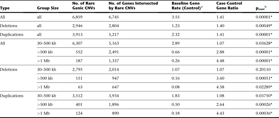
assessed. Consistent with our previous data, we observed
that compared to control subjects, affected subjects had
an increased burden in the number of genes affected by
rare CNVs (1.41-fold increase, empirical p
¼
1
3
10
5;
Table 1
). This enrichment was apparent for both deletions
and duplications and remained after we controlled for
potential case-control differences (
Table 1
). Similar
find-ings were obtained when each stage was considered
sepa-rately (
Tables S3
A–S3C).
Array- and exome-based studies have revealed a
substan-tial contribution of de novo variation to ASD risk,
19prompting us to assess this further. After screening 2,096
trios (of all ancestries), we found 102 rare de novo CNVs
in 99 affected subjects (three of whom had two events;
Table S4
). Overall, 4.7% of trios had at least one de novo
difference (1.2-fold for deletions and 2.8-fold for
duplica-tions, p
¼
0.02). Furthermore, de novo CNVs in simplex
families intersected 4.0-fold more genes than did CNVs
in controls
4,31(1.8-fold after size correction, p
¼
0.01).
There were no significant differences between subjects
from simplex families and those from multiplex families
in the frequency (5% and 4.2%, respectively) or gene
con-tent (n
¼
18.7 and 18.8, respectively) of de novo CNVs.
Similarly, no significant difference was found between
males and females in the size (1.17 and 1.2 Mb,
respec-tively) or gene content (n
¼
18 and 17.3, respectively) of
de novo CNVs. For 85 of 102 de novo events, it was possible
to determine the parent of origin, and roughly equal
numbers of events originated on the paternal allele (n
¼
45) and the maternal allele (n
¼
40) (
Tables S5
A–S5H).
Taken together, our data indicate that there is an increased
burden of de novo events in ASD-affected subjects. The
clinical relevance of de novo CNVs in ASD is confirmed
by the fact that among 102 such events identified, half
(n
¼
46) are considered etiologically relevant, including
40 loci known to be involved in ASD and ID (see below).
We replicated previous observations, such as a de novo
deletion intersecting
PTCHD1AS
in a male (adding to the
evidence that both
PTCHD1
and
PTCHD1AS
contribute to
ASD risk
14) and de novo events involving the miRNA
miR137
(MIM 614304) in 1p21.2–p21.3 in two subjects.
Mi-crodeletions of
miR137
have been reported in ASD,
39ID,
40and schizophrenia.
41Examples of ASD candidate genes
identified by small de novo CNVs include
SETD5
,
DTNA
(MIM 601239), and
LSAMP
(MIM 603241) (
Supplemental
Data
section ‘‘Highlighted Genes,’’
Figures S9
,
S10
, and
S14
).
CNV Burden in Autosomal-Dominant or X-Linked
Genes and Loci Implicated in ASD and/or ID
At least 124 genes and 55 genomic loci have been
impli-cated in ASD to date (
Tables S6
A and S6B; updated from
Betancur
18), all of which have also been implicated in ID.
In addition, we compiled a list of genes and loci that
have been implicated in ID, but not yet in ASD (
Tables
S6
C–S6D). When we analyzed samples of inferred
European ancestry, we found that 4% (87/2,147) of
ASD-affected subjects had CNVs overlapping
autosomal-domi-nant or X-linked genes and loci implicated in ASD and/or
ID; this percentage was significantly higher than that in
controls (OR
¼
4.09, 95% confidence interval [CI]
¼
2.64–6.32, p
¼
5.7
3
10
12;
Figure 1
A). We further
classi-fied these events into pathogenic, uncertain, or benign
according to the American College of Medical Genetics
guidelines.
32Pathogenic (or clinically significant) CNVs
were identified in 2.8% (60/2,147) of affected subjects
(OR
¼
12.62, 95% CI
¼
5.44–29.27, p
¼
2.74
3
10
15),
and pathogenic deletions showed a striking estimated OR
of 23.13 (95% CI
¼
5.57–96.08, p
¼
2.6
3
10
11;
Figure 1
B). Furthermore, the enrichment of pathogenic
[image:5.603.57.547.59.266.2]CNVs overlapping genes involved in ASD and/or ID was
independently observed when the data were broken
down by stages: 2.6% (25/979) of affected subjects in stage
1 (OR
¼
7.61, p
¼
1.22
3
10
5) carried pathogenic CNVs,
whereas 3.0% (35/1,168) in stage 2 (OR
¼
6.47, p
¼
2.89
3
10
7) carried pathogenic CNVs. Some of these
CNVs (e.g.,
NRXN1
deletion, 1q21 duplications [MIM
612475], and 16p11.2 duplications) were seen in a small
fraction of control subjects, consistent with their variable
Table 1. Genome-wide Burden of Genes Intersected by Rare CNVs in a Combined Sample of 2,147 European ASD Affected Subjects and 2,640 European Control Subjects
Type Group Size
No. of Rare Genic CNVs
No. of Genes Intersected by Rare CNVs
Baseline Gene
Rate (Control)a Case-ControlGene Ratio p corrb
All all 6,859 6,745 3.55 1.41 0.00001*
Deletions all 2,946 2,804 1.23 1.40 0.00049*
Duplications all 3,913 5,217 2.32 1.41 0.00001*
All 30–500 kb 6,307 5,163 2.89 1.07 0.03628*
>500 kb 552 2,491 0.66 2.88 0.00001*
>1 Mb 187 1,337 0.26 4.48 0.00001*
Deletions 30–500 kb 2,795 2,014 1.07 1.07 0.20110
>500 kb 151 947 0.16 3.60 0.00051*
>1 Mb 63 647 0.08 4.58 0.02289*
Duplications 30–500 kb 3,512 3,934 1.83 1.08 0.03750*
>500 kb 401 1,896 0.50 2.64 0.00026*
>1 Mb 124 890 0.18 4.43 0.00036*
Rare CNVs in samples of European ancestry were defined asR30 kb in size and present in the total sample set at a frequency<1%. Gene coordinates were defined by the RefSeq boundaries plus a 10 kb region on either side. All genomic analyses used UCSC Genome Browser hg18. *Significant differences (p%0.05) are indicated.
aThe baseline gene rate (control) is defined as the average number of genes intersected by CNVs per control subject. bGenome-wide p values were estimated in 100,000 permutations (one sided) and additionally corrected (p
expressivity and/or incomplete penetrance. Among the
affected subjects with pathogenic CNVs, 63% (38/60)
car-ried de novo events (
Figure 1
C), including two subjects
with two pathogenic events each.
When we further considered affected subjects of all
ancestries (n
¼
2,446) and included chromosome
abnor-malities (>7.5 Mb), select large rare de novo events, and
select experimentally validated smaller CNVs (<30 kb),
we identified pathogenic CNVs in ~3.3% of individuals
with unexplained ASD (84 pathogenic events in 82/2,446
subjects;
Figures 2
A and
S1
A–S1C;
Tables S7
A and S7B).
This most likely represents an underestimate of the true
etiologic yield, given that many of the subjects were
assessed with clinical diagnostic methods and excluded
if positive; similarly, those individuals with known
con-genital malformations or dysmorphic features were not
enrolled. Interestingly, 83% (64/77 [5 without
informa-tion]) of carriers of pathogenic CNVs were nonsyndromic
(i.e., ASD without reported accompanying physical or
neurological abnormalities), and 57% (44/77 [5 without
information]) had no ID (
Figure 2
B). The fraction of
sub-jects with ID among carriers of pathogenic CNVs (42%)
was not significantly different from the fraction of ID
among all affected subjects (46%).
Inheritance data showed that 64% (54/84) of pathogenic
CNVs were de novo events (59% were deletions, and 41%
were duplications) and that the remaining (36%) were
in-herited, including seven X-linked CNVs maternally
trans-mitted to males and 23 (13 maternal and 10 paternal
[27%]) on autosomes (
Figure 2
C). Pathogenic deletions
tended to be smaller than duplications (
Figure 2
D). As
ex-pected, pathogenic de novo events were on average
signif-icantly larger than inherited ones (3.14 Mb—excluding
three affected subjects with whole-chromosome
aneu-ploidy—versus 1.44 Mb, respectively). We also observed
that the proportion of females was significantly increased
among carriers of highly penetrant pathogenic CNVs
(male-to-female ratio of 2:1 versus 6:1 among all affected
subjects; two-tailed Fisher’s exact test p
¼
0.017;
Figure 2
E).
In contrast, the male-to-female ratio among individuals
with CNVs associated with variable expressivity was 6:1.
Pathogenic CNVs included well-characterized highly
penetrant disorders associated with de novo CNVs, such
as Phelan-McDermid syndrome (MIM 606232, 22q13.3
deletion including
SHANK3
[MIM 606230]),
Smith-Mage-nis syndrome (MIM 182290, 17p11.2 deletion including
All CNV Pathogenic Uncertain BenignAll CNV Pathogenic Uncertain Benign 0.0
0.2 0.4 0.6 0.8 1.0 1.2 1.4 1.6 1.8
Pathogenic deletions
Pathogenic duplications
Percentage of individuals with CNV
Percentage of individuals with CNV
A
C
B
OR = 4.1 [2.6-6.3] p = 5.7×10⁻¹²
OR = 12.6 [5.4-29.3] p = 2.7×10⁻¹⁵
OR = 1.7 [0.9-3.2] p = 0.14
OR = 1.2 [0.4-4.3] p = 0.76
OR = 23.1 [5.6-96.1] p = 2.6×10⁻¹¹
OR = 7.1 [2.5-20.7] p = 2.0×10⁻⁵
0.0 0.5 1.0 1.5 2.0 2.5 3.0 3.5 4.0 4.5
0.0 0.1 0.2 0.3 0.4 0.5 0.6 0.7
Affected
Control
Affected Control
[image:6.603.58.291.31.604.2]De novo CNV fraction in affected subjects
Figure 1. CNV Burden in Genes and Loci Implicated in ASD
and/or ID
CNV data from 2,147 European affected subjects and 2,640 Euro-pean control subjects were analyzed for overlap with genes and loci implicated in ASD and/or ID (results including non-European affected and control subjects are shown inFigure S1). Only CNVs affecting autosomal-dominant and X-linked dominant genes or loci in both genders (132 genes, 56 loci), as well as X-linked reces-sive genes or loci in males (52 genes, 2 loci), were considered (‘‘all CNV’’). ExonicR30 kb CNVs affecting an ASD- and/or
ID-associ-ated gene or overlapping at least 50% of the target loci were selected for further analysis. Rare CNVs were divided into three categories—pathogenic, uncertain clinical significance, or benign—without regard to affected status.
(A) Percentage of individuals with CNVs overlapping genes and loci implicated in ASD and/or ID (‘‘all CNV’’), pathogenic CNVs, uncertain CNVs, or benign CNVs; and OR in affected and control subjects.
(B) Percentage of individuals with pathogenic deletions or duplica-tions and OR in affected and control subjects.
<30 kb 30-100 kb 100-500 kb 500 kb-1 Mb 1-5 Mb 5-10 Mb >10 Mb Deletions Duplications
Pathogenic CNV (%)
CNV size
B
D
Probands with pathogenic CNV (%)
no ID nonsyndromic
C
de novo inherited de novo inherited
Percentage of CNV
pathogenic CNV all CNV
Deletions Duplications
All probands All pathogenic CNV
Highly penetrant CNV
CNV with variable expressivity/ incomplete penetrance
Males Females
Gender distribution (%)
Probands with autosomal pathogenic CNV
E
p = 0.017Genomic disorders, recurrent breakpoints
(n=3, 1.6-4.5 Mb) Chromosomal abnormalities
Down syndrome (n=1, dn)
Terminal 1q duplication syndrome (n=1, dn) CNV disrupting ASD and/or ID genes
NRXN1 exonic deletion (n=8, 4 dn, 4 inh)
SHANK2 exonic deletion (n=3, 3 dn)
SHANK3 exonic deletion (n=1, dn)
DMD exonic deletion (n=2, XL mat)
DMD exonic duplication (n=1, XL mat)
SYNGAP1 exonic deletion (n=1, dn)
ARID1B exonic deletion (n=1, dn)
HDAC4 exonic deletion (n=1, inh)
PTCHD1 exonic deletion (n=1, XL mat)
Genomic disorders, nonrecurrent breakpoints
Ring chromosome 8 syndrome (n=1, dn)
A
Unbalanced translocation (n=2, 1 dn, 1 inh)
Kleefstra syndrome (9q34.3 deletion) (n=1, dn) Jacobsen syndrome (11q deletion) (n=1, dn)
Phelan-McDermid syndrome (22q13 deletion) (n=3, 3 dn) Terminal 9p deletion (n=1, dn)
1q21.1 deletion syndrome (n=1, dn) 1q21.1 duplication syndrome (n=4, 3 dn, 1 inh) Williams syndrome (7q11.23 deletion) (n=1, dn)
15q11-q13 duplication syndrome (n=7, 5 dn, 2 inh; origin: 6 mat, 1 pat) 15q13.3 deletion syndrome (n=4, 1 dn, 3 inh)
Distal 15q25 deletion syndrome (n=1, inh)
16p11.2 deletion syndrome (n=5, 4 dn, 1 inh) 16p11.2 duplication syndrome (n=4, 2 dn, 2 inh) 16p13.11 deletion syndrome (n=3, 3 inh)
Smith-Magenis syndrome (17p11.2 deletion) (n=2, 2 dn) 17q12 duplication syndrome (n=1, inh)
22q11 deletion syndrome (DiGeorge syndrome) (n=2, 2 dn) 22q11 duplication syndrome (n=5, 2 dn, 3 inh)
Xq28 duplication including GDI1 (n=2, 1 dn, 1 XL mat)
NRXN1 intragenic duplication (n=1, dn)
IL1RAPL1 intragenic duplication (n=1, XL mat)
CASK partial duplication (n=1, XL mat)
CHD2 exonic deletion (n=1, dn) 10q11.21-q11.23 deletion (n=2, 1 dn, 1 inh)
XYY syndrome (n=2, 2 dn)
Rare, large, de novo CNV
100
0 20 40 60 80
100
0 20 40 60 80
0 5 10 15 20 25 30
0 20 40 60 80 100
57 15 7 41 7
6:1
4:1
2:1
6:1
[image:7.603.57.545.42.549.2]2114 332 14
Figure 2. All Pathogenic CNVs Identified in Affected Subjects
CNVs overlapping genes and loci implicated in ASD and/or ID in 2,446 affected subjects irrespective of ancestry, plus chromosomal ab-normalities, other large rare de novo events, and further experimentally validated CNVs<30 kb. Pathogenic CNVs identified in affected subjects (84 CNVs in 82 probands) were divided into different categories: CNVs disrupting genes implicated in ASD and/or ID, genomic disorders with recurrent breakpoints, genomic disorders with nonrecurrent breakpoints, chromosomal abnormalities, and other rare, large de novo CNVs.
(A) Pie chart displaying the proportion for each of these categories. The number of events and inheritance are in parentheses. (B) Percentage of probands with no ID or with nonsyndromic ASD among carriers of pathogenic CNVs.
(C) Distribution of de novo and inherited deletions and duplications in all CNVs versus in pathogenic CNVs in affected subjects. (D) Size distribution of pathogenic CNVs.
RAI1
[MIM 607642]), Kleefstra syndrome (MIM 610253,
9q34.3 deletion including
EHMT1
[MIM 607001]),
Wil-liams syndrome (MIM 194050, 7q11.23 deletion), and
large chromosomal abnormalities (
Figure 2
A;
Table S7
B).
Recurrent deleterious CNVs mediated by segmental
dupli-cations affecting 12 distinct regions were identified in
44 individuals. For example, two unrelated males were
found to harbor Xq28 duplications (MIM 300815), one
de novo and one maternal, corresponding to a ~0.3 Mb
segmental-duplication-mediated gain (153.2–153.5 Mb),
which was previously reported in X-linked ID.
42GDI1
(MIM 300104), mutations of which are linked to ID, is
the most likely gene involved (
Figure S8
). Thus, our
find-ings implicate abnormal
GDI1
dosage in ASD.
Interest-ingly, one AGP proband with the duplication had autism
and a normal IQ, whereas the second had a borderline IQ
(72) (see
Table S8
for phenotype information of all affected
subjects with pathogenic CNVs). Some other findings
include a 1.7 Mb de novo deletion encompassing
ARID1B
(MIM 614556), recently implicated in ID and Coffin-Siris
syndrome (MIM 135900), and a small maternally inherited
intragenic deletion of
HDAC4
(MIM 605314), involved
in
brachydactyly-mental-retardation
syndrome
(MIM
600430;
Figure S7
). Although many 2q37 deletions have
been described in ASD, the deletion found in our proband
directly implicates
HDAC4
haploinsufficiency in autism.
In
Table S9
, we analyzed data across three ASD cohorts,
including a total of 5,106 nonoverlapping affected subjects
and 3,512 control subjects from the AGP, the Simons
Simplex Collection, and the Autism Genetic Resource
Exchange (AGRE), for 17 loci and genes commonly reported
as implicated in ASD. The most frequent deletions involved
16p11.2 and
NRXN1
, accounting for 0.31% and 0.32% of
affected subjects, respectively. Typical 15q11–q13
duplica-tions of the imprinted PWS-AS critical region were found
in 0.25% (13/5,106) of affected subjects, reaffirming this
re-gion’s importance in ASD. The majority of these
duplica-tions were of maternal origin, but two were paternally
derived (one without information;
Table S9
). Although
paternally derived duplications appear to have incomplete
penetrance in comparison to maternal ones, there have
been several cases reported in subjects with ASD.
43FMRP Targets, PSD Genes, and Other Neuronal Genes
Are Implicated in ASD
We expanded our analysis to lists of genes important for
neurological function, such as highly-brain-expressed
genes, PSD genes,
33genes implicated in neurological
dis-eases,
44,45genes with a high pHI,
35and FMRP targets,
34the latter of which have been reported to be enriched in
de novo LoF SNVs.
7Our analysis focused on exonic events,
and deletions and duplications were analyzed separately
(
Figures 3
A and 3B).
FMRP targets (n
¼
842) and PSD genes (n
¼
1,453)
car-ried a significant excess of both deletions and duplications
in affected subjects (
Figures 3
A and 3B). Five percent (73/
1,486) of affected subjects with exonic CNVs, including
52 subjects with genes not previously implicated in ASD
and/or ID, carried deletions overlapping one or more
FMRP targets, yielding 43 ASD candidate genes (
Figure 3
A;
Table S10
). Given that the lists of FMRP targets and PSD
genes shared 279 genes, we performed conditional
ana-lyses showing that the excess of affected subjects carrying
deletions overlapping PSD genes was independent of the
signal in FMRP targets (OR
¼
2.62, 95% CI
¼
1.62–4.32,
p
¼
2.24
3
10
5) and represented 4% of subjects with
exonic events (59/1,486) or 3% after exclusion of
pathogenic events (p
¼
0.007). Notably, females were
over-represented among affected subjects carrying exonic
deletions overlapping FMRP targets (17 females in 73
affected subjects, 1.98-fold more than males, p
¼
0.022,
95% CI
¼
1.06–3.52).
Brain-expressed genes showed significant excess in
affected versus control subjects for deletions only (OR
¼
1.89, 95% CI
¼
1.51–2.37, Fisher’s exact test p
¼
2.6
3
10
8;
Figures 3
A and 3B). Similarly, deletions (and not
duplications) overlapping genes implicated in dominant
neurological diseases and orthologous genes associated
with abnormal phenotypes in heterozygous knockout
mice conferred significant increase in ASD risk (OR
¼
2.94, 95% CI
¼
1.76–4.93, p
¼
2.5
3
10
5). Many of the
genes implicated in dominant diseases have been related
to loss of function or haploinsufficiency, previously
sug-gested to be more frequent and penetrant when deletions
rather than duplications are involved.
46Accordingly, we
detected an excess of affected subjects carrying deletions
overlapping genes with a high pHI (>0.35) (OR
¼
1.41,
95% CI
¼
1.13–1.76, p
¼
0.002).
Increased Multigene Burden in ASD-Affected Subjects
We tested whether multiple genes within a CNV or across
unlinked genetic lesions in the same individual might act
in concert to increase risk of ASD. In logit modeling, the
number of genes overlapped by CNVs, the average
brain-expression value for those genes, and the deletion or
dupli-cation status were used as predictors with the case-control
status as the outcome (
Figures 3
C and 3D).
We found that ASD risk increased (as measured by the
predicted OR) as the numbers of deleted brain-expressed
genes increased (generalized linear model chi-square
good-ness of fit, p
¼
3.2
3
10
5and p
¼
4.7
3
10
7, respectively;
Figures 3
C and 3D). These results were consistent across
removal of both de novo and inherited pathogenic CNVs)
(
Tables S11
A–S11E). Thus, it is likely that de novo and
pathogenic CNVs contribute to risk by altering the
expres-sion of more than one gene, suggesting that genetic
inter-actions between these genes can underlie ASD risk.
Network Analysis Links Exonic Deletions to
Neurodevelopmental Processes
We performed a gene-set enrichment analysis
6on our
expanded sample set after refining our criteria to consider
only exonic events (see
Supplemental Data
for details)
and found only deletions to be significantly enriched in
gene sets in affected versus control subjects (
Figure 4
A).
We found 86 significantly enriched gene sets, including
MAPK signaling components and neuronal synaptic
functions and processes, in 42.5% (335/789) of affected
subjects with exonic deletions (
Figure 4
A;
Tables S12
A–
S12D). Enrichment of synaptic functioning has also been
reported among inherited events in the AGRE families
47and among de novo events in the Simons Simplex
Collec-tion.
36Enriched sets delineate candidate genes disrupted
by deletions not found in control subjects; these genes
notably include those in the KEGG glutamatergic pathway
(e.g.,
GRIK2
[MIM 138244],
GRM5
[MIM 604102],
SHANK1
AffectedControl
% of subjects
0 10 20 30 40 50
***
*** ***
*** ***
** **
0 5 10
OR=1
OR
Deletions
A
Highly brain expressed Not brain expressed(control genes)
Postsynaptic density genes FMRP
targets
Phenotypes (HPO/MPO)
Autosomal dominant phenotypes (HPO/MPO)Haploinsuf
ficiency >0.15
Haploinsuf ficiency >0.35
Haploinsuf ficiency >0.55
% of subjects
1 2 3 4 5 6 7 8 9 10
Multigene hit: Duplications
0 5 10 15 20 25 30 35
Affected Control
Predicted OR (
*
)
1.0 1.5
* * *
* *
* *
* *
*
1.1 1.2 1.3 1.4
D
Number of gene hits in brain-expressed genes
1 2 3 4 5 6 7 8 9 10
Multigene hit: Deletions
% of subjects
Affected Control
0 5 10 15 20 25 30 35
Predicted OR (
*
)
1.0 1.5
* * * *
* *
* *
* *
1.1 1.2 1.3 1.4
C
Number of gene hits in brain-expressed genes
Affected Control
% of subjects
0 10 20 30 40 50
0 5 10
OR=1
** ***
OR
Duplications
B
Highly brain expressedNot brain expressed(control genes)
Phenotypes (HPO/MPO)
Autosomal dominant phenotypes (HPO/MPO) FMRP
targets
[image:9.603.93.511.37.393.2]Postsynaptic density genes
Figure 3. Enrichment of Functional Gene Sets Affected by Rare Exonic CNVs in Affected versus Control Subjects
Overrepresentation of deletions (A) and duplications (B) in various functional gene sets. ORs, with 95% CIs, and the percentages of affected subjects (n¼1,486) and control subjects (n¼1,820) with exonic CNVs overlapping genes are given for the following gene sets: (1) highly-brain-expressed genes (log(RPKM)>4.5, BrainSpan; n¼5,610); (2) functionally characterized control genes not ex-pressed in the brain (log(RPKM)<1, BrainSpan; n¼5,410); (3) PSD genes (n¼1,453);33(4) FMRP interactors (n¼842);34(5) genes associated with neurological phenotypes compiled from the HPO and MPO (n¼3,112); (6) genes as described in (5) but filtered for auto-somal-dominant genes (n¼739); and (7) genes grouped by their pHI35into three subgroups: pHI>0.15 (n¼8,862), pHI>0.35 (n¼
4,136,) and pHI>0.55 (n¼2,214). Genes with a pHI>0.35 were considered haploinsufficient. The p values for affected and control subjects were estimated with two-tailed Fisher’s exact tests (*p<0.01, **p<0.001, ***p<0.0001).
UBE3A DBT NRXN1 PARK2 GALR1 NRXN3 TRIP12 NTSR2 CBS UBE2W LIMK1 ARHGAP32 SYNJ2 PLCB1 MSI2 RIMS1 SYNCRIP STAC2 SNX14 CYFIP1 RAP1GDS1 SH3GL3 CUL2 PARD3 DLGAP2 GRID2 SHANK2 PSMC2 ARHGDIA SMAP1 CYP7B1 RPL19 STEAP3 RNASE6 PCYT1A RER1 LARGE PSMB1 RNASE1 STAU2 WDR4 ERCC6 BNC1 ZNF516 ZSCAN2 DDX53 DBI CHD1L WTAP CUTA PMPCB SMC2 SMARCA2 CHD2 CEP70 KCNH8 DTNA BRMS1L SETD5 UHRF1 ZEB2 KLF13 EHMT1 SCAND2 SCAPER BAZ1B SNRPN RTCD1 KAT2B SKI TICAM1 MAPK11 TSSK2 RALGAPA1 PRKAB2 PPP3CB TAOK2 MAPK12 ESR2 FLNA RFX3 IKBKG CACNA1B DPP6 SNX9 TNK2 SYNGAP1 ANKS1B IL1RL1 PTPRD TUBGCP5 CRKL LSAMP MAPK7 TBP RAB5A MAX MAPK3 BHLHE22 ATOH1 SREBF1 CDK2 CAMK2G ABCA1 SOD2 PIK3CB ETS1 ABL1 CSNK1D CHRNA7 MAPK8 CDK4 RAC3 FDR 5, 10, 15, 20%
ASD & ID
ID only Development & Cell proliferation Cell motility Cell projection Neural development Axonogenesis MAPK & other signalling Neuronal synapse Endocytosis Membrane organization
Cell adhesion molecules
Growth MAPK Neurotrophin signaling Insulin pathway Glutamergic synapse Cholinergic synapse Vascular development Cardiac development Pos. reg. Cell proliferation Embryonic development Amino acid peptidase Early endosome Glycosyl transferase Cytoskeleton organization SH3 domain Neg. reg. Cell proliferation/ Cell cycle Cell motility
A
B
MAPK & other signallingChromatin remodeling & Transcription regulation Unbiased
Postnatally biased
Prenatally biased
Very low expression/not listed
Satellite synaptic cluster Synaptic
signalling/components
ID ASD & ID
[image:10.603.82.521.35.659.2]Dominant ASD and/or ID genes
Figure 4. Functional ASD Maps
(A) Gene-set enrichment for rare exonic deletions (de novo and inherited) in affected versus control subjects. Enrichment results were mapped as a network of gene sets (nodes) related by mutual overlap (edges). Node size is proportional to the gene-set size, and edge thick-ness scales with the number of genes overlapping between sets. Only gene sets enriched in affected subjects with a FDR%20% are
[MIM 604999],
SHANK2
[MIM 603290], and
SHANK3
[MIM 606230];
Figure S2
A and
Tables S13
A–S13D), the
KEGG cholinergic pathway (e.g.,
KCNJ12
[MIM 602323],
CHAT
[MIM 118490], and
SLC18A3
[MIM 600336], the
latter two of which are within a recurrent 10q11.21–
q11.23 deletion, recently reported in individuals with
ID and ASD;
48Figure S2
B and
Tables S13
A–S13D), or in
both pathways (e.g.,
GNG13
[MIM 607298],
PRKACB
[MIM 176892],
PLCB1
[MIM 607120],
CAMK2G
[MIM
602123 ], and
PPP3CB
[MIM 114106]). When analyzing
human homologs of mouse genes, we also found
enrichment of phenotypes mostly related to the brain,
including abnormal telencephalon morphology, neuron
morphology, behavior, and nervous system physiology
(
Table S12
D).
Genes within De Novo CNVs Cluster in a Gene
Network
De novo deletions (52 in European affected subjects) were
found to be significantly enriched within each gene set or
pathway cluster (for 85 of the 86 gene sets or pathways), as
well as across clusters (chi-square test p
¼
9.8
3
10
9)
(
Table S12
B), prompting us to search for enriched
biolog-ical functions within de novo events separately. Taking
into account our observations of a significant multigene
burden in ASD subjects (
Figures 3
C and 3D), we analyzed
de novo events by using NETBAG
36to identify up to two
ASD candidate genes per CNV among 102 de novo events
in 99 subjects. NETBAG identifies networks of genes under
the premise that if genomic regions are perturbed by
genetic variants associated with the same phenotype,
they will contain genes forming connected clusters. The
NETBAG analysis resulted in a network of 113 genes
(global cluster p value
¼
0.02;
Figure 4
B). Ten genes have
been previously implicated in autosomal-dominant or
X-linked forms of ASD and ID (
UBE3A
[MIM 601623],
NRXN1
,
SHANK2
,
EHMT1
,
SYNGAP1
[MIM 603384], and
SMARCA2
[MIM 600014]) or ID only (
ZEB2
[MIM
605802],
FLNA
[MIM 300017],
SKI
[MIM 164780], and
IKBKG
[MIM 300248]). On the basis of cumulative
evi-dence from various sources, an additional 68% (67/98)
are likely to affect ASD risk (
Tables S14
A–S14E); 27/67 of
these are either FMRP targets or PSD genes. Compared to
all other genes within de novo CNVs (or deletion CNVs
only), genes in the network exhibited a significantly
higher pHI (Wilcoxon rank sum p
¼
7.07
3
10
8), and
55% (59/107 [6 without information]) had a pHI
>
0.35.
A similar NETBAG analysis of de novo CNVs in control
subjects did not yield significant results.
36We further characterized the biological processes related
to the NETBAG cluster (
Tables S14
B–S14E;
Figures 4
B and
S3
) and found a significant enrichment (false-discovery
rate [FDR]
<
10%) of genes involved in chromatin and
transcription regulation, MAPK signaling, and synaptic
signaling and components (
Figure 4
B). We recapitulated
many of the results of our gene-set analysis (
Figure 4
A),
notably for synapse functions and processes, and also
iden-tified genes involved in chromatin and transcription
regulation. The latter category included a high-risk gene
associated with ASD, the chromatin gene
CHD2
(MIM
602119), which is affected by a de novo 83 kb deletion in
a male with ASD, mild ID, and dysmorphic features
including micrognathia and protruding ears. His
ASD-affected brother has mild ID, similar dysmorphic features,
and epilepsy with onset at age 9 years and carries the same
deletion, which removes the first six exons of
CHD2
.
Neither parent carries the deletion, suggesting germline
mosaicism (the deletion arose on the paternal
chromo-some). De novo SNVs in
CHD2
have been reported in an
ASD subject and in several individuals with a broad
spectrum of neurodevelopmental disorders, including ID
and epileptic encephalopathy
8,49,50(
Figure S6
). Two other
genes in the chromodomain family have been linked to
neurodevelopmental disorders:
CHD7
(MIM 608892) in
CHARGE syndrome (MIM 214800) and
CHD8
(MIM
610528) in ASD. Another example is
TRIP12
(MIM
604506), encoding an E3 ubiquitin ligase that can regulate
chromatin function to maintain genome integrity (
Figure
S12
). The chromatin and transcription module showed a
predominance of genes with a prenatally biased expression
profile (
Figures 4
B and
S4
).
De Novo CNVs and LoF SNVs Converge on Functional
Gene Networks
We expanded our analysis to genes altered by both our
de novo CNVs (
Figure 5
A) and de novo LoF SNVs compiled
from four exome sequencing studies in ASD.
7–10Eleven
genes affected by de novo CNVs (
NRXN1
,
SHANK2
,
ARID1B
,
RIMS1
[MIM
606629],
TRIP12
,
SMARCC2
[MIM 601734],
DLL1
[MIM 606582],
TM4SF19
,
MLL3
[MIM 606833],
PHF2
[MIM 604351], and
CSTF2T
[MIM
611968]) were found to be altered by de novo LoF SNVs
among autism cohorts, and three of them (
NRXN1
,
SHANK2
, and
RIMS1
) were selected by NETBAG (
Figure 4
B).
Chromatin modification & Transcription regulation
Neuronal development & Axon guidance
MAPK & other signalling
113 genes dn CNV (NETBAG)
(64 used, 56.6%)
58 34 1 3 4 2 3 AFF2 DMD MECP2 RIMS1 NRXN1 SHANK2 CHD8 ARID1B DYRK1A SCN2A EHMT1 SYNGAP1 SMARCA2 42 4
122 genes LoF dn SNV
(41 used, 33.6%)
92 known ASD genes, dominant (AD or XLD) or XLR in males
(54 used, 58.7%)
31 genes LoF XL SNVs in males
[image:12.603.59.543.37.628.2](7 used, 22.6%)
A
B
ARHGDIA FGFR2 ITGA5 ARHGAP32 SPATA13 EPHB2L1CAM SYNCRIP MAPK8 CRKL PARD3 CACNA1B CACNA1C RIMS1 CDK2 SYNJ2 DLGAP2 CDKL5 NLGN4X SNRPN SHANK2 TSC2 TSC1 PTEN RAB5A TNK2 MCF2 SNX9 OCRL IL1RAPL1 SVIL NLGN3 NRXN1 TBR1 NRXN3 CASK PTPRD ANKRD11 IQGAP2 RAC3 CSDE1 PIK3CB MAP2K1 PTPN11 HRAS NF1 SYNGAP1 ABL1 BRAF PLCB1 GRIN2B RAP1GDS1KRAS YWHAE PARK2 AFF2 SPAST DYRK1A CUBN SCN2A LRP2 HAUS7 MED12 FMR1 CYFIP1 SH3GL3 NCKAP1 MED13L RER1 LIMK1 ABCA1 CREBBP MDM2 SYN1 FLNA ETS1 RPS6KA3 MAPK3 CAMK2G ANK2 KAT2B KLF13 TBP SRCAP NFIA IKBKG PPM1D TBC1D23 PRKAB2 PHF8 SMC2 KDM6A SETD2 ZNF292 KIAA0232 ATP1B1 BAZ1B SMARCC2 VCP SOD2 FTSJ1 PSMB1 SMARCA2 NIPBL UPF3BPSMC2 CDK4 SMC1A ARID1B PIR ZMYND11 CHD2 CHD8 NFIX DTNA DMD DRP2 EHMT1 BCL11A MAX STAU2 PAFAH1B1 DCX MAPK11 CBX4 DLL1 TAOK2 MAPK7 CACNA1F SREBF1 SHANK3 MEF2C ESR2 MAPK12 DST PPP3CB TCF3 FOXG1 ATOH1 MECP2 GARNL1 CNOT3 PAX5 SMAD4 SKI STXBP1 SCN1A CUL3 CUL2 BRWD1 ERCC6Figure 5. Genes Affected by CNVs and SNVs Converge on Functional Gene Networks
(A) Venn diagram showing the overview of 151 genes resulting from a DAPPLE analysis of 336 unique genes. A similar diagram of DAPPLE input genes is shown inFigure S5. For the DAPPLE analysis, we compiled the following lists of genes: (1) 113 genes identified from our de novo CNVs by NETBAG; (2) 122 genes with de novo LoF SNVs from four published exome sequencing studies;7–10(3) 31
genes with hemizygous LoF SNVs on the X chromosome of male ASD subjects and not observed in male control subjects;16and (4) 92
ASD-implicated genes previously described as autosomal dominant, X-linked dominant, or X-linked recessive in males.18
Of the 11 genes, only
TM4SF19
(3q29) belongs to a
known locus associated with a recurrent genomic disorder.
Despite the limited overlap observed among genes altered
by de novo CNVs and LoF SNVs, there is reinforcing
evidence of the role of
NRXN1
and
SHANK2
in ASD (
Fig-ure 5
A). In addition,
RIMS1
, altered by both a de novo
duplication and a LoF SNV (
Figure 5
A) and encoding a
brain-specific synaptic Rab3a-binding protein, emerges as
an ASD candidate gene.
RIMS1
has a regulatory role in
syn-aptic-vesicle exocytosis modulating synaptic transmission
and plasticity.
51Three other genes affected by de novo
CNVs (also picked by NETBAG)—
CHD2
,
SYNGAP1
, and
SYNCRIP
—are also affected by LoF de novo SNVs in
ID,
50,52and two (
SYNGAP1
and
DPYD
[MIM 612779]) are
altered in schizophrenia.
53CHD2
(discussed above) and
SYNGAP1
(encoding a Ras/Rap GTP-activating protein)
are both involved in autosomal-dominant ID, ASD, and
epilepsy.
54Under the assumption that different genes harboring
suspected causative mutations for the same disorder often
physically interact, we sought to evaluate protein-protein
interactions (PPIs) encoded by genes known to be
impli-cated in ASD and genes affected by rare CNVs or SNVs
(data drawn from our de novo CNVs and published
de novo ASD LoF SNVs;
Figures 5
A, 5B, and
S5
). The union
set of 336 unique genes (
Table S15
) analyzed by DAPPLE
37resulted in a network of direct PPIs encoded by 151 genes
(
Figure 5
A) from each of the three main lists: 54/92
(58.7%) genes involved in ASD, 64/113 (56.6%) de novo
CNV genes, and 41/122 (33.6%) de novo LoF SNV genes.
The number of direct PPIs was 1.5-fold higher than
ex-pected (p
<
0.001,
Figure 5
B), suggesting that many of
the de novo CNV or SNV genes and ASD-implicated genes
function cooperatively. Overrepresentation analysis
iden-tified convergent functional themes related to neuronal
development and axon guidance, signaling pathways,
and chromatin and transcription regulation. Overall, these
findings are consistent among the three different types of
analyses shown in
Figures 4
A, 4B, and
5
.
Although 54 genes were previously implicated in ASD, the
DAPPLE analysis singled out an additional 97 CNV or SNV
high-confidence candidate genes (
Figure 5
B;
Table S16
).
We found that compared to the 54 ASD-implicated genes,
the newly selected 97 CNV or SNV genes had a comparably
high pHI (median pHI
¼
0.58,
Figure 6
A). This is consistent
with the observation that ASD subjects have more deletions
with haploinsufficient genes than do controls (
Figure 6
B).
Furthermore, similar to genes with known disease-causing
mutations (
Figure 6
C), those genes have high
functional-indispensability scores and a comparable high degree of
centrality (i.e., high number of direct neighbors) and
num-ber of networks in which they are involved (
Figures 6
D and
6E). Compared to the genome average, they are also among
the top 75% of more-conserved genes (on the basis of
Genomic Evolutionary Rate Profiling scores and PhyloP)
and are highly expressed in the brain. Interestingly, 39 of
the 97 genes are either FMRP-related or PSD genes (of the
initial 151 genes identified by DAPPLE, 51 are FMRP
interac-tors and 24 are PSD genes). Thus, despite little overlap in
genes, the strong interconnectedness between the resulting
networks identifies pathways through which the effects of
distinct mutations might converge.
Discussion
We used multiple approaches to prioritize key candidate
ASD-associated genes disrupted by CNVs and further
iden-tified biological relationships and common pathways
shared among those genes. Our data (1) confirm excess
burden of genome-wide rare genic CNVs in an
indepen-dent set of ASD subjects versus control subjects; (2) further
reveal an extreme degree of etiological heterogeneity (36
different genetic loci were found among 82 individuals
with pathogenic CNVs); (3) confirm the contribution of
de novo CNVs to the etiology of autism and highlight
the contribution of inherited pathogenic imbalances
(36%); (4) show an increased proportion of females among
carriers of highly penetrant pathogenic CNVs, as well as
among carriers of deletions affecting FMRP targets; (5)
show no significant difference in the frequency of de
novo CNVs between simplex and multiplex families; (6)
show that both deletions and duplications involving
FMRP targets and PSD genes increase ASD risk; (7) show
evidence of multigene contributions to ASD; (8) show
that ASD-associated deletions impair synapse function
and neurodevelopmental processes; (9) implicate
chro-matin and transcription regulation genes in ASD in a
network analysis of de novo CNVs; and (10) show that
genes affected by de novo CNVs and de novo LoF SNVs
converge on functional gene networks.
Importantly, when considering highly penetrant
patho-genic CNVs, we found a 2:1 male-to-female ratio (deviating
from the overall ratio of 6:1 in the study sample). In
contrast, the ratio was unchanged among carriers of
CNVs characterized by variable expressivity and/or
incom-plete penetrance. Moreover, among affected subjects,
females were twice as likely as males to have exonic
dele-tions involving FMRP targets. Given the sex bias of ASD
toward males, it has been suggested that females require
A
B
C
D
E
Wilcoxon rank sum
ASD and/or ID
p = 3.20 x 10⁻⁹ p = 1.60 x 10⁻¹³
Wilcoxon rank sum
p = 3.20 x 10⁻⁹
Wilcoxon rank sum
p = 1.80 x 10⁻¹¹ p = 7.70 x 10⁻¹¹
dn
dn
dn dn
[image:14.603.56.546.33.635.2]Affected
Figure 6. Functional Metrics for Various Gene Sets Derived from CNV and SNV Studies, as well as HI Scores for Genic Deletions
a higher genetic load to express ASD,
56and our
founda-tional data support this general hypothesis. This same
phe-nomenon has recently been shown for
SHANK1
57and the
16p13.11 CNV.
58We observed significant enrichment of both deletion
and duplication events overlapping FMRP and PSD targets,
indicating that altered dosage of these genes can underlie
ASD susceptibility. This is consistent with evidence that
FMRP targets belong to multiple signaling and
intercon-nected pathways such as PI3K-Akt-TSC-PTEN-mTOR and
PI3K-RAS-MAPK,
59,60which have been linked to ASD
through both underexpression and overexpression of
genes in these pathways. Although both deletions and
duplications can contribute to risk, we found that
dele-tions can have a stronger impact when
highly-brain-expressed genes, genes conferring dominant phenotypes
in humans and mice, or genes with a high pHI are
consid-ered. We also found that ASD risk increases as a function
of the number of brain-expressed genes affected by rare
de novo and pathogenic CNVs, consistent with an additive
model of risk underlying ASD etiology.
We developed an expanded and extensively
intercon-nected network of high-confidence ASD candidate genes
by integrating protein products from CNV and SNV genes
and ASD-implicated genes. Overall, these results
demon-strate that genes involved in ASD participate in a wide
array of processes, from neuronal development and axon
guidance to MAPK and other kinase signaling cascades
(including the PI3K-Akt-mTOR and PI3K-RAS-MAPK
pathways) to chromatin modification and transcription
regulation. An increasing number of genes involved in
chromatin structure and epigenetic regulation have been
implicated in a variety of developmental disorders.
61Other
chromatin regulator genes, such as
MBD5
(MIM 611472)
and
KMT2D
(MIM 602113), have been implicated in ID
and ASD, highlighting the need to further study this
category of genes as ASD risk factors.
In addition to underlining important pathways, our
results highlight specific genes in ASD risk. Whereas the
majority of the 97 genes in the CNV or SNV network
(not including the genes already known to be involved
in ASD) most likely act via haploinsufficiency, a few are
affected by duplications. One example is the duplication
of
PIK3CB
(MIM 602925), which is likely to increase its
expression and thus lead to excessive phosphatidylinositol
3-kinase (PI3K) activity. PI3K, which is regulated by
FMRP,
59is elevated in FXS mouse knockouts,
62,63and
downregulation of this pathway has been shown to
have therapeutic effect in ASD and FXS mouse models.
RAC3
(MIM 602050), another example of a gene affected
by duplication, encodes a Rho family GTPase that
en-hances neuritogenesis and neurite branching when
over-expressed.
64Our findings implicate many ASD candidate genes
altered by de novo, inherited, or X-linked CNVs (e.g.,
SETD5
,
miR137
, and
HDAC9
[MIM 606543];
Supplemental
Data
section ‘‘Highlighted Genes’’) or altered by both
de novo CNVs and LoF SNVs (e.g.,
RIMS1
,
TRIP12
, and
DLL1
;
Figures 4
B and
5
). Taken together, our results suggest
that rare variants affecting ASD risk in the population
collectively encompass hundreds of genes. Despite this
heterogeneity, many genes converge in interconnected
functional modules, providing diagnostic and therapeutic
targets.
Supplemental Data
Supplemental Data include 18 figures, 19 tables, and Supple-mental Acknowledgments and can be found with this article online athttp://dx.doi.org/10.1016/j.ajhg.2014.03.018.
The median pHI for HPO het was selected as the threshold to differentiate between dominant and recessive genes (red horizontal line). Genes implicated in ASD and ID were further annotated into dominant (dom), recessive (rec), or X-linked (XL) genes. Other abbreviations are as follows: dn, de novo; allg; all genes; DEL, deletion; ‘‘1g-NBG 69g,’’ 69 genes selected by NETBAG analysis of 102 de novo CNVs with up to one gene per each CNV region; ‘‘2g-NBG 113g,’’ 113 genes selected by NETBAG analysis with up to two genes per CNV (as depicted inFigure 4B); ‘‘2g-NBG DEL/disr 80g,’’ subset of NETBAG genes completely overlapped or disrupted by deletions (no duplications were considered); ‘‘ASD (CompStudies) dn SNV 122g,’’ 122 genes affected by de novo LoF SNVs from ASD exome sequencing studies; ‘‘ID (CompStudies) dn SNV 32g,’’ 32 genes affected by de novo LoF SNVs from ID exome sequencing studies; ‘‘SCZ (Xu2012) dn SNV 22g,’’ 22 genes affected by de novo LoF SNVs from schizophrenia exome sequencing studies; ‘‘ASD (Lim2013) rec SNV 49g,’’ 49 genes affected by hemizygous LoF SNVs on the X chromosome of ASD males; ‘‘ID (Najmabadi2011) rec SNV 73g,’’ 73 genes hit by recessive SNVs in consanguineous ID-affected families; ‘‘pre-DAPPLE ASD input 336g,’’ 336 DAPPLE input genes; ‘‘336g minus 151g excluded 185g,’’ 185 genes not used by DAPPLE; ‘‘DAPPLE ASD direct-PPI 151g,’’ 151 genes depicted in the network ofFigure 5B; ‘‘DAPPLE minus 54 known genes 97g,’’ 97 genes depicted in the network ofFigure 5B (and listed inTable S16), not including the 54 genes previously implicated in ASD (yellow nodes); and ‘‘DAPPLE known genes only 54g,’’ 54 genes known to be involved in ASD.
(B) LOD scores of the probability that at least one gene within a rare deletion will cause haploinsufficiency were calculated for affected and control subjects. Deletion-based LOD scores are plotted as a function of the number of genes in each event for rare genic deletions in affected and control subjects. The p value for the difference in the slope of the two regression lines is indicated.
(C) Box plots with the distribution of predicted functional indispensability scores for gene categories from Khurana et al.55(LoF-tolerant genes, neutral genes, genes with known mutations as listed in the Human Genome Mutation Database, and essential genes [i.e., genes in which LoF mutations result in infertility or death before puberty]) and CNV or SNV genes from