Conjugated linoleic acid suppresses IRF3 activation via modulation of CD14
☆
Jennifer K. Dowling
a, Claire E. McCoy
b, Sarah L. Doyle
b, Nadia BenLarbi
a, Mary Canavan
a,
Luke A. O'Neill
b, Christine E. Loscher
a,⁎
a
Immunomodulation Group, School of Biotechnology, Dublin City University, Dublin 9, Ireland b
School of Biochemistry and Immunology, Trinity College Dublin, Dublin, Ireland
Received 21 October 2011; received in revised form 31 May 2012; accepted 8 June 2012
Abstract
Polyunsaturated fatty acids (PUFA) can modulate the immune response, however the mechanism by which they exert this effect remains unclear. Previous studies have clearly demonstrated that thecis-9, trans-11isomer of conjugated linoleic acid (c9,t11-CLA), found predominantly in beef and dairy products, can modulate the response of immune cells to the toll-like receptor (TLR) 4 ligand, lipopolysaccharide (LPS). This study aimed to investigate further the mechanism by which these effects are mediated. Treatment of macrophages with c9,t11-CLA significantly decreased CD14 expression and partially blocked its association with lipid rafts following stimulation with LPS. Furthermore the c9,t11-CLA isomer inhibited both nuclear factor-κB (NF-κB) and IRF3 activation following TLR4 ligation while eicosapentaenoic acid (EPA) only suppressed NF-κB activation. Given that the ability of LPS to activate IRF3 downstream of TLR4 depends on internalisation of the TLR4 complex and involves CD14, we examined TLR4 endocytosis. Indeed the internalisation of TLR4 to early endosomes following activation with LPS was markedly inhibited in c9,t11-CLA treated cells. These effects were not seen with the n-3 fatty acid, EPA, which was used as a comparison. Our data demonstrates that c9,t11-CLA inhibits IRF3 activation via its effects on CD14 expression and localisation. This results in a decrease in the endocytosis of TLR4 which is necessary for IRF3 activation, revealing a novel mechanism by which this PUFA exerts its anti-inflammatory effects.
© 2012 Elsevier Inc. All rights reserved.
Keywords:Conjugated linoleic acid; IRF3; NFkappaB; CD14
1. Introduction
It is well established that nutrition plays an important role in
health and that dietary fatty acids are essential components of the
diet
[1]
. These include polyunsaturated fatty acids (PUFA)
eicosa-pentaenoic acid (EPA) and docosahexaenoic acid (DHA), commonly
found in high fat
fi
sh and marine mammals
[2]
, which have been
shown to have bene
fi
cial effects in immune-mediated diseases such
as rheumatoid arthritis, in
fl
ammatory bowel disease, systemic lupus
erythematosus and asthma
[3
–
5]
. Recent studies indicate strong
immunomodulatory potential for another dietary fatty acid,
conju-gated linoleic acid (CLA)
[6]
. CLAs are a group of positional and
stereoisomers of conjugated dienoic derivatives of linoleic acid and
the
cis
-9,
trans
-11 CLA isomer (c9,t11-CLA) is a naturally occurring
dietary form of CLA, predominantly found in foods derived from
ruminants such as meat and dairy
[7]
. c9,t11-CLA has been shown
to suppress proin
fl
ammatory cytokine production in a number of
cell types. Indeed, we previously reported that c9,t11-CLA
modu-lates the activation of dendritic cells characterised by a decrease in
production of interleukin (IL)-12 and an increase in IL-10
[8]
.
Furthermore, studies in animal models have demonstrated that
feeding CLA has bene
fi
cial effects in endotoxin-induced anorexia
and on mucosal damage in experimental colitis
[9,10]
.
While the bene
fi
cial effects of PUFA are clear, the exact
mechanisms through which they exert such effects have still to be
elucidated. Numerous studies focus on the cumulative effects of
PUFA on eicosanoid production, gene expression and transcription
factor activation
[11
–
13]
. PUFA are known to modulate activation of
nuclear factor-
κ
B (NF-
κ
B)
[13]
and peroxisome proliferator activated
receptors, PPARs
[14]
. Speci
fi
cally, EPA and DHA have been shown to
suppress NF-
κ
B activation in LPS-stimulated macrophages
[15,16]
.
This suppressive effect on NF-
κ
B in PUFA-treated cells results in
decreased secretion of pro-in
fl
ammatory cytokines
[17
–
19]
, which
may partly explain their overall anti-in
fl
ammatory actions. Babcock
et al have demonstrated that EPA suppresses tumor necrosis factor
α
(TNF
α
) production in murine macrophages
[20]
, and we have
previously reported that c9,t11-CLA suppressed NF-
κ
B in adipose
tissue from mice fed on diets of CLA with a concomitant suppression
of TNF
α
[21]
. In addition, studies employing human macrophage
models report that EPA and DHA inhibit TNF
α
, IL-6 and IL-1
β
production
[22,23]
. We have also demonstrated the suppressive
effects of EPA and DHA on NF-
κ
B and subsequent TNF
α
, 6 and
IL-1
β
production in human macrophage
[24,25]
.
Journal of Nutritional Biochemistry xx (2012) xxx
–
xxx
☆ This work was supported by funds from the Irish Research Council for
Science, Engineering and Technology and Science Foundation Ireland.
⁎ Corresponding author.
E-mail address:[email protected](C.E. Loscher).
A study showing that DHA fails to inhibit NF-
κ
B activation
in the presence of dominant negatives mutants and
constitu-tively active components of upstream signalling, MyD88 and
AKT
[26]
suggests that PUFA may exert their effects at the cell
membrane. Indeed, PUFA greatly in
fl
uence membrane composition,
size and distribution
[27
–
29]
. Signi
fi
cantly, there is a role for
membrane microdomains or
“
lipid rafts
”
in immune cell signalling
[30,31]
and the impact of PUFA on these domains is well reported
[32
–
34]
. T-cell activation has been linked with the speci
fi
c
recruitment of signalling complexes to lipid rafts
[35]
. Changes to
the fatty acid composition of these rafts is associated with a
decrease in the translocation of PKC to lipid rafts, a key molecule
regulating CD4
+T-cell activation
[27,36]
. Importantly, toll-like
receptor (TLR) 2
[37]
, TLR4 and its associated molecule CD14
[38]
are recruited to lipid rafts following stimulation with their
respective ligands.
Activation of TLR4 initiates downstream signalling which in turn
activates NF-
κ
B and IRF3 via MyD88 dependant and independent
pathways, respectively
[39,40]
. Activation of the MyD88 dependant
pathway is mediated at the plasma membrane, while induction of
IRF via the MyD88-independent pathway is dependant on the
endo-cytosis of TLR4, a process requiring CD14
[41,42]
.
In the present study, we test the hypothesis that c9,t11-CLA
may exert its anti-in
fl
ammatory effects partly at the plasma
membrane by in
fl
uencing CD14. We demonstrate that c9,t11-CLA
modulates CD14 by altering its localisation on the membrane and
association to lipid rafts in response to LPS, which was not found to
be the case with EPA which was used for comparison. CLA
treatment also decreased the internalisation of TLR4 and its
localisation to early endosomes in LPS stimulated cells, an event
known to be dependent on CD14. Subsequently it suppressed IRF3
activation which could be reversed following overexpression of
CD14. These
fi
ndings provide greater insight into the
anti-in
fl
ammatory mechanisms used by the c9,t11- CLA isomer while
also highlighting a selective inhibition of the in
fl
ammatory
transcription factor IRF3.
2. Materials and methods
2.1. Plasmids and reagents
c9,t11-CLA was purchased from Alexis Chemicals (Cayman Chemicals). Eicosa-pentaenoic acid (EPA), docosahexaenoic acid (DHA) and lauric acid (LA) were all purchased from Sigma-Aldrich. Escherichia coli LPS (serotype R515) was purchased from Alexis Biochemicals. The c9,t11-CLA isomer, EPA, DHA and LA were dissolved in sterile DMSO (Sigma-Aldrich) and stored at −20°C in the dark. The interferon stimulated response element (ISRE) and NF-κB luciferase plasmids were purchased from Clontech and the pcDNA3.1 empty vector was from Invitrogen.
2.2. Cell culture and transient transfection
J774A.1 were purchased from the ECACC and maintained in RPMI supplemented with 10% fetal calf serum and 1% penicillin/streptomycin solution (v/v). HEK293Tand HEK-293TLR4-CD14-MD-2 were grown in DMEM supplemented with 10% fetal calf serum and 1% penicillin/streptomycin solution (v/v) with the addition of 50μg/ml Hygrogold (Invitrogen) and 1μg/ml Blasticidin (Invitrogen) to maintain expression of TLR4, CD14 and MD-2. U373 astrocytoma cells and those stably transfected with CD14 (U373-CD14) were a kind gift from Katherine Fitzgerald (University of Massachusetts Medical School, Worcester, MA, USA) and were grown in complete DMEM with the addition of 250 μg/ml neomycin analog G418 to maintain CD14 expression. For luciferase assays, cell lines were seeded in 24-well plates and incubated overnight prior to transfection using geneJuice transfection reagent (Novagen, Madison, WI, USA).
2.3. Western Blotting
Pre-stained protein molecular weight marker (Bio-Rad laboratories) and protein samples were resolved on 10% sodium dodecyl sulfate-polyacrylamide gel electro-phoresis (SDS-PAGE) gels and transferred onto nitrocellulose membranes. Membranes were blocked in 5% (w/v) nonfat dried milk in TBS-T and incubated overnight at 4°C with either anti-CD14 (AbCam), anti-FL-1 (BD), or anti-β-actin (Sigma) antibodies. Membranes were washed and incubated for 2 h at room temperature with peroxidase-conjugated anti-mouse or anti-rabbit IgG (Sigma-Aldrich) before being developed by enhanced chemiluminescence (Cell Signaling Technology).
2.4. Flow cytometry
J774 macrophages were treated with PUFA (as indicated in thefigure legends) and left resting or stimulated with LPS (100 ng/ml) for 6 h and 12 h. Cells were slightly agitated into suspension and the surface expression of CD14 was assessed using an anti-mouse CD14-FITC (BD) and appropriately labelled isotype-matched antibody control (data not shown for isotype). Immunofluorescence analysis was performed on a BD FACSCalibur using CellQuest software.
2.5. Membrane fractionation
Macrophages were treated with PUFA (as indicated in the figure legends), seeded at 2×10⁵
cell/ml in a six-well plate and stimulated with LPS (100 ng/ml) for 0 and 2 h. Following stimulation cells were resuspended in 300μl membrane fractionation buffer (MFB; 20 mM Tris, pH 7.5, 10 mM MgCl2, 1 mM EDTA, 250 μM sucrose, 200μM PMSF) containing protease inhibitors (1μg/ml aprotinin, 1μg/ ml leupeptin, 100μM sodium orthovanidate and 0.1 mM PMSF). Cells were lysed with 50 strokes of a dounce homogenizer (Sigma) and spun in thick wall polycarbonate Beckman tubes at 425,000 g for 1 h at 4°C. The resulting supernatant (cytosolic fraction) was removed to a fresh tube, and the pellet (membrane fraction) was resuspended in 60μl of sample buffer [125 mM Tris, 1M Tris HCl pH 6.8, 10% glycerol, 2% SDS, 0.05 % (w/v) bromophenol blue and 0.25 M dithiothreitol]. Protein within the cytosolic fraction was concentrated by MeOH/ chloroform precipitation and resuspended in 60μl sample buffer. Equal volumes of samples were run on 10 % (v/v) SDS-PAGE gels.
2.6. Localisation of CD14 by confocal microscopy
J774 cells were grown in c9,t11-CLA (50μM) or DMSO (vehicle control) for 7 days. Cells were then plated on cover slips (1×10⁵cells/ml) and left to rest
[image:3.594.87.530.414.695.2]over-night. The following day cells were left unstimulated or treated with LPS (100 ng/ml) as indicated. Cells were incubated with a CellMask plasma membrane stain (Deep Red C10046, Molecular Probes, Invitrogen) at 2.5μg/ml for 3 min at 37°C. After membrane staining, cells were washed with cold phosphate-buffered saline (PBS) solution three times andfixed with warm 3.75% formaldehyde in PBS at 37°C for 10 min. Samples were washed 3×5 min in PBS-baths, cover slips were incubated with
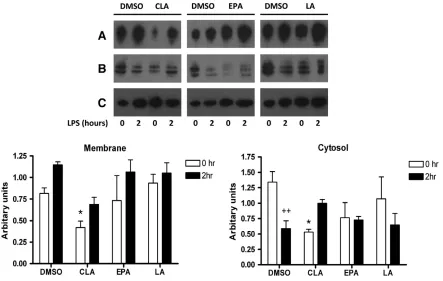
Fig. 2. c9,t11-CLA modulates membrane and cytosolic CD14 in J774 macrophages. Cells were cultured for 7 days with either DMSO (vehicle control), c9,t11-CLA (50μM), EPA (25μM) or LA (50μM). Cells were plated at 2×10⁵cell/ml, left to rest overnight and left unstimulated or stimulated with LPS (100 ng/ml) for 2 h. Cell lysates were harvested and membrane
fractionation performed. Western blotting was used to determine the expression of CD14 in the cytosolic and membrane fractions of cells treated with DMSO, c9,t11-CLA, EPA and LA. Total cellular levels ofβ-actin were used as a loading control. Immunoblots shown are representative of 3 experiments. Densitometric analysis was carried out on immunoblots and CD14 is expressed as arbitrary units (± S.E.M.) after normalising toβ-actin. *Pb.05, ANOVA, comparing DMSO vs. PUFA-treated groups at 0 h,++
100 mM glycine PBS-1.2%fish gelatin blocking buffer and incubated with a rabbit polyclonal anti-CD14 (M305, Santa Cruz) over night at 4°C. The next day, cell preparations were washed and incubated with an AlexaFluor 488 donkey anti-rabbit secondary antibody (Molecular Probes, Invitrogen) for 1 h at 37°C. Finally, cover slips were washed and mounted on slides with antifade medium (Dako). Slide preparations were observed using a Zeiss Axio Observer. Z1 equipped with a Zeiss 710 and ConfoCor 3 laser scanning confocal head (Carl Zeiss, Germany). Images were analysed using ZEN 2008 software.
2.7. Lipid Raft Isolation
For the isolation of lipid rafts 1×10⁷
of DMSO or PUFA-treated cells were stimulated with 100 ng/ml LPS for 30 min prior to membrane preparation. Cells were then washed, pelleted and resuspended in 2 ml membrane extraction buffer (MEB; 20 mM MES, 150 Mm NaCl, pH 6.5) containing 5 mM iodoacetamide, 1 mM PMSF, 1μg/ml aprotinin and 1μg/ml leupeptin and left on ice for 30 min. For mechanical cell disruption, lysates were freeze-thawed in liquid nitrogen three times followed by 40 strokes with a dounce homogeniser and passage through a 25G 1-in. syringe (BD Microlance). Lysates were spun at 100,000 g for 1 h at 4°C. The membrane pellet was resuspended in 500μl MEB containing 0.5 % (v/v) Triton X-`100. For the isolation of lipid rafts the membrane preparation was mixed with an equal volume (500μl) of 90 % (w/v) sucrose/MEB (with protease inhibitors as described) and applied to a discontinuous sucrose gradient (30% (w/v) sucrose/MEB, 5% (w/v) sucrose/MEB).
Samples were spun for 18 h at 175,000 g in a TH-641 swinging bucket rotor in a Sorvall WX ultracentrifuge. After centrifugation 1 ml fractions were carefully collected from the top of the gradient and designated fractions 1–11. A light refracting band (typically in fractions 5–6) represented fractions enriched for lipid rafts. Aflotillin-1 antibody (BD) was used to confirm this.
2.8. Luciferase assays
For ISRE and NF-κB luciferase assays, 75 ng of ISRE/NF-κB luciferase plasmid, 30 ng ofRenillaluciferase, and empty pcDNA3.1 vector made up to a total of 220 ng of DNA were transfected into each well. Empty pcDNA3.1 vector alone was also transfected into cells and as expected gave no detectable response. Cells were left to rest overnight and then stimulated with LPS (100 ng/ml) as indicated in the figure legends. After stimulation cells were lysed in 100μl of passive lysis buffer (Promega, Southampton, UK) for 15 min. Firefly luciferase activity was assayed by the addition of 40μl of luciferase assay mix (20 mm Tricine, 1.07 mm (MgCO3)4Mg(OH)2·5H2O,
2.67 MgSO4, 0.1 m EDTA, 33.3 mm dithiothreitol, 270 mm coenzyme A, 470 mm
[image:4.594.136.450.292.707.2]luciferin, 530 mm ATP) to 20μl of the lysed sample.Renillaluciferase was read by the addition of 40μl of a 1:1000 dilution of Coelentrazine (Argus Fine Chemicals) in phosphate-buffered saline. Luminescence was read using the Reporter microplate luminometer (Turner Designs). TheRenillaluciferase plasmid was used to normalize for transfection efficiency in all experiments.
2.9. Endocytosis of TLR4 by confocal microscopy
PUFA treated HEK293-TLR4-MD-2-CD14 cells were plated on poly-L-lysine-treated glass cover slips and left to rest overnight. For the investigation of TLR4 and EEA1
localisation, TLR4-YFP and EEA1-CFP constructs were kind gifts from Douglas Gollenbock (University of Massachusetts Medical School, Worcester, MA, USA) and Terje Espeviks (Norwegian University of Science and Technology, Trondheim, Norway), respectively. Cells were transfected with TLR4-YFP (0.75μg) and EEA1-CFP (0.25μg) using geneJuice according to manufacturer's instructions. Cells were left to rest for 24 h before stimulation with 250 ng/ml LPS for 7.5 and 15 min. Cell preparations were washed,fixed and mounted to glass coverslips and analysed using an Olympus FluoView FV1000 and FV1000 Viewer Software version 1.7.
2.10. Statistics
One-way analysis of variance (ANOVA) was used to determine significant differences between conditions. When this indicated significance (Pb.05), post hoc Student-Newmann-Keul test analysis was used to determine which conditions were significantly different from each other.
3. Results
3.1. c9,t11-CLA modulates CD14 expression in macrophages
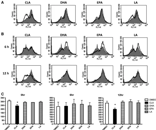
[image:5.594.57.284.127.644.2]The ligation of LPS with TLR4 activates downstream signalling
pathways that initiate in
fl
ammatory responses and CD14 is
important in this process. Therefore, we examined whether the
c9,t11-CLA isomer could modulate the expression of CD14 using
fl
ow cytometry. We examined the surface expression of CD14
following LPS stimulation of J774 macrophages pre-treated with
either c9,t11-CLA, EPA or DHA. c9,t11-CLA suppressed the surface
expression of CD14 in both resting macrophages (
Fig. 1
A) and in
LPS-stimulated macrophages (
Fig. 1
B). This suppressive effect was
Fig. 4. c9,t11-CLA decreases expression of CD14 in lipid rafts in J774 macrophage following stimulation with LPS. Cells were cultured for 7 days with either DMSO (vehicle control), c9,t11-CLA (50μM) or EPA (25μM). 1×10⁷
cells were stimulated with LPS (100ng/ml) for 30 min. Cells were then lysed with buffer containing 0.5% Triton X-100 and layered on the bottom of a sucrose gradient. Following centrifugation 1 ml fractions were collected and analysed by SDS-PAGE. Lipid raft fractions were identified by localisation of constitutive raft protein flotilin-1 (FL-1) in fractions 5 and 6. Similarly, fractions were analysed for CD14 content by SDS-PAGE using a CD14 specific antibody (AbCam). Immunoblots shown are representative of 3 experiments. Densitometric analysis was carried out on immunoblots and CD14 is expressed as arbitrary units (± S.E.M.) after normalising to FL-1. **Pb.01, ***Pb.001, determined by one-way ANOVA test comparing all groups.
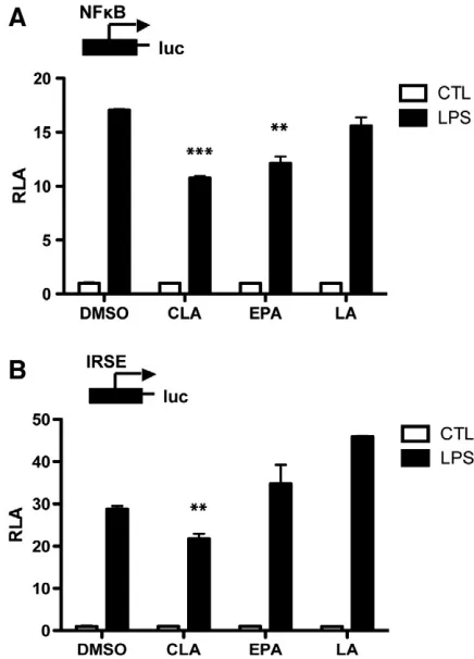
Fig. 5. c9,t11-CLA suppresses NF-κB and IRF3. HEK-TLR4-CD14-MD-2 cells were cultured for 7 days with either DMSO (vehicle control), c9,t11-CLA (50μM),EPA (25
μM) or LA (50μM). Subsequently, cells were transiently transfected with either an
[image:5.594.333.551.380.683.2]not evident in either DHA or EPA-treated cells, indeed they
slightly increased CD14 expression following activation of
macro-phages with LPS.
3.2. c9,t11-CLA modulates CD14 localisation following stimulation
with LPS
In order to con
fi
rm the effect of c9,t11-CLA on membrane
expression of CD14 we isolated both the membrane and cytosolic
fractions of J774 macrophages treated with either c9,t11-CLA, EPA
or LA and used western blotting to determine the levels of CD14
on the membrane and in the cytosol at early points after LPS
stimulation. We also used confocal microscopy to further establish
the location of CD14. Exposure of DMSO control cells to LPS
increased CD14 levels in membrane fractions which was
concom-itant with a decrease in cytosolic CD14 (
Fig. 2
A). Pre-treatment of
cells with c9,t11-CLA resulted in a decreased level of membrane
CD14 in both resting cells and 2 h post stimulation with LPS
(
Fig. 2
A). In contrast to DMSO, the c9,t11-CLA-treated cells showed
an increase in cytosolic CD14 after LPS stimulation. Neither EPA
nor LA suppressed membrane levels of CD14 (
Fig. 2
B and C) when
normalised to beta-actin levels. Indeed the levels of membrane
CD14 are increased by EPA, which concurs with the
fl
ow
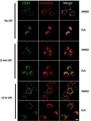
cytometry data. Furthermore, investigation by confocal microscopy
using double-staining of DMSO or c9,t11-CLA treated cells with
anti-CD14 and the plasma membrane marker, CellMask,
demon-strated that the cellular traf
fi
cking of CD14 in the presence of c9,
t11-CLA was altered.
Fig. 3
shows that in resting DMSO-treated
cells CD14 clearly localises at the plasma membrane. In contrast,
c9,t11-CLA-treated cells had signi
fi
cantly reduced cell surface
expression of CD14 and it was primarily con
fi
ned to the
cytoplasm. Following 15 min of stimulation with LPS, CD14 leaves
the membrane and moves into the cytoplasm in DMSO-treated
cells. This movement is not obvious in the c9,t11-CLA-treated cells
due to the low levels of CD14 on the membrane in these cells.
After a longer exposure to LPS (12 h) CD14 re-circulated to the
cell surface in DMSO-treated cells which was reduced in
c9,t11-CLA-treated cells.
3.3. c9-t11- CLA decreases the expression of CD14 in lipid rafts
The recruitment of CD14 to lipid rafts following activation of cells
with LPS is an important event
[38,43]
. Our data clearly demonstrates
that c9,t11-CLA suppresses expression of CD14 at the membrane;
therefore, we examined whether c9,t11-CLA altered the expression of
CD14 in lipid rafts following LPS stimulation. We used EPA as a
comparison given that it did not suppress CD14 expression. Anti-FL-1
was used as a marker for lipid rafts. Fractions 4
–
6 were designated to
be lipid raft fractions with the expression of FL-1 predominant in
these samples.
Fig. 4
demonstrates the presence of CD14 in raft
fractions following LPS stimulation of DMSO-treated cells. The levels
of CD14 in these lipid raft fractions was decreased in
c9,t11-CLA-treated cells which was not seen with EPA-c9,t11-CLA-treated cell.
3.4. c9,t11-CLA selectively inhibits IRF3 activation downstream of TLR4
Given the role of CD14 in endocytosis of TLR4 and subsequent
signalling to IRF3, we next examined whether c9,t11-CLA could
modulate activation of IRF3. It is well established that dietary fatty
acids can suppress NF-
κ
B; therefore, we also examined the effect of
c9,t11-CLA on this transcription factor. EPA was again used as a
comparison as it does not suppress CD14 expression. Treatment of
HEK-TLR4-MD2-CD14 cells with EPA and c9,t11-CLA resulted in
sup-pression of NF-
κ
B following LPS stimulation (
P
b
.01;
P
b
.001;
Fig. 5
A).
Furthermore, treatment with c9,t11-CLA suppressed LPS-induced
IRF3 activation (
P
b
.01;
Fig. 5
B) which was indicated by its effect on
the ISRE plasmid. In contrast, treatment of cells with EPA had no
effect on IRF3 activation.
3.5. Overexpression of CD14 reverses the inhibitory effect of c9,t11-CLA
on IRF3 activation in response to LPS
Our data demonstrated that c9,t11-CLA selectively suppressed the
surface expression of CD14 and subsequently inhibited activation of
IRF3. CD14 is necessary for endocytosis of the TLR4 complex and
subsequent activation of IRF3
[41,42]
. Therefore, in order to
deter-mine whether the effect of c9,t11-CLA on CD14 expression could be
responsible for its effect on IRF3 activation, we employed U373 cell
lines, one of which stably overexpressed CD14. Cells were exposed to
c9,t11-CLA and then LPS-induced IRF3 activation was measured. c9,
t11-CLA suppressed activation of IRF3 in U373 cells (
Fig. 6
A;
P
b
.001);
however, this inhibitory effect was abrogated in U373 stably over
expressing CD14 (
Fig. 6
B).
3.6. c9,t11-CLA decreases endocytosis of TLR4 following stimulation
with LPS
[image:6.594.317.531.424.684.2]Given the requirement of CD14 for endocytosis of the TLR4
com-plex and subsequent activation of IRF3
[41,42]
we assessed whether
CLA could alter endocytosis of the TLR4 complex following LPS
stimulation. Again we compared this to EPA, one of the PUFA which
did not have a suppressive effect on either CD14 or IRF3 activation.
PUFA treated HEK-TLR4-MD2-CD14 cells were transiently transfected
with TLR4-YFP and EEA1-CFP (early endosomal antigen) constructs.
Localisation of TLR4 with EEA1 was examined following stimulation
with LPS for 7.5 and 15 min. As expected, following 7.5 min
Fig. 7. c9,t11-CLA decreases endocytosis of TLR4 following LPS stimulation. HEK-TLR4-MD-2-CD14 cells were cultured for 7 days in either DMSO (vehicle control), c9,t11-CLA (50μM) (EPA (25μM) or LA (50μM). Subsequently, cells were transiently transfected with TLR4-YFP (green) and EEA1-CFP (red) plasmids. Endocytosis of TLR4 and its localisation relevant to early endosomal marker EEA1 was assessed using confocal microscopy following stimulation with LPS for 7.5 min (B) and 15 min (C) compared to resting cells (A). Colocalisation of TLR4 and EEA1 is shown in the composite image (yellow). Results shown are representative of three experiments.
7
Dowling
et
al.
/
Journal
of
Nutritional
Biochemistr
y
xx
(2012)
xxx
–
stimulation with LPS in DMSO-treated cells, TLR4 was rapidly
endocytosed and is almost completely inside the cell with early
endosome formation and co-localisation of EEA1 with TLR4 being
quite prominent (
Fig. 7
A and B, top panel). After 15 min stimulation
there appears to be less co-localisation of the two molecules and less
endosomes present (
Fig. 7
C, top panel). In direct comparison to
DMSO, c9,t11-CLA-treated cells displayed distinct patterns of TLR4
localisation and endocytosis. Upon stimulation more TLR4 appears to
be retained at the plasma membrane in these cells (
Fig. 7
B and C,
second panels down). Conversely, almost all TLR4 was observed to be
inside cells and co-localised with EEA1 in EPA treated cells stimulated
with LPS (
Fig. 7
B, third panel down). In addition, at 15 min
stimu-lation there is a distinct movement of TLR4 away from endosomes in
these cells (
Fig. 7
C, third panel down). It was noted that more
co-localisation was observed at this time point in EPA treated cells
compared to DMSO (
Fig. 7
B, third panel down). Cells treated with the
saturated fat, LA, displayed patterns of TLR4 endocytosis and early
endosome formation similar to those of DMSO-treated cells (
Fig. 7
B
and C, bottom panels).
4. Discussion
In this study we explored whether the CLA isomer, c9,t11-CLA,
exerts its anti-in
fl
ammatory actions via the TLR4 receptor complex.
We observed suppressed expression of CD14 both on the cell
membrane and in lipid rafts following stimulation with LPS in c9,
t11-CLA-treated cells. This effect on CD14 resulted in a decrease in
endocytosis of TLR4 to early endosomes following activation with
LPS and subsequent suppression of IRF3 activation. These effects
were not seen with EPA, which was assessed as a comparison, and
suggests a novel mechanism by which c9,t11-CLA exerts its
anti-in
fl
ammatory effects.
Activation of TLR4 at the plasma membrane initiates downstream
signalling which in turn activates NF-
κ
B and IRF3 via
MyD88-dependent and -inMyD88-dependent pathways, respectively
[39,40]
.
Know-ing that the incorporation of PUFA to the plasma membrane modi
fi
es
its
fl
uidity and indeed its function via receptors, proteins and
enzymes bound to it, we
fi
rstly examined the effect of the
c9,t11-CLA isomer on the components of the TLR4 complex. CD14 is a 56 kDa,
GPI-anchored protein lacking a transmembrane and intracellular
domain and acts as an accessory molecule for both TLR4 and TLR2
signalling
[41,44]
. c9,t11-CLA was the only fatty acid to suppress
surface expression of CD14 in both resting and LPS-activated
macro-phages. Furthermore, analysis by western blot revealed suppressed
levels of CD14 at the membrane in c9,t11-CLA treated cells which
was concomitant with enhanced levels of CD14 in the cytosol
sug-gesting that CD14 may be retained in the cytosol in these cells. This
fi
nding was supported by confocal microscopy which showed that in
c9,t11-CLA-treated cells, CD14 was mainly con
fi
ned to the
cyto-plasm in both resting cells and after 15 min of stimulation with LPS,
as well as after 30 min, 1 h and 6 h LPS (data not shown). It was
only after prolonged stimulation, some but not all of CD14 was seen
to return to the plasma membrane. In contrast EPA did not alter
CD14 in this way. These
fi
ndings suggested a distinct mechanism by
which c9,t11-CLA may exert its anti-in
fl
ammatory effects.
CD14 is recruited to lipid rafts in order to complex with TLR4 and
this is a crucial element in subsequent signalling by the complex
[30,38,45,46]
. Therefore we examined whether c9,t11-CLA altered the
localisation of CD14 in these lipid rafts and whether this had
consequences for endocytosis of TLR4. We found CD14 to be markedly
suppressed in lipid raft fractions isolated from LPS-stimulated
macrophages treated with c9,t11-CLA. On the other hand,
macro-phages treated with the n-3 PUFA, EPA for comparison, displayed
enhanced levels of CD14 in lipid raft fractions compared to control.
There is currently no other published data available on the effects of
c9,t11-CLA on lipid rafts. The most studied fatty acid to date is DHA
which has been shown to modulate the recruitment of TLR4 into lipid
rafts
[47]
and also modify the clustering and size of lipid rafts
[48]
.
GPI-anchored proteins are known to contain two saturated fatty
acid chains in their phosphatidylinositol moiety allowing them to be
incorporated to lipid rafts
[49]
. The remodelling of the GPI anchor
takes place between the ER and Golgi with the unsaturated sn-2 chain
being removed and a saturated one added. This process is mediated
by post GPI-attachment proteins (PGAP2 and PGAP3). An increase in
unsaturated fatty acids following exposure of the cells to c9,t11-CLA
may affect the availability of saturated fatty acids for this process.
Furthermore, S-acylation of proteins with heterogeneous unsaturated
fatty acids has been proposed as a mechanism by which cells regulate
signal transduction by altering the association of proteins with rafts
[50,51]
. Our
fi
nding that c9,t11-CLA reduced the localization of CD14
in lipid rafts, may suggest that this fatty acid could interfere with
these processes.
In agreement with a number of published studies, here we have
also observed a suppressive effects of c9,t11-CLA and EPA on NF-
κ
B
[17
–
19]
. We previously demonstrated that c9,t11-CLA can suppress
NF-
κ
B in vitro in dendritic cells
[8]
and in vivo in adipose tissue from
mice fed on diets of CLA
[21]
. TLR4 signalling is unique in its ability to
activate both NF-
κ
B and IRF3
[39,40]
. Interestingly, our data
demonstrates a novel and selective inhibition of IRF3 in
HEK-TLR4-CD14-MD2 cells exposed to c9,t11-CLA but not to EPA. This is the
fi
rst
study to demonstrate a suppressive effect of a dietary fatty acid on
IRF3 in any cell type. Given that activation of IRF3 downstream of
TLR4 requires endocytosis of the TLR4 complex
[42]
and is a process
dependent on CD14
[52]
, our
fi
ndings suggested that the distinct
modulation of CD14 by c9,t11-CLA could be responsible for the
subsequent suppression of IRF3 we observed. Further evidence is
provided by the experiments using U373 cells which demonstrated
that the inhibitory effect of c9,t11-CLA on IRF3 was completely
reversed by over-expressing CD14. Our data on the effect of
c9,t11-CLA on CD14 is consistent with our recent study demonstrating that
bone-marrow derived dendritic cells isolated from mice fed on a
high-CLA diet show decreased levels of CD14
[53]
.
In order to assess whether the suppression of CD14 was
associated with reduced endocytosis of TLR4, which would explain
the decrease in IRF3 activation, confocal microscopy was employed
examining TLR4 expression at the membrane and in early
endosomes in HEK-TLR4-MD2-CD14 cells treated with c9,t11-CLA,
EPA or LA. In this study we demonstrate retention of TLR4 on the
plasma membrane following stimulation with LPS in c9,t11-CLA
treated cells indicating a decrease in endocytosis of TLR4 compared
to the DMSO control. This was not the case in EPA-treated cells
which were used as a comparison due to the fact that EPA did not
decrease CD14 expression or IRF3 activation. This data reveals a
novel mechanism which explains how c9,t11-CLA may exert its
anti-in
fl
ammatory effects.
Previously, research has focussed on downstream signalling
components
[8,53]
and suggested these as key targets through
which PUFA exert their effects. This study demonstrates a novel
mechanism through which c9,t11-CLA exerts its effects at the
membrane. With emerging roles for CD14 in the exacerbation of
infection and in
fl
ammatory disease our
fi
nding with regard to
c9,t11-CLA may have implications for the possible use of this fatty acid as a
complementary treatment.
References
[1] Wan JM, Haw MP, Blackburn GL. Nutrition, immune function, and inflammation: an overview. Proc Nutr Soc 1989;48:315–35.
[3] MacLean CH, Mojica WA, Morton SC, Pencharz J, Hasenfeld Garland R, Tu W, et al. Effects of omega-3 fatty acids on lipids and glycemic control in type II diabetes and the metabolic syndrome and on inflammatory bowel disease, rheumatoid arthritis, renal disease, systemic lupus erythematosus, and osteoporosis. Evid Rep Technol Assess (Summ) 2004(89):1–4.
[4] Schachter HM, Reisman J, Tran K, Dales B, Kourad K, Barnes D, et al. Health effects of omega-3 fatty acids on asthma. Evid Rep Technol Assess (Summ) 2004(91): 1–7.
[5] Fritsche K. Fatty acids as modulators of the immune response. Annu Rev Nutr 2006;26:45–73.
[6] Roche HM, Noone E, Gibney AN. Conjugated linoleic acid: a novel therapeutic nutrient? Nutr Res Rev 2001;14:173–88.
[7] Belury MA. Dietary conjugated linoleic acid in health: physiological effects and mechanisms of action. Annu Rev Nutr 2002;22:505–31.
[8] Loscher CE, Draper E, Leavy O, Kelleher D, Mills KH, Roche HM. Conjugated linoleic acid suppresses NF-kappa B activation and IL-12 production in dendritic cells through ERK-mediated IL-10 induction. J Immunol 2005;175:4990–8. [9] MacDonald HB. Conjugated linoleic acid and disease prevention: a review of
current knowledge. J Am Coll Nutr 2000;19:111S–8S.
[10] Bassaganya-Riera J, Reynolds K, Martino-Catt S, Cui Y, Hennighausen L, Gonzalez F, et al. Activation of PPAR gamma and delta by conjugated linoleic acid mediates protection from experimental inflammatory bowel disease. Gastroenterology 2004;127:777–91.
[11] Schmitz G, Ecker J. The opposing effects of n-3 and n-6 fatty acids. Prog Lipid Res 2008;47:147–55.
[12] Calder PC. Polyunsaturated fatty acids and inflammation. Prostaglandins Leukot Essent Fatty Acids 2006;75:197–202.
[13] Sampath H, Ntambi JM. Polyunsaturated fatty acid regulation of genes of lipid metabolism. Annu Rev Nutr 2005;25:317–40.
[14] Jump DB. Dietary polyunsaturated fatty acids and regulation of gene transcription. Curr Opin Lipidol 2002;13:155–64.
[15] Lee JY, Plakidas A, Lee WH, Heikkinen A, Chanmugam P, Bray G, et al. Differential modulation of Toll-like receptors by fatty acids: preferential inhibition by n-3 polyunsaturated fatty acids. J Lipid Rese 2003;44:479–86.
[16] Zhao Y, Joshi-Barve S, Barve S, Chen LH. Eicosapentaenoic acid prevents LPS-induced TNF-alpha expression by preventing NF-kappaB activation. J Am Coll Nutr 2004;23:71–8.
[17] Huang F, Wei H, Luo H, Jiang S, Peng J. EPA inhibits the inhibitor of kappaBalpha (IkappaBalpha)/NF-kappaB/muscle RING finger 1 pathway in C2C12 myotubes in a PPARgamma-dependent manner. Br J Nutr 2011;105:348–56.
[18] Draper E, Reynolds CM, Canavan M, Mills KH, Loscher CE, Roche HM. Omega-3 fatty acids attenuate dendritic cell function via NF-kappaB independent of PPARgamma. J Nutr Biochem 2011;22:784–90.
[19] Schumann J, Fuhrmann H. Impairment of NFkappaB activity by unsaturated fatty acids. Int Immunopharmacol 2010;10:978–84.
[20] Babcock TA, Helton WS, Hong D, Espat NJ. Omega-3 fatty acid lipid emulsion reduces LPS-stimulated macrophage TNF-alpha production. Surg Infect (Larchmt) 2002;3:145–9.
[21] Moloney F, Toomey S, Noone E, Nugent A, Allan B, Loscher CE, et al. Antidiabetic effects ofcis-9,trans-11-conjugated linoleic acid may be mediated via anti-inflammatory effects in white adipose tissue. Diabetes 2007;56:574–82. [22] Chu AJ, Walton MA, Prasad JK, Seto A. Blockade by polyunsaturated n-3 fatty acids
of endotoxin-induced monocytic tissue factor activation is mediated by the depressed receptor expression in THP-1 cells. J Surg Res 1999;87:217–24. [23] Goua M, Mulgrew S, Frank J, Rees D, Sneddon AA, Wahle KW. Regulation of
adhesion molecule expression in human endothelial and smooth muscle cells by omega-3 fatty acids and conjugated linoleic acids: involvement of the transcrip-tion factor NF-kappaB? Prostaglandins Leukot Essent Fatty Acids 2008;78:33–43. [24] Weldon SM, Mullen AC, Loscher CE, Hurley LA, Roche HM. Docosahexaenoic acid induces an anti-inflammatory profile in lipopolysaccharide-stimulated human THP-1 macrophages more effectively than eicosapentaenoic acid. J Nutr Biochem 2007;18:250–8.
[25] Mullen A, Loscher CE, Roche HM. Anti-inflammatory effects of EPA and DHA are dependent upon time and dose–response elements associated with LPS stimulation in THP-1-derived macrophages. J Nutr Biochem 2009.
[26] Lee JY, Ye J, Gao Z, Youn HS, Lee WH, Zhao L, et al. Reciprocal modulation of Toll-like receptor-4 signaling pathways involving MyD88 and phosphatidylinositol 3-kinase/AKT by saturated and polyunsaturated fatty acids. J Biol Chem 2003;278: 37041–51.
[27] Chapkin RS, Wang N, Fan YY, Lupton JR, Prior IA. Docosahexaenoic acid alters the size and distribution of cell surface microdomains. Biochim Biophys Acta 2008;1778:466–71.
[28] Li Q, Wang M, Tan L, Wang C, Ma J, Li N, et al. Docosahexaenoic acid changes lipid composition and interleukin-2 receptor signaling in membrane rafts. J Lipid Res 2005;46:1904–13.
[29] Ma DW, Seo J, Switzer KC, Fan YY, McMurray DN, Lupton JR, et al. n-3 PUFA and membrane microdomains: a new frontier in bioactive lipid research. J Nutr Biochem 2004;15:700–6.
[30] Dykstra M, Cherukuri A, Sohn HW, Tzeng SJ, Pierce SK. Location is everything: lipid rafts and immune cell signaling. Annu Rev Immunol 2003; 21:457–81.
[31] Goebel J, Forrest K, Flynn D, Rao R, Roszman TL. Lipid rafts, major histocompat-ibility complex molecules, and immune regulation. Hum Immunol 2002;63: 813–20.
[32] Ruth MR, Proctor SD, Field CJ. Feeding long-chain n-3 polyunsaturated fatty acids to obese leptin receptor-deficient JCR:LA- cp rats modifies immune function and lipid-raft fatty acid composition. Br J Nutr 2009;101:1341–50.
[33] Martin V, Fabelo N, Santpere G, Puig B, Marin R, Ferrer I, et al. Lipid alterations in lipid rafts from Alzheimer's disease human brain cortex. J Alzheimers Dis 2010; 19:489–502.
[34] Kim W, Fan YY, Barhoumi R, Smith R, McMurray DN, Chapkin RS. n-3 polyunsaturated fatty acids suppress the localization and activation of signaling proteins at the immunological synapse in murine CD4 + T cells by affecting lipid raft formation. J Immunol 2008;181:6236–43.
[35] Horejsi V, Drbal K, Cebecauer M, Cerny J, Brdicka T, Angelisova P, et al. GPI-microdomains: a role in signalling via immunoreceptors. Immunol Today 1999; 20:356–61.
[36] Fan YY, Ly LH, Barhoumi R, McMurray DN, Chapkin RS. Dietary docosahexaenoic acid suppresses T cell protein kinase C theta lipid raft recruitment and IL-2 production. J Immunol 2004;173:6151–60.
[37] Soong G, Reddy B, Sokol S, Adamo R, Prince A. TLR2 is mobilized into an apical lipid raft receptor complex to signal infection in airway epithelial cells. J Clin Invest 2004;113:1482–9.
[38] Triantafilou M, Miyake K, Golenbock DT, Triantafilou K. Mediators of innate immune recognition of bacteria concentrate in lipid rafts and facilitate lipopolysaccharide-induced cell activation. J Cell Sci 2002;115:2603–11. [39] Brikos C, O'Neill LA. Signalling of toll-like receptors. Handb Exp Pharmacol 2008;
183:21–50.
[40] Akira S, Takeda K. Toll-like receptor signalling. Nat Rev Immunol 2004;4:499–511. [41] Jiang ZF, Georgel P, Du X, Shamel L, Sovath S, Mudd S, et al. CD14 is required for
MyD88-independent LPS signaling. Nat Immunol 2005;6:565–70.
[42] Kagan JC, Su T, Horng T, Chow A, Akira S, Medzhitov R. TRAM couples endo-cytosis of Toll-like receptor 4 to the induction of interferon-[beta]. Nat Immunol 2008;9:361.
[43] Pfeiffer A, Bottcher A, Orso E, Kapinsky M, Nagy P, Bodnar A, et al. Lipopolysac-charide and ceramide docking to CD14 provokes ligand-specific receptor clustering in rafts. Eur J Immunol 2001;31:3153–64.
[44] Wright SD, Ramos RA, Tobias PS, Ulevitch RJ, Mathison JC. CD14, a receptor for complexes of lipopolysaccharide (LPS) and LPS binding protein. Science 1990; 249:1431–3.
[45] Triantafilou M, Brandenburg K, Kusumoto S, Fukase K, Mackie A, Seydel U, et al. Combinational clustering of receptors following stimulation by bacterial products determines lipopolysaccharide responses. Biochem J 2004;381:527–36. [46] Dai Q, Zhang J, Pruett SB. Ethanol alters cellular activation and CD14 partitioning
in lipid rafts. Biochem Biophys Res Commun 2005;332:37–42.
[47] Wong SW, Kwon MJ, Choi AM, Kim HP, Nakahira K, Hwang DH. Fatty acids modulate Toll-like receptor 4 activation through regulation of receptor dimer-ization and recruitment into lipid rafts in a reactive oxygen species-dependent manner. J Biol Chem 2009;284:27384–92.
[48] Shaikh SR, Rockett BD, Salameh M, Carraway K. Docosahexaenoic acid modifies the clustering and size of lipid rafts and the lateral organization and surface expression of MHC class I of EL4 cells. J Nutr 2009;139:1632–9.
[49] Maeda Y, Tashima Y, Houjou T, Fujita M, Yoko-o T, Jigami Y, et al. Fatty acid remodeling of GPI-anchored proteins is required for their raft association. Mol Biol Cell 2007;18:1497–506.
[50] Liang X, Nazarian A, Erdjument-Bromage H, Bornmann W, Tempst P, Resh MD. Heterogeneous fatty acylation of Src family kinases with polyunsaturated fatty acids regulates raft localization and signal transduction. J Biol Chem 2001;276: 30987–94.
[51] Webb Y, Hermida-Matsumoto L, Resh MD. Inhibition of protein palmitoylation, raft localization, and T cell signaling by 2-bromopalmitate and polyunsaturated fatty acids. J Biol Chem 2000;275:261–70.
[52] Shuto T, Kato K, Mori Y, Viriyakosol S, Oba M, Furuta T, et al. Membrane-anchored CD14 is required for LPS-induced TLR4 endocytosis in TLR4/MD-2/CD14 overexpressing CHO cells. Biochem Biophys Res Commun 2005;338: 1402–9.