Thesis by
Eun Jung Choi
In Partial Fulfillment of the Requirements
for the Degree of
Doctor of Philosophy
California Institute of Technology
Pasadena, California
2006
Acknowledgements
I would like to thank my advisor, Dr. Stephen Mayo, for his support throughout my graduate studies. He is a brilliant scientist, and during my stay in his lab, I learned a lot from him. I am grateful for the opportunity I had in learning about the exciting field of computational protein design.
My committee members, Dr. Pamela Bjorkman, Dr. David Chan and Dr. Bill Goddard, always gave me helpful advice at my annual committee meetings. They cared not only about my research, but also about my long-term goals. I always looked forward to these meetings.
I thank everyone in the Mayo lab, Alex, Barry, Ben, Christina, Dan, Darryl, Deepshika, Eric, Geoff, Heidi, Jennifer, JJ, Julia, Karin, Kyle, Oscar, Mary, Matthew, Peter, Possu, Premal, Sarah, Scott, Shannon, Shira, Tom, and Welison for providing a stimulating but comfortable research environment. Needless to say, Cynthia and Rhonda are the two most indispensable people in the lab. Marie spent endless hours editing my papers and this thesis would not have seen the light of day were it not for her efforts. Jessica M. deserves special mention because it was her fabulous idea to collaborate together. It was a pleasure working with her.
I owe a lot to my valuable friends whom I was lucky to have met during my stay at
Caltech. Hyun Jee, In Jung, Jessica E., Ji Young, Keum Joo, Kyung Ah, Mi Sook, Sarina and all of the other people, both past and present, who were always there for me, thank you so much. I would especially like to thank Caglar for his endless capacity to help on anything and everything. Ann Marie, Daphne and Frosso, thanks for being such good housemates.
Abstract
Success in computational design of proteins requires a good understanding of the physical properties that determine the protein structure. However, computational protein design also provides us the opportunities to test and improve our current knowledge of protein structure. In our lab, the “protein design cycle” is used to improve the energy function and increase our knowledge of protein chemistry. This strategy utilizes a cyclic protocol where first, a modification that is predicted to improve the result is applied to the energy function. Next, proteins are simulated using this modified function. These molecules are then synthesized and analyzed to see if our simulation results correlate with the physical properties of the molecules. The new knowledge from the analysis is incorporated into the next design in the form of a new modification and the whole cycle starts again. Using this technique, an energy function highly successful in designing protein stability has been acquired in our lab.
A similar approach was used to determine whether the introduction of a backbone entropy term allows us to incorporate proline into the pool of amino acids we consider in designs. Using ORBIT, several proline mutants of protein G were simulated and synthesized. The correlation between the experimental results and computational results was analyzed. From this analysis, we learned that the entropic benefit of a proline mutation is usually small compared to the enthalpic disadvantages. However, including a backbone entropy term did increase the correlation between the rank order of the experimental stabilities and computational energies.
Table of Contents
Acknowledgements... iii
Abstract ...iv
Table of Contents...vi
List of Tables and Figures ... viii
Chapter 1. Introduction...1
Chapter 2. Generation and analysis of proline mutants in protein G ...17
Chapter 3. Analysis of computationally designed TRAF6 binding peptides...44
Chapter 4. Computational design and biochemical characterization of non-specific lipid transfer protein variants for biosensor applications...69
Appendix A.
List of Tables and Figures
Chapter 1.
Figure 1. Protein design and protein folding problems...13 Figure 2. Potential energy function terms for ORBIT...15
Chapter 2.
Table 1. Midpoint of thermal unfolding transition, free energy of folding at 25°C and computed energy for Gβ1 variants...32
Figure 3. Views of the 10 positions in Gβ1 mutated to proline... 33 Figure 4. Correlation between calculated energies, using the Street and Mayo
solvation model, and experimental results...35
Figure 5. Correlation between calculated energies, using the Lazaridis and Karplus solvation model, and experimental results... 38
Figure 6. CD data...41
Chapter 3.
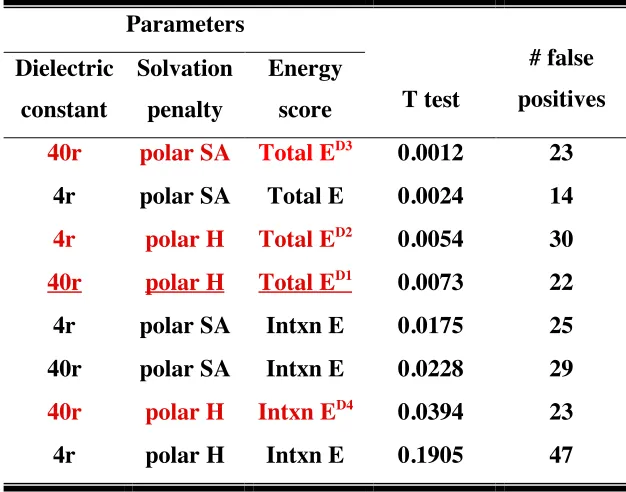
Table 2. Parameters used to fit experimental binding assay data with computational energies, T test scores and the number of false positives...61
Figure 7. Ribbon and molecular surface representation of TRAF-C domain of TRAF6 bound to CD40 peptide...64
Figure 8. Anisotropy data of all the peptides... 66
Chapter 4.
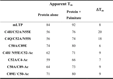
Table 5. Apparent Tms of mLTP and designed variants...86 Figure 9. Ribbon diagram of mLTP and the designed variants of each disulfide....87 Figure 10. Wavelength scans of mLTP and designed variants... 89 Figure 11. Thermal denaturations of mLTP and designed variants...91 Figure 12. Circular dichroism wavelength scans of the four protein-acrylodan conjugates...93
Figure 13. Titration of C52A/C4-Ac with palmitate monitored by fluorescence emission...95
Chapter 5.
Figure 14. Structures of 1SM3 and its mutants showing the mutated residues...116
Appendix A.
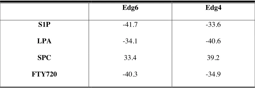
Table 6. Computational binding energies of Edg6 and Edg4 to S1P, LPA, SPC and FTY720...135
Chapter 1
The idea of protein design debuted in the early 1980s (Drexler 1981; Pabo 1983). The protein design problem, which is the search for an amino acid sequence that folds into the desired structure, was thought of as the inverse of the protein folding problem, which aims to find the correct fold for a given amino acid sequence. Unlike the protein folding problem where there is a single solution fold per sequence, multiple solution sequences can be found for each target fold in the protein design problem (Figure 1). Assisted by the increase in computational power and developments in molecular biology, drastic progress has been made in the field of protein design. Today, proteins are engineered for increased stability and solubility, and for new or altered functionalities.
Computational protein design methods use force fields to select the optimal sequence of amino acids for a target fold. They employ an energy potential, statistically significant conformations of amino acids called rotamers and a selection algorithm, which screens through the combinatorial complexity of the sequence space to obtain the lowest energy sequences. In ORBIT (Optimization of Rotamers By Iterative Techniques), the protein design program developed in our lab, the energy function is comprised of 5 terms: van der Waals, hydrogen bonding, electrostatic, solvation and entropy. The energy function is empirical, but based on fundamental physical principles. A simple explanation of each of the terms will be given below.
Description of the energy function in ORBIT
are still able to produce stable and folded mutants, which points to the importance of the van der Waals term (Desjarlais and Handel 1995; Dahiyat and Mayo 1996; 1997b; Lazar et al. 1997).
Hydrogen bonds are part electrostatic and part covalent interactions that have been shown to be important in the design of enzymatic activities, binding sites and full sequence designs (Dahiyat and Mayo 1997a; Looger et al. 2003). An angle-dependent, 12-10 hydrogen-bond potential is used in ORBIT (Figure 2B).
Electrostatics, like hydrogen bonds, is thought to be more important in the specificity and functionality of proteins rather than their stability (Tanford et al. 1962). Coulomb’s law is used to describe the electrostatic interactions in ORBIT (Figure 2C). Our selection algorithm requires interactions to be described in a pairwise decomposable manner, and Coulomb’s law satisfies this requirement. The Coulombic term is scaled down through a distance dependent dielectric constant. This reflects the small contribution of electrostatic interactions to protein stability.
When designing for binding and catalysis, electrostatic interactions in the active site become important. For this purpose, a more accurate electrostatic function based on physical properties is needed. Several different methods for continuum electrostatics were recently modified to fit the need of protein design protocols. These include the Poisson-Boltzman equation (Marshall et al. 2005), the Tanford-Kirkwood model (Havranek and Harbury 1999), and the Generalized Born model (Pokala and Handel 2004).
hydrophobic effect that providesthe major driving force for proteinfolding (Kauzmann 1959); and (2) the solvent screens the electrostatic interactions. ORBIT utilizes a surface area based solvation method (Figure 2D) (Street and Mayo 1998). This method penalizes polar surface area burial and nonpolar surface area exposure, and benefits nonpolar surface area burial. Recently, the Lazaridis and Karplus excluded volume model (LK model) (Lazaridis and Karplus 1999) was successfully incorporated into ORBIT. Unlike the surface area based solvation method, which is computationally expensive, the LK model assumes that the solvation free energy can be decomposed into a sum of group contributions that makes it computationally efficient. Ideally, an electrostatic term should be able to describe all electrostatic interactions including hydrogen bonds and solvation. Unfortunately, the implementation of such a description in protein design is intractable at the moment, due to limitations in the selection algorithms and the available computational power.
design protocol (Koehl and Delarue 1994). Although these results show an improvement in accuracy through the inclusion of a side chain entropy term, other studies have found no improvement (Dahiyat and Mayo 1996; Kortemme and Baker 2002; Hu and Kuhlman 2006). Certain assumptions made in the entropy calculations might be responsible for these contradictory results. In some methods, the conformational freedom is assumed to be completely restricted in the folded state, while in other methods the unfolded state is assumed to be completely unfolded and has no structure.
Even though an explicit entropy term is not included in ORBIT, there are negative design terms that reflect some portions of the entropy term. For instance, the nonpolar exposure penalty represents the hydrophobic effect that results from changes in the entropy of the protein and water molecules during folding. An effect commonly seen in proteins designed in the absence of an entropy term is the increase in the number of methionines introduced into the core and the number of long positively-charged residues on the surface. Our lab has made successful attempts to overcome these unnatural effects. For the methionines, a penalty of 8kcal/mol was applied for any methionines that were considered in the design. To reduce the amount of large positively charged residues on the surface, we employed a baseline strategy in which the distribution of residues placed on the protein surface was biased to be similar to the distribution obtained from a statistical survey of the PDB database.
providing a stabilizing effect. In chapter 2, the effect of proline on protein stability is analyzed by mutational studies on protein G. It is shown that prolines can both stabilize and destabilize the protein depending on their local environment. This reconfirms the general understanding that mutations to proline are stabilizing because of entropic factors, although this behavior can be masked and even reversed by destabilizing enthalpic changes. In the study, ORBIT was shown to predict the stability of the proline mutants considerably well, likely due to the dominance of enthalpic contributions over entropic contributions. Including a backbone entropy term with a proper weighing factor, however, did increase the correlation between the experimental and the calculated energies.
Applications of computational protein design
Computational protein design has two major applications: it is a convenient test for our understanding of the physical properties of proteins; another is the more obvious application of designing novel proteins for various purposes. Since it would be impossible to list all of the applications of computational protein design here, I will discuss three applications related to the projects in this thesis.
Desjarlais 2001; Sood and Baker 2006). In chapter 3, we used ORBIT, which utilizes a fixed backbone, to design a high affinity peptide for TRAF6 protein. All of our designed peptides had affinities similar to the CD40 peptide. Surprisingly, the best peptide designed had about a two-fold increase in its affinity, similar to the increase in affinity of the peptides designed with flexible backbones. Thus, from our results it is not clear whether the backbone flexibility is necessary for high affinity peptide design, although in protein-peptide docking studies, the flexibility of both peptide and protein was necessary to achieve good correlation between calculated binding energies and experimental affinities (Liu et al. 2004).
and hydrophilic, to accommodate their small and hydrophilic natural ligands. Although PBPs were designed to recognize small non-native hydrophilic ligands, it would be an extremely difficult task to design the PBP binding site to accept large hydrophobic ligands and still retain their native fold. To expand the chemical scope of target ligands, a protein that naturally binds to large hydrophobic molecules, the maize LTP, was selected as a scaffold for biosensor design. To achieve this goal, the four disulfide bonds which exist in all LTPs were designed out individually and the fluorophore attached LTP variant, which shows the largest fluorescence intensity change after ligand binding, was selected as a putative biosensor scaffold. With this scaffold, it will be possible to design biosensors for hydrophobic ligands of interest.
Engineered proteins and peptides have high potential for use as therapeutics for a variety of disease. This is because the design of protein therapeutics integrates the traditional goals of protein design that include stability, solubility and binding specificity, with biological and clinical parameters that include half-life, immunogenicity, toxicity and degradation susceptibility (Lazar et al. 2003). Antibodies are epitope-specific, which enable them to easily be used as therapeutics. By binding to specific antigens, antibodies can block interactions; tag target molecules; lock proteins into specific conformation, and even act as catalysts (Stocks 2004). Recently, the Fc region of an anti-tumor antibody was designed using ORBIT for affinity and specificity for Fcγ receptors, increasing
References
Dahiyat, B.I., and Mayo, S.L. 1996. Protein design automation. Protein Sci 5: 895-903. Dahiyat, B.I., and Mayo, S.L. 1997a. De novo protein design: fully automated sequence
selection. Science 278: 82-87.
Dahiyat, B.I., and Mayo, S.L. 1997b. Probing the role of packing specificity in protein design. Proc Natl Acad Sci USA 94: 10172-10177.
Desjarlais, J.R., and Handel, T.M. 1995. De novo design of the hydrophobic cores of proteins. Protein Sci 4: 2006-2018.
Drexler, K.E. 1981. Molecular engineering: An approach to the development of general capabilities for molecular manipulation. Proc Natl Acad Sci U S A 78: 5275-5278. Dwyer, M.A., and Hellinga, H.W. 2004. Periplasmic binding proteins: a versatile
superfamily for protein engineering. Curr Opin Struct Biol 14: 495-504.
Filikov, A.V., Hayes, R.J., Luo, P., Stark, D.M., Chan, C., Kundu, A., and Dahiyat, B.I. 2002. Computational stabilization of human growth hormone. Protein Sci 11: 1452-1461.
Gilardi, G., Zhou, L.Q., Hibbert, L., and Cass, A.E. 1994. Engineering the maltose binding protein for reagentless fluorescence sensing. Anal Chem 66: 3840-3847. Havranek, J.J., and Harbury, P.B. 1999. Tanford-Kirkwood electrostatics for protein
modeling. Proc Natl Acad Sci U S A 96: 11145-11150.
Hu, X., and Kuhlman, B. 2006. Protein design simulations suggest that side-chain conformational entropy is not a strong determinant of amino acid environmental preferences. Proteins 62: 739-748.
Kauzmann, W. 1959. Some factors in the interpretation of protein denaturation. Adv Protein Chem 14: 1-63.
Koehl, P., and Delarue, M. 1994. Application of a self-consistent mean field theory to predict protein side-chains conformation and estimate their conformational entropy. J Mol Biol 239: 249-275.
Kortemme, T., and Baker, D. 2002. A simple physical model for binding energy hot spots in protein-protein complexes. Proc Natl Acad Sci U S A 99: 14116-14121.
Kryshtafovych, A., Venclovas, C., Fidelis, K., and Moult, J. 2005. Progress over the first decade of CASP experiments. Proteins 61 Suppl 7: 225-236.
Lazar, G.A., Dang, W., Karki, S., Vafa, O., Peng, J.S., Hyun, L., Chan, C., Chung, H.S., Eivazi, A., Yoder, S.C., et al. 2006. Engineered antibody Fc variants with enhanced effector function. Proc Natl Acad Sci U S A 103: 4005-4010.
Lazar, G.A., Desjarlais, J.R., and Handel, T.M. 1997. De novo design of the hydrophobic core of ubiquitin. Protein Sci 6: 1167-1178.
Lazar, G.A., Marshall, S.A., Plecs, J.J., Mayo, S.L., and Desjarlais, J.R. 2003. Designing proteins for therapeutic applications. Curr Opin Struct Biol 13: 513-518.
Liu, Z., Dominy, B.N., and Shakhnovich, E.I. 2004. Structural mining: self-consistent
design on flexible protein-peptide docking and transferable binding affinity potential.
J Am Chem Soc 126: 8515-8528.
Looger, L.L., Dwyer, M.A., Smith, J.J., and Hellinga, H.W. 2003. Computational design
of receptor and sensor proteins with novel functions. Nature 423: 185-190.
Marshall, S.A., Vizcarra, C.L., and Mayo, S.L. 2005. One- and two-body decomposable
Poisson-Boltzmann methods for protein design calculations. Protein Sci 14: 1293. Pabo, C. 1983. Molecular technology. Designing proteins and peptides. Nature 301: 200. Pokala, N., and Handel, T.M. 2004. Energy functions for protein design I: efficient and
accurate continuum electrostatics and solvation. Protein Sci 13: 925-936.
Sood, V.D., and Baker, D. 2006. Recapitulation and design of protein binding peptide
structures and sequences. J Mol Biol 357: 917-927.
Stocks, M.R. 2004. Intrabodies: production and promise. Drug Discov Today 9: 960-966. Street, A.G., and Mayo, S.L. 1998. Pairwise calculation of protein solvent-accessible
surface areas. Fold Des 3: 253-258.
Tanford, C., Buckley, C.E., 3rd, De, P.K., and Lively, E.P. 1962. Effect of ethylene
glycol on the conformation of gama-globulin and beta-lactoglobulin. J Biol Chem
237: 1168-1171.
Vaidehi, N., Floriano, W.B., Trabanino, R., Hall, S.E., Freddolino, P., Choi, E.J.,
Zamanakos, G., and Goddard, W.A., 3rd. 2002. Prediction of structure and function
of G protein-coupled receptors. Proc Natl Acad Sci U S A 99: 12622-12627.
A. Lennard-Jones 12-6 potential for van der Waals interaction
B. Hydrogen bond 12-10 potential
C. Coulombic function for electrostatic interaction
Chapter 2
Generation and analysis of proline mutants in protein G
The text of this chapter is adapted from the publication
Abstract
The pyrrolidine ring of the amino acid proline reduces the conformational
freedom of the protein backbone in its unfolded form and thus, enhances protein stability.
The strategy of inserting proline into regions of the protein where it does not perturb the
structure has been utilized to stabilize many different proteins including enzymes.
However, most of these efforts have been based on trial and error, rather than rational
design. Here, we try to understand proline’s effect on protein stability by introducing
proline mutations into various regions of the B1 domain of Streptococcal protein G.
Using two different solvation models, we also applied the ORBIT computational protein
design program to determine the extent to which it could predict the stabilizing and
destabilizing effects of prolines. The use of a surface area dependent solvation model
resulted in a modest correlation between the experimental free energy of folding and
computed energies; on the other hand, the use of a Gaussian solvent exclusion model led
to significant positive correlation. By including a backbone conformational entropy term
to the computational energies, we increased the statistical significance of the correlation
Introduction
Proline is the only naturally occurring amino acid in which the side chain is
bonded to the backbone nitrogen, forming a five-membered pyrrolidine ring. This
pyrrolidine ring restricts the rotation of the N-Cα bond, decreasing the backbone
conformational entropy of the unfolded form of the protein relative to other naturally
occurring amino acids. This allows proline substitution to increase the stability of a
protein by decreasing the entropic difference between the unfolded and the folded form,
thereby increasing the free energy difference (Nemethy et al. 1966; Matthews et al.
1987). Based on this concept, different residues in various proteins have been mutated to
prolines, resulting in increased stability (Matthews et al. 1987; Watanabeet al. 1994).
On the other hand, prolines are also notorious for destabilizing proteins. It is well
known that prolines, located internally in α-helices or β-sheets, break these secondary
structures, thus destabilizing the protein. Two main factors cause prolines to break
secondary structures. One is the absence of the hydrogen on the amide nitrogen, which
prohibits prolines from acting as a donor in a hydrogen bond. Another is the steric
constraints placed on the proline and the neighboring residues by the pyrrolidine ring,
hindering secondary structure formation. The phi and psi angles preferred by prolines are
far from the typical range of those for β-sheets and thus, distort the strand significantly.
In addition, steric restriction drives the residue preceding proline to prefer the β
conformation, thus limiting the occurrence of prolines in α-helices.
Because of the contradictory effects described above, stabilization of proteins by
proline incorporation has typically been achieved by trial and error. The general
behavior can be masked and even reversed by destabilizing enthalpic changes. Thus,
protein stabilization by prolines has been achieved by placing prolines in relatively
solvent-exposed locations where they would not disturb the stabilizing interactions of the
protein, for example in loops and turns or the first turn of an α-helix (Watanabe and
Suzuki 1998). Here we examine the effects of proline mutations in protein G and attempt
to “predict” these effects using computational tools with the aim of reasonably
incorporating the consideration of proline in computational protein design efforts.
Materials and methods
Mutagenesis and protein purification
Mutants of the B1 domain of protein G (Gβ1) were constructed by inverse
polymerase chain reaction in plasmid pET11A, expressed using BL21 (DE3) cells and
purified as previously described (Malakauskas and Mayo 1998). Two forms of Gβ1, 56
and 57 residue species, result due to incomplete processing of the N-terminal methionine.
In this study, the 56-residue species of Gβ1 mutants and wild type were used. Molecular
weights were verified by mass spectrometry.
Circular dichroism analysis
Circular dichroism (CD) data were collected on an Aviv 62DS spectrometer
equipped with a thermoelectric unit. Thermal denaturation experiments were monitored
at 218 nm from 1°C to 99°C by 1°C increments with an equilibration time of 1.5 minutes
using 50 µM protein in 50 mM sodium phosphate at pH 5.5. The midpoint of the thermal
curve (Minor and Kim 1994). Guanidinium chloride denaturations were performed at
25°C using 5 µM protein in 50 mM sodium phosphate at pH 5.5. Data were collected for
5 minutes and averaged. Free energies of folding (ΔGf) and error estimates were obtained
by fitting the denaturation data to a two-state transition model (Santoro and Bolen 1988)
using Kaleidagraph (Synergy Software). Chemical and thermal melting curves for protein
G and its variants are presented in Figure 6.
Computational analysis
The crystal structure of wild-type Gβ1 (PDB entry 1pga) was used as the starting
template for energy calculations. Explicit hydrogens were added using MolProbity
(Lovellet al. 2003) and the structure was energy minimized for fifty steps to remove any
steric clashes (Mayo et al. 1990). For each mutant, proline was substituted at the selected
position and the protein design program, ORBIT (Optimization of Rotamers By Iterative
Techniques) (Dahiyat et al. 1997; Dahiyat and Mayo 1997a; b; Street and Mayo 1998;
Pierce et al. 2000), was used to optimize the structure (selecting the optimal rotamer for
proline as well as for all the other residues in the protein) and to calculate energies.
Solvation energies were calculated using either the method of Street and Mayo (1998) or
Lazaridis and Karplus (1999).
Results and discussion
Proline mutants of Gβ1
Prolines tend to have a phi angle of approximately -63°, while the psi angle
region) (MacArthur and Thornton 1991). We selected ten Gβ1 residues with phi and psi
angles compatible with proline for mutation: Thr2, Gly9, Lys10, Val21, Ala23, Ala24,
Thr25, Val29, Asp36 and Ala48. In order to explore the effect of prolines in different
structural environments, these residues were selected from various regions of the protein
(Figure 3). Thr2 and Gly9 are located in a β-strand. Ala23, Ala24 and Thr25 are the first
three N-terminal residues on the α-helix, Val29 is in the middle of the helix, and Asp36
is the C-terminal residue of the helix. The remaining residues are located in the loops and
turns connecting the secondary structural elements. The phi and psi angles of the
preceding residue of Thr2, Gly9, Lys10, Val21 and Ala23 are in the β region of the
Ramachandran map. Residues preceding proline prefer the β region because their Cβ and
amide nitrogen sterically conflict with the Cδ of proline (Schimmel and Flory 1968;
Matthews et al. 1987; Hurley et al. 1992).
Stability studies and analysis
The stability of each of the mutants was determined by performing thermal and
chemical denaturation experiments (Table 1 and Figure 6). The far UV CD spectra before
and after thermal denaturation indicate that all mutants except for K10P, T25P and V29P
fold reversibly (data not shown). The post-transition region of the melting curves for
V21P and V21P/A23P extends beyond the experimental range of 99°C, which leads to
large estimated errors.
As expected from the fact that proline is a well-known secondary structure
breaker, most of the proline mutants were less stable than the wild-type protein. An
increase of 6°C compared to the wild-type protein. This value agrees well with the
expected energy of stabilization generated by the entropic difference between Val and
Pro in the unfolded state. According to the method of Nemethy et al. (1966) a Val to Pro
mutation should increase stability by 0.5 kcal/mol, while the method of Stites and Pranata
(1995) suggests an increase of 0.3 kcal/mol at 25°C. Comparisons between mesophilic
and thermophilic proteins and mutational studies indicate that proline residues located in
loops help increase the rigidity of the loop and thus, increase the stability of the protein
(Vieille and Zeikus 1996). Val21 is the first residue in a two-residue loop, which
connects one of the edge strands of the β-sheet and the N-terminus of the α-helix. It is
solvent-exposed and does not interact with other residues; thus, mutation to proline does
not disturb any energetically favorable interaction. Given these observations, it is not
surprising that the increase in stability for V21P is close to the expected value.
Two residues, Lys10 and Ala48, are located in the i+1 position of a β-turn. The
fact that proline is the most favored residue for the i+1 position of β-turns has been
rationalized by analysis of protein structures. These studies reveal that prolines have phi
angles that are favored in that position (Hutchinson and Thornton 1994). In addition,
mutational studies report an increase in thermostability with proline substitution in this
position (Watanabe and Suzuki 1998). In our case, these two proline mutants are slightly
destabilizing compared to the wild-type protein. This may be partly due to the loss of a
hydrogen bond between the amide nitrogen of Ala48 and the carboxylate of Asp46, and
for Lys10, the loss of electrostatic interactions with negatively charged residues in the
Prolines occur in the first turn of α-helices with high frequency. It has been
suggested that proline residues are not destabilizing in this position because the amide
hydrogens in the first turn do not make backbone/backbone hydrogen bonds within the
helix (von Heijne 1991). Within the first turn, prolines exist predominantly at the N1
position because steric clashes will result if the preceding residue is in a helical
conformation (Yun et al. 1991; Cochran et al. 2001). In our experiments, proline
mutation at the N2 position (A24P) was more destabilizing than mutation at the N1
position (A23P), consistent with previous observations. However, both proline mutations
were slightly destabilizing. Contrary to some mutational studies, but consistent with our
results for A23P and A24P as well as for K10P and A48P, peptide helicity measurements
demonstrated that prolines do not stabilize N terminal residues of α-helices or the i+1
residue of β-turns. Instead, it was suggested that prolines occur frequently in certain
locations because they are tolerated rather than stabilizing (Cochran et al. 2001). Proline
substitution at Thr25, which is located in the N3 position, was highly destabilizing with
ΔΔGf of 2.8 kcal/mol and Tm decrease of 21°C compared to the wild-type protein. The
amide nitrogen of residues in the first turn of an α-helix frequently makes hydrogen
bonds to a nearby side chain such as the N-cap residue in order to satisfy its hydrogen
bond donor potential. This is particularly true for the amide nitrogen of an N3 residue
which has frequently been observed to form a hydrogen bond to the N-cap side chain
(Penel et al. 1999). In the wild-type Gβ1 structure, the backbone and side chain of Thr25
make extensive hydrogen bonds with the carboxylate group of the N-cap residue, Asp22.
Substituting proline at this position eliminates this stabilizing interaction, thus
destabilizing the protein.
It has been reported that the cost of introducing a proline into an α-helix is about
3.4 kcal/mol (Oneil and Degrado 1990; Yun et al. 1991). Consistent with this, our Val29
to proline mutation destabilized the protein by 3.5 kcal/mol and decreased the Tm by
23°C.
Proline substitution in the β-sheet at Thr2 and Gly9 destabilizes the protein by
distorting the secondary structure of the β-sheet. The far UV CD spectra of these two
mutants deviate significantly from the wild-type spectrum, suggesting a change in
secondary structure content (data not shown).
Introduction of increasing numbers of prolines, up to nine, additively increased
the stability of oligo–1,6-glucosidase (Watanabe et al. 1994). To explore whether the
stabilizing/destabilizing effects of our proline mutants were additive, we constructed a
double mutant, V21P/A23P. To a first approximation, the effect was additive, resulting in
a near zero effect on stability, as can be seen in Table 1.
Comparison of computational and experimental energies
The energy of each of the mutants and the wild-type protein was calculated by
substituting proline at the respective positions and optimizing all side chains with
ORBIT. We used two different methods of calculating solvation energy, a surface area
dependent solvation model from Street and Mayo (1998) and a Gaussian solvent
exclusion model from Lazaridis and Karplus (1999). The energies are reported in Table
mutated region after minimization of the optimized structure determined by ORBIT
and/or exhibited large deviations of their far UV CD spectra compared to the wild type.
Since ORBIT utilizes the static backbone of the wild-type crystal structure for its
calculations, the ORBIT energy of these mutants is not likely to reflect the energy of the
true structure. Thus, they were not considered in the correlation analysis between the
calculated ORBIT energy (Ecalc) and ΔGf obtained by experiment. However, we would
like to point out that ORBIT does predict the destabilizing effects of three (G9P, V29P,
D36P) out of the four mutants excluded. These mutants all have very high computed
energies, due to large van der Waals clashes between the side chain and the backbone.
We propose that this is the reason for these mutants’ destabilization and deviation of their
CD spectra.
Excluding the mutants mentioned above, the agreement between the ORBIT
energy difference between the mutant (P) and the wild type (WT) (ΔEcalc(P-WT)), and the
experimentally determined free energy difference (ΔΔGf(P-WT)) resulted in an R
2 values
of 0.79 using the Street and Mayo (SM) solvation model (Figure 4A), and 0.94 using the
Lazaridis and Karplus (LK) solvation model (Figure 5A). Correlation between Tm and
ORBIT energy resulted in R2 values of 0.78 and 0.79, for the SM and LK models,
respectively (Figure 4B and Figure 5B). In order to estimate the prediction error of
ORBIT energies more accurately and to consider whether the correlation for the data set
is dominated by the result for T25P, we used the “leave-one-out” cross validation method
on the free energy data set. The cross validation estimate of prediction error was 14.5 for
the SM method, while the LK method gave a significantly lower value of 0.12. Thus, the
greater correlation to experimental ΔGu and a lower estimate of prediction error compared
to the surface area dependent solvation model based energies, suggesting that the LK
model performs better in describing the proline mutants. Overall, despite some false
positives, ORBIT is reasonably predictive in ranking the stabilities of the various mutants
as indicated by Spearman’s rank correlation coefficients of 0.75 (P < 0.01) and 0.93 (P <
0.01) for the SM and LK free energy correlations, respectively (data not shown).
In our hands, prolines have typically not been included in the set of amino acids
that ORBIT considers in protein design calculations because the potential energy function
used in ORBIT does not include a conformational entropy term. We tested whether
including a backbone conformational entropy term to the computational energy increases
the rank correlation between the experimental stabilities and computational energies.
Using the backbone entropy scale from Stites and Pranata (1995), the weighting factor for
the entropy term was determined by optimizing for the rank correlation between the
experimental and computational results. For computational energies calculated with the
LK model, a weighting factor in the range of 2.9 to 12.9 gave a rank correlation of 0.96
(P < 0.01). For those calculated with the surface area based method, a weighting factor in
the range of 15.01 to 15.2 also gave a rank correlation of 0.96 (P < 0.01).
As described above, proline stabilizes a protein by decreasing the backbone
conformational entropy of the unfolded state. ORBIT predicts the stability of proline
mutations reasonably well without an entropic term. This is likely due to the dominance
of enthalpic contributions over entropic contributions in protein stability modulation by
prolines, which overshadows the missing entropic term in the energy function.
the correlation between computational energy and experimental energy, especially for the
References
Cochran, D.A., Penel, S., and Doig, A.J. 2001. Effect of the N1 residue on the stability of
the alpha-helix for all 20 amino acids. Protein Sci10: 463-470.
Dahiyat, B.I., Gordon, D.B., and Mayo, S.L. 1997. Automated design of the surface
positions of protein helices. Protein Sci6: 1333-1337.
Dahiyat, B.I., and Mayo, S.L. 1996. Protein design automation. Protein Sci5: 895-903.
Dahiyat, B.I., and Mayo, S.L. 1997. De novo protein design: fully automated sequence
selection. Science278: 82-87.
Dahiyat, B.I., and Mayo, S.L. 1997. Probing the role of packing specificity in protein
design. Proc Natl Acad Sci U S A94: 10172-10177.
Gronenborn, A.M., Filpula, D.R., Essig, N.Z., Achari, A., Whitlow, M., Wingfield, P.T.,
and Clore, G.M. 1991. A novel, highly stable fold of the immunoglobulin binding
domain of streptococcal protein G. Science253: 657-661.
Humphrey, W., Dalke, A., and Schulten, K. 1996. VMD: Visual molecular dynamics. J Molec Graphics14: 33-38.
Hurley, J.H., Mason, D.A., and Matthews, B.W. 1992. Flexible-geometry conformational
energy maps for the amino acid residue preceding a proline. Biopolymers 32:
1443-1446.
Hutchinson, E.G., and Thornton, J.M. 1994. A revised set of potentials for beta-turn
formation in proteins. Protein Sci3: 2207-2216.
Lazaridis, T., and Karplus, M. 1999. Effective energy function for proteins in solution.
Proteins35: 133-152.
M.G., Richardson, J.S., and Richardson, D.C. 2003. Structure validation by C alpha
geometry: phi, psi and C beta deviation. Proteins50: 437-450.
MacArthur, M.W., and Thornton, J.M. 1991. Influence of proline residues on protein
conformation. J Mol Biol218: 397-412.
Malakauskas, S.M., and Mayo, S.L. 1998. Design, structure and stability of a
hyperthermophilic protein variant. Nat Struct Biol5: 470-475.
Matthews, B.W., Nicholson, H., and Becktel, W.J. 1987. Enhanced protein
thermostability from site-directed mutations that decrease the entropy of unfolding.
Proc Natl Acad Sci U S A84: 6663-6667.
Mayo, S.L., Olafson, B.D., and Goddard, W.A. 1990. Dreiding - a generic force-field for
molecular simulations. J Phys Chem-Us94: 8897-8909.
Minor, D.L., Jr., and Kim, P.S. 1994. Measurement of the beta-sheet-forming
propensities of amino acids. Nature367: 660-663.
Nemethy, G., Leach, S., and Scheraga, H. 1966. The influence of amino acid side chains
on the free energy of helix-coil transitions. J Phys Chem70: 998-1004.
Oneil, K.T., and Degrado, W.F. 1990. A Thermodynamic scale for the helix-forming
tendencies of the commonly occurring amino-acids. Science250: 646-651.
Penel, S., Hughes, E., and Doig, A.J. 1999. Side-chain structures in the first turn of the
alpha-helix. J Mol Biol287: 127-143.
Pierce, N.A., Spriet, J.A., Desmet, J., and Mayo, S.L. 2000. Conformational splitting: A
more powerful criterion for dead- end elimination. J. Comput. Chem.21: 999-1009.
Santoro, M.M., and Bolen, D.W. 1988. Unfolding free energy changes determined by the
alpha-chymotrypsin using different denaturants. Biochemistry27: 8063-8068.
Schimmel, P.R., and Flory, P.J. 1968. Conformational energies and configurational
statistics of copolypeptides containing L-proline. J Mol Biol34: 105-120.
Street, A.G., and Mayo, S.L. 1998. Pairwise calculation of protein solvent-accessible
surface areas. Fold Des 3: 253-258.
Vieille, C., and Zeikus, J.G. 1996. Thermozymes: Identifying molecular determinants of
protein structural and functional stability. Trends Biotechnol.14: 183-190.
von Heijne, G. 1991. Proline kinks in transmembrane alpha-helices. J Mol Biol218:
499-503.
Watanabe, K., Masuda, T., Ohashi, H., Mihara, H., and Suzuki, Y. 1994. Multiple proline
substitutions cumulatively thermostabilize Bacillus cereus ATCC7064
oligo-1,6-glucosidase. Irrefragable proof supporting the proline rule. Eur J Biochem226:
277-283.
Watanabe, K., and Suzuki, Y. 1998. Protein thermostabilization by proline substitutions.
J Mol Catal B4: 167-180.
Yun, R.H., Anderson, A., and Hermans, J. 1991. Proline in alpha-helix - stability and
Table 1. Midpoint of thermal unfolding transition (Tm), free energy of folding (ΔGf)
at 25°C, and computed energy for Gβ1 variants (errors determined from non-linear
fits)
Name Tm
(ºC)
∆Gf
(kcal/mol)
∆∆Gf
(kcal/mol)
Ecalc (SM) 1
(kcal/mol)
Ecalc (LK) 2
(kcal/mol) wild type 89.6 ± 2.6 -5.9 ± 0.4 -90.1 -72.2
T2P 83.0 ± 1.2 -3.2 ± 0.3 2.7 -90.5 -72.9 G9P 72.6 ± 0.7 -3.5 ± 0.3 2.4 120.2 146.9 K10P 81.2 ± 1.1 -5.7 ± 0.4 0.2 -89.0 -71.9 V21P 95.8 ± 14.3 -6.4 ± 0.4 -0.5 -90.5 -73.2 A23P 88.2 ± 1.9 -5.6 ± 0.5 0.3 -93.5 -72.7 A24P 85.0 ± 0.8 -5.4 ± 0.3 0.5 -90.1 -71.2 T25P 68.7 ± 0.4 -3.1 ± 0.2 2.8 -77.5 -60.4 V29P 67.0 ± 0.5 -2.4 ± 0.2 3.5 1464.7 1487.1 D36P 68.6 ± 0.8 -2.8 ± 0.3 3.1 3310.3 3330.0 A48P 82.8 ± 0.7 -5.2 ± 0.3 0.7 -86.4 -70.8 V21P/A23P 96.5 ± 28.4 -5.8 ± 0.4 0.1 -93.9 -73.8
1
ORBIT energy using Street and Mayo (1998) solvation model.
2
Figure 3. Views of the 10 positions in Gβ1 mutated to proline. These structural figures
K10
G9
T2 A48
T25 D36
V29
V21 A23
A24
Figure 4. Correlation between calculated energies, using the Street and Mayo (1998)
solvation model, and experimental results. (A) Comparison of experimental (∆∆Gf) and
calculated (Ecalc) stability change between mutant and the wild type. (B) Comparison of
-1
0
1
2
3
-4
0
4
8
12
ΔΔ
G
f(kcal/mol)
Δ
E
calc
(kcal/mol)
T25P
K10P
A48P
A24P
V21P
A23P
V21P/A23P
A
65
70
75
80
85
90
95
100
-95
-90
-85
-80
-75
E
calc
(kcal/mol)
T25P
K10P
A48P
wildtype
V21P
V21P/A23P
A23P
A24P
T
m(°C )
R
2= 0.78
Figure 5. Correlation between calculated energies, using the Lazaridis and Karplus
(1999) solvation model, and experimental results. (A) Comparison of experimental
(∆∆Gf) and calculated (Ecalc) stability change between mutant and the wild type. (B)
-1
0
1
2
3
-4
0
4
8
12
ΔΔ
G
f(kcal/mol)
Δ
E
calc
(kcal/mol)
T25P
K10P
A48P
A24P
V21P
A23P
65
70
75
80
85
90
95
100
-75
-70
-65
-60
E
calc
(kcal/mol)
T25P
K10P
A48P
wildtype
V21P
V21P/A23P
A23P
A24P
T
m(°C )
R
2= 0.79
Figure 6. CD data. (A) Thermal and (B) guanidinium chloride denaturation curves for
A 0 0.2 0.4 0.6 0.8 1
40 48 56 64 72 80 88 96
T2P Fraction unfolded Temperature (°C) 0 0.2 0.4 0.6 0.8 1
40 48 56 64 72 80 88 96
G9P Fraction unfolded Temperature (°C) 0 0.2 0.4 0.6 0.8 1
40 48 56 64 72 80 88 96 K10P Fraction unfolded Temperature (°C) 0 0.2 0.4 0.6 0.8 1
40 48 56 64 72 80 88 96 V21P Fraction unfolded Temperature (°C) 0 0.2 0.4 0.6 0.8 1
40 48 56 64 72 80 88 96 A23P Fraction unfolded Temperature (°C) 0 0.2 0.4 0.6 0.8 1
40 48 56 64 72 80 88 96
A24P Fraction unfolded Temperature (°C) 0 0.2 0.4 0.6 0.8 1
40 48 56 64 72 80 88 96
T25P Fraction unfolded Temperature (°C) 0 0.2 0.4 0.6 0.8 1
40 48 56 64 72 80 88 96 V29P Fraction unfolded Temperature (°C) 0 0.2 0.4 0.6 0.8 1
40 48 56 64 72 80 88 96 D36P Fraction unfolded Temperature (°C) 0 0.2 0.4 0.6 0.8 1
40 48 56 64 72 80 88 96
A48P Fraction unfolded Temperature (°C) 0 0.2 0.4 0.6 0.8 1
40 48 56 64 72 80 88 96 V21P A23P Fraction unfolded Temperature (°C) 0 0.2 0.4 0.6 0.8 1
40 48 56 64 72 80 88 96
wildtype
Fraction
unfolded
B 0 0.2 0.4 0.6 0.8 1
0 1 2 3 4 5 6
T2P Fraction unfolded GuHCl (M) 0 0.2 0.4 0.6 0.8 1
0 1 2 3 4 5 6
G9P Fraction unfolded GuHCl (M) 0 0.2 0.4 0.6 0.8 1
0 1 2 3 4 5 6
K10P Fraction unfolded GuHCl (M) 0 0.2 0.4 0.6 0.8 1
0 1 2 3 4 5 6
V21P Fraction unfolded GuHCl (M) 0 0.2 0.4 0.6 0.8 1
0 1 2 3 4 5 6
A23P Fraction unfolded GuHCl (M) 0 0.2 0.4 0.6 0.8 1
0 1 2 3 4 5 6
A24P Fraction unfolded GuHCl (M) 0 0.2 0.4 0.6 0.8 1
0 1 2 3 4 5 6
T25P Fraction unfolded GuHCl (M) 0 0.2 0.4 0.6 0.8 1
0 1 2 3 4 5 6
V29P Fraction unfolded GuHCl (M) 0 0.2 0.4 0.6 0.8 1
0 1 2 3 4 5 6
D36P Fraction unfolded GuHCl (M) 0 0.2 0.4 0.6 0.8 1
0 1 2 3 4 5 6
A48P Fraction unfolded GuHCl (M) 0 0.2 0.4 0.6 0.8 1
0 1 2 3 4 5 6
V21P A23P Fraction unfolded GuHCl (M) 0 0.2 0.4 0.6 0.8 1
0 1 2 3 4 5 6
wildtype
Fraction
unfolded
Chapter 3
Analysis of computationally designed TRAF6 binding
Abstract
Members of the tumor necrosis factor receptor (TNFR) associated factor (TRAF)
family bind to a variety of cell surface receptors and are involved in adaptive and innate
immunity, stress response and tissue homeostasis. Starting with the TRAF6-CD40
peptide complex crystal structure, we used the ORBIT computational protein design
software to design peptides that would bind to TRAF6 with high affinity. A fluorescence
anisotropy binding assay was developed and used to measure binding affinities. All
designed peptides had binding affinities similar to the CD40 peptide, with the best
Introduction
Tumor necrosis factor receptor (TNFR) associated factors (TRAFs) bind to a
variety of cell surface receptors, acting as adaptors in the activation of diverse down
stream molecules. They are involved in a wide range of biological functions, including
adaptive and innate immunity, stress response and tissue homeostasis (Chung et al.
2002). To date, seven members of the TRAF family have been identified in mammals:
TRAF1-7. All TRAF family proteins, excluding TRAF7, contain a TRAF domain at the
carboxy terminus, which is further divided into an amino terminal TRAF-N domain and a
highly conserved carboxy terminal TRAF-C domain. The TRAF-N domain forms a
coiled coil and is involved in oligomerization, while the TRAF-C domain forms an
antiparallel β sandwich and interacts with upstream receptors (Figure 7) (Park et al. 1999;
Ye et al. 2002a).
TRAF6 mediates signal transduction for members of both the TNFR superfamily
and the interleukin-1 receptor (IL-1R)/Toll-like receptor (TLR) superfamily. Among the
TNFR superfamily members, the interaction of TRAF6 with CD40 and RANK has been
studied extensively. TRAF6 plays an important role in CD40-induced activation of
antigen presenting cells and interacts with RANK and thus, affects dendritic cell survival
and osteoclast differentiation. TRAF6 differs from the other members of the TRAF
family in many ways. It has a unique recognition motif (Pro-X-Glu-X-X-aromatic/acidic
residue) and a different mode of ligand binding. Comparison of TRAF6 and TRAF2
crystal structures reveals completely different ligand binding sites. The direction of the
bound peptides differs by 40° and TRAF6 ligands assume an extended beta
unique in that it activates transcription factors through different downstream components
and is the only TRAF known to activate the Src tyrosine kinase family. TRAF6 plays a
critical role in (1) adaptive immunity by interacting with CD40 and RANK; (2) bone
resorption by interacting with RANK; and (3) innate immunity by interacting with
members of the IL-1R/TLR superfamily. TRAF6 has also been implicated in the
development of epidermal appendices and central nervous system development (Wu and
Arron 2003).
Recently, Wu and co-workers determined the crystal structures of the TRAF-C
domain of TRAF6 alone and in complex with receptor peptide ligands. The authors
showed that the peptide ligands block TRAF6 mediated signal transduction in RAW264.7
cells and primary mouse derived monocytes. These cells differentiate into multinucleated,
tartrate resistant acid phosphatase (TRAP)-positive osteoclasts when stimulated with
TRANCE, the ligand for RANK. When co-treated with TRAF6 binding peptide ligands,
they observed a dose-dependent decrease of TRAP-positive osteoclasts (Ye et al. 2002a).
These results suggest that high affinity peptide ligands of TRAF6 may prove useful in the
treatment of osteoporosis.
With this in mind, we used the ORBIT (Optimization of Rotamers By Iterative
Techniques) protein design software to design several peptide ligands for TRAF6. The
binding affinities of the peptides were determined using anisotropy from the fluorophores
attached to the peptides. This method was used instead of isothermal titration calorimetry
(ITC), which has been employed by others, because the aggregation-prone characteristics
of the protein gave inconsistent results in our ITC experiments. The sensitivity of
ITC experiment required 10-fold excess. All our designed peptides exhibited binding
affinities similar to the CD40 peptide, suggesting that the CD40 peptide is highly
optimized for the wide binding groove of the TRAF6 protein. Our best design showed a
two-fold improvement in TRAF6 binding affinity compared to the CD40 peptide. Since
TRAF6 depends on avidity based affinity enhancement (Ye and Wu 2000), we predict
that in the context of the full trimer protein receptor, our best design will show over an
eight-fold enhancement in TRAF6 binding affinity compared to CD40.
Materials and methods
Protein purification and peptide synthesis
TRAF6 constructs (residues 333-508) containing part of the TRAF-N domain and
all of the TRAF-C domain were obtained from Dr. Hao Wu (Cornell Medical College)
and was previously described (Ye et al. 2002b). The constructs were expressed in BL21
(DE3) cells and induced overnight at 20°C. The recombinant proteins were purified by
Ni2+
affinity chromatography and gel filtration using a Superdex 200 column (GE
Healthcare). All peptides were chemically synthesized at the Biopolymer Synthesis
Center, California Institute of Technology. All peptides used in the fluorescence assay
were synthesized with fluorescein attached to the amino terminus and an amide group
attached to the carboxy terminus. The peptides used in ITC experiments were acetylated
at the amino terminus and amidated at the carboxy terminus to mimic the intact protein.
The molecular mass of each peptide was verified by matrix assisted laser desorption
this study is a variant from wild-type, with an Asn to Asp mutation at the P2 position to
enhance affinity to TRAF6.
Computational methods
The 1.8 Å resolution crystal structure of TRAF6 in complex with the nine-residue
human CD40 peptide (PDB ID: 1LB6) was used as the initial structure for ORBIT.
Explicit hydrogens were added using MolProbity (Lovell et al. 2003) and the structures
were minimized for 50 steps to remove any steric clashes. For all designs, ORBIT was
used to calculate the energies and predict the global minimum energy conformation
(GMEC) sequence (Dahiyat et al. 1997; Dahiyat and Mayo 1997a; b; Street and Mayo
1998; Pierce et al. 2000). We used a large backbone-dependent rotamer library, which
was expanded about the χ1 and χ2 dihedral angles by one standard deviation. On the
nine-residue CD40 peptide, three consensus residues (P-X-E-X-X-F) and the first residue,
which did not make direct contact with TRAF6, were excluded from the design; the other
five residues were allowed to change identity. On the TRAF6 protein, residues within 12
Å of the peptide were allowed to change conformation, and the rest were held fixed.
Interaction energies were calculated by using a Monte Carlo search algorithm. 1000 best
(lowest energy) sequences were obtained by using the GMEC as the starting structure.
The apo-TRAF6 and the peptide ligand were separated in each of these 1000 protein
peptide complexes, and their individual energies were calculated and subtracted from the
energy of the complex to obtain the interaction energy. The sequences were resorted by
interaction energy and the top ranking sequences were inspected visually. ORBIT
intermolecular. To circumvent this problem, we used a modified energy function that
biases toward intermolecular interactions (Shifman and Mayo 2003).
Isothermal titration calorimetry (ITC) and fluorescence anisotropy measurement
ITC data were obtained using a micro calorimetry system from MicroCal
(Amherst, Ma). All titration experiments were done at 20°C. 5 mM peptide ligand was
injected into 1.5 ml of 0.03 ~ 1.1 mM TRAF6 in 3 ~ 5 µl volumes. The titration data
were analyzed with the ORIGIN software (MicroCal). The constant heat at the end of the
experiment was assumed to be the control background heat and was subtracted out from
the raw data before fitting. Nonconstraint fitting was performed for all the samples to
extract the binding affinity (Kd).
Fluorescence anisotropy data were obtained using a fluorometer from Photon
Technology International (Lawrenceville, NJ). Experiments were done at room
temperature or at 4°C. Serial dilutions of TRAF6 were prepared with 12.5 nM of peptide
at final volume of 1 ml. All samples were incubated overnight at 4°C and measurements
were taken immediately thereafter. Anisotropy measurements were taken for 1 minute
and averaged. The average anisotropy and the concentration of TRAF6 were plotted and
fit to a binding equation by nonlinear least squares analysis (Lundblad et al. 1996) using
Results and discussion
Parameterization of ORBIT for the design of high affinity peptides
ORBIT was developed and optimized for stabilization of single isolated proteins
with emphasis on hydrophobic packing. We therefore predicted that the default
parameters used for protein stabilization would not be optimal for increasing the affinity
between a peptide and a protein, especially for TRAF6 and CD40 peptide, where there is
little hydrophobic contact. To determine the best combination of ORBIT parameters for
this system, we analyzed the results of an experimental colorimetric binding assay in
which all the binding site residues were singly substituted to all 20 amino acids (Pullen et
al. 1999). Peptide sequences from the assay were divided into two categories: those that
bind to TRAF6 (binder), and those that do not (nonbinder). We tried different force field
parameter values to calculate the energies of all the peptides used in the experimental
assay by ORBIT and looked at how well the energies correlated with the experimental
results. We varied the distance dependent dielectric constant (4r or 40r), and used surface
area based solvation with either penalties for polar hydrogen burial or polar surface area
burial. We calculated two energies: total energy and interaction energy, defined as the
sum of all enthalpic interaction energies between the protein and the peptide. The
goodness of fit between the experimental data and the computed energies was determined
using two scores: T test between the mean energies of the binders and the nonbinders,
and the number of false positives. False positives were defined as nonbinders with total
energy or interaction energy better than the median of the binders’ energies. The results
were first sorted by the T score, then the number of false positives were taken into
interaction energies, and in contrast to our prediction, the default parameter set used in
designing isolated proteins ranked very well. We selected four peptides to test
experimentally: three were obtained from calculations with parameters which had
high-ranking T scores and were selected based on total energies (D1 to D3), and one was
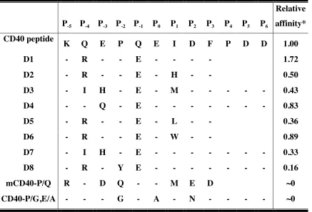
predicted using interaction energies (D4) (Table 2).
Binding affinity measurements
Previously, affinities of various peptides to TRAF6 were determined by ITC (Ye
et al. 2002a). Unfortunately, in our hands, TRAF6 had limited solubility and aggregated
during the ITC run, giving us inconsistent binding results that were, on average, an order
of magnitude better than the published values. We therefore developed a fluorescence
anisotropy assay to determine the binding affinity of the designed peptides (Figure 8 and
Table 3). We synthesized four of the peptides ORBIT predicted using the parameters
described above (D1 to D4, Table 3) and measured their binding affinities. Designed
peptides D3 and D4 were overly hydrophobic, so additional PDD sequence was added to
their C terminus to improve solubility. All affinities reported in Table 3 are relative to the
CD40 peptide of the same length as the peptide tested (9-residue or 12-residue).
Surprisingly, the Kd values we obtained by fluorescence anisotropy were similar to the Kd
values obtained with ITC, and were an order of magnitude better than the Kd value
previously reported using ITC (Ye et al. 2002a) (Table 4). Nevertheless, we are confident
of the relative affinities from our anisotropy measurements because the standard
deviations from multiple experiments are very small (Table 4). Our anisotropy assay was
negative control peptides used were a mouse CD40 peptide with the Pro at P-3 position
mutated to Gln (mCD40-P/Q) and human CD40 peptide with the Pro at P-3 position
mutated to Gly and Glu at P0 position mutated to Ala (CD40-P/G,E/A) (Table 3).
Previously, mCD40-P/Q was shown to have about half the activity of wild-type mouse
CD40 peptide in an NF-κB reporter activity assay, and CD40-P/G,E/A was shown in
vitro to have no affinity for TRAF6 (Pullen et al. 1999; Ye et al. 2002a). In our
anisotropy assay, both show little or no binding to TRAF6 (Figure 8).
Other design trials
In the initial designs, our best peptide (D1) exhibited about a two-fold affinity
enhancement compared to the CD40 peptide sequence. This improvement encouraged us
to try to design a better binder using the knowledge gained from the first four peptides.
We decided to use ORBIT’s bias function , described in the methods section, to favor the
intermolecular interactions between the peptide side chains and the TRAF6 side chains.
Using the parameters for D1, we applied a 2-fold bias and a 4-fold bias to the
intermolecular optimization, which resulted in the two sequences, D5 and D6 (Table 3).
Both are one mutation away from D1, at the P1 position: instead of an Ile at P1, D5 has a
Leu, and D6 has a Trp. Interestingly, the exact same sequences are obtained when the
parameters for D2 are biased 2-fold and 4-fold. We expected D5 to have less affinity for
TRAF6 because Ile has higher beta strand propensity than Leu. The ligand peptide binds
to TRAF6 in an extended beta strand conformation and a Leu instead of Ile should
destabilize the peptide in the beta strand conformation. As predicted, D5 has significantly
indicated that D6, however, might bind tighter. This data was used above in determining
the ORBIT parameters, in which all 20 amino acids were substituted in all positions of
the peptide and binding was observed using membrane binding assays. In their assay,
substituting Trp for Ile resulted in 4-fold better binding to TRAF6. In our anisotropy
assay, D6 affinity was similar to the wild-type sequence and less than D1. This might be
explained by the fact that Pullen and co-workers used a crude colorimetric plate assay
while we used purified proteins in a solution state assay. Also, their Trp mutation is in the
context of the naturally occurring wild-type sequence, while our designed peptide has
two other residues different from the naturally occurring CD40 peptide.
Comparing the D1, D2, D5 and D6 sequences indicates that among the residues
tested at P1, Ile is the most stable. Met, on the other hand, was thought to be destabilizing
because of the large entropic loss upon binding. We therefore synthesized and tested
another peptide (D7) with the same sequence as D3, but with Ile instead of Met at P1. Ile
turned out to be less optimal than Met in the context of D3, resulting in decreased binding
affinity.
Another strategy we tried was to design one of the conserved sequences. When
the peptide residues are classified as core, boundary or surface (Dahiyat and Mayo
1997a), only the Pro located in the P-2 position is classified as core. This suggested that
P–2 is the anchoring position in the TRAF6-peptide interaction, and that it makes a large
contribution to the binding affinity. It has been shown that hydrophobic interactions are
important in affinity while polar interactions are important for specificity (Clackson and
Wells 1995). Taking this into consideration, the P-2 residue, which is the only completely
affinity of the peptide. To determine whether a larger hydrophobic side chain at P-2 would
be allowed, we ran three side chain replacement calculations substituting the Pro to Phe,
Tyr and Trp. There were no significant steric clashes with all three substitutions when the
VDW scale factor for D1 design (0.9) was used. Trp and Phe had high nonpolar
exposure, while the hydroxyl group of Tyr made a favorable hydrogen bond with Asp451
of TRAF6. Thus, we decided to test out a peptide with the same sequence as D1 except
for a Pro to Tyr substitution at P-2 (D8). Unfortunately, the Pro to Tyr mutation decreased
the binding affinity to 9% of D1. This implies that Tyr might be sterically clashing with
TRAF6, which did not show in our side chain replacement calculation because of the
small VDW scale factor used.
Affinity versus avidity
It has been pointed out that the low affinity nature of TRAF-receptor ligand
binding ensures that TRAFs bind to their receptors only when the receptors are trimerized
and active (Ye and Wu 2000). The structural characteristics of the binding site, which
forms a wide and shallow groove, implies a relatively flat binding energy landscape that
allows many different sequences to attach, however with low affinity. In addition, the
peptide ligand assumes an extended beta conformation when fitting into this binding site,
allowing only half of the residues of the peptide to face the protein and be involved in
direct contact (Figure 7) The other side chains are facing away from the protein and thus,
are highly exposed to the solvent, which may decrease their hydrogen bonding and
electrostatic interaction contribution to the binding energy. The fact that the majority of
structure are between backbones and that only one interface residue is fully buried
implies that the binding affinity between the peptide and protein will be nontrivial to
optimize. We have tried to overcome this difficulty by using a biased energy function to
optimize specifically for interactions between the side chains of the peptide and TRAF6,
but this did not result in a high affinity peptide (D5 and D6). It could be that nature has
designed the binding interface of TRAF6 and its receptor to maintain low affinity so that
affinity can be achieved through avidity. The fact that the affinities of naturally occurring
TRAF6 binding domains are all in the micromolar range (Ye et al. 2002a), even though
the sequence diversity is very high in the nonconsensus positions, also supports this
hypothesis.
The dynamic nature of proteins is not represented in our design protocol, which
employs a fixed backbone. Flexible backbones have been successfully used to design
single proteins (Harbury et al. 1998; Larson et al. 2002; Kraemer-Pecore et al. 2003;
Kuhlman et al. 2003). Recently, several groups have also designed protein-binding
peptides using this method (Wollacott and Desjarlais 2001; Sood and Baker 2006). In
these studies, a flexible backbone was necessary to obtain the sequence diversity of
experimentally verified ligands. This indicates that a flexible backbone is desirable for
successful design and specificity prediction. It would be interesting to see whether a
flexible backbone will improve the design effort for the TRAF6 and CD40 peptide
system. On the other hand, some results imply that a flexible backbone is not necessary,
and that allowing side chain flexibility or decreasing the van der Waals radii scale of the
atoms to reduce the effects of using discrete rotamers and a fixed backbone is enough to
our design, which used a large rotamer library with expansion about both χ1 and χ2, and
used 0.9 van der Waals scale factor, should give an optimal sequence, even though
backbone flexibility was not included. Even with a flexible backbone, the affinity
increase for the best designed peptide for the dystrophin protein and Mdm2 were very