Analyte(s) Matrix Aminoglycosides Bovine kidney
Benzimidazoles Beef liver
Benzimidazoles Swine muscle
Beta-agonists Bovine liver
Carbofuran Corn
Chloramphenicol Milk
Chlorsulfuron Milk
Chlorsulon Milk
Clenbuterol Bovine liver
Furazolidone Chicken muscle
Furazolidone Milk
Furazolidone Swine muscle
Ivermectin Fish muscle
Ivermectin Milk
Ivermectin Bovine liver
Moxidectin Bovine tissues
Nicarbazin Animal tissues
Oxolinic acid Catfish
Oxytetracycline Catfish muscle
PCBs Fish
Pesticides Beef fat
Pesticides Catfish muscle
Pesticides Crayfish
Pesticides Fish
Pesticides Fruit, vegetables
Pesticides Milk
Pesticides Oysters
Sulfa drugs Chicken tissues
Sulfadimethoxine Catfish muscle Sulfadimethoxine Catfish, plasma
Sulfonamides Infant formula
Sulfonamides Milk
Sulfonamides Salmon muscle
Sulfonamides Swine muscle
Sulfonamides Eggs
Tetracyclines Milk
with such methods on several levels and should be considered as an alternative when pursuing new ana-lytical methodology. This is especially the case for solid or semi-solid biological materials.
ridges. Sorbent Selection for Solid-Phase Extraction.
Further Reading
Barker SA (1992) Application of matrix solid-phase dispersion (MSPD) to the extraction and subsequent analysis of drug residues in animal tissues. In: Agarwal VK (ed.)Analysis of Antibiotic Residues in Food Prod-ucts of Animal Origin, pp. 119}132. New York: Plenum Press.
Barker SA and Floyd ZE (1996) Matrix solid-phase disper-sion (MSPD): implications for the design of new bonded-phase surface chemistries. In: Pesek JJ, Matyska MT and AbuelaRya RR (eds)Chemically ModiTed Sur-faces: Recent Developments, pp. 66}71. Cambridge, UK: Royal Society of Chemistry.
Barker SA and Long AR (1992) Tissue drug residue extrac-tion and monitoring by matrix solid-phase dispersion (MSPD)-HPLC analysis.Journal of Liquid Chromatog-raphy15: 2071}2089.
Barker SA, Long AR and Hines ME (1993) The disruption and fractionation of biological materials by matrix solid-phase dispersion. Journal of Chromatography 629: 23}34.
Barker SA, Long AR and Short CR (1989) Isolation of drug residues from tissues by solid-phase dispersion.Journal of Chromatography475: 353}361.
Crouch MC and Barker SA (1997) Analysis of toxic wastes in tissues from aquatic species: applications of matrix solid-phase dispersion.Journal of Chromatography774: 287}309.
US Patent C 5 272 094. Issued 21 December (1993) A bonded-phase matrix dispersion and extraction process. Isolation of drugs, and drug residues, from biological specimens, and tissues (Dr Steven A Barker; co-patent applicant, Dr Austin R Long, Louisiana State University).
SOLID-PHASE MICROEXTRACTION
Biomedical Applications
H. Kataoka, Okayama University, Tsushima, Okayama, Japan
H. L. Lord and J. Pawliszyn,
University of Waterloo, Ontario, Canada
Copyright^ 2000 Academic Press
[image:1.568.52.279.83.466.2]is required in many circumstances, such as clinical control for diagnosis and treatment of diseases, dop-ing control, forensic analysis and toxicology. Al-though high efRciency instruments have been developed, most analytical instruments cannot handle the sample matrix directly. Therefore, sample preparation is very important to achieve a practical and reliable method for the analysis of complex ma-trices such as biological samples. In general, over 80% of analysis time is spent on sampling and sample preparation steps such as extraction, concentration and isolation of analytes. However, previous sample preparation techniques, such as liquid}liquid extrac-tion and solid-phase extracextrac-tion, have their problems. These techniques are generally time-consuming and require large volumes of samples and solvents. For example, a long sample preparation time limits the number of samples that can be analysed, and multi-step procedures are prone to loss of analytes. Further-more, the use of a large amount of solvent inSuences trace analysis, and also causes environmental pollution and health concerns. Ideally, sample preparation tech-niques should be fast, easy to use, inexpensive and compatible with a range of analytical instruments.
Solid-phase microextraction (SPME), developed by Pawliszyn and co-workers in 1990, is a new sample preparation technique using a fused-silicaRbre that is coated on the outside with an appropriate stationary phase. The analyte in the sample is directly extracted onto theRbre coating. The method saves preparation time, solvent purchase and disposal costs, and can improve the detection limits. It has been used routine-ly in combination with gas chromatography (GC) and GC/mass spectrometry (GC/MS), and successfully applied to a wide variety of compounds, especially for the extraction of volatile and semi-volatile organic pollutants from water samples. SPME was also intro-duced for direct coupling with high performance liquid chromatography (HPLC) and LC/MS in order to analyse weakly volatile or thermally labile com-pounds not amenable to GC or GC/MS. The SPME/HPLC interface, equipped with a special de-sorption chamber, is utilized for solvent dede-sorption prior to HPLC analysis, instead of thermal desorption in the injection port of the GC. Moreover, a new SPME/HPLC system known as in-tube SPME, was recently developed using an open-tubular fused-silica capillary column as the SPME device in place of the SPMERbre. In-tube SPME is suitable for automa-tion, and automated sample handling procedures not only shorten the total analysis time, but also usually provide better accuracy and precision relative to manual techniques.
In this article, we review SPME techniques coupled with various analytical instruments and the
applications of these techniques to drug analysis. The review consists of two main parts. In the Rrst part, general aspects of SPME techniques are surveyed for Rbre and in-tube SPME methods coupled with vari-ous instruments. In the second part, applications of the SPME methods in drug analysis are considered according to the drug type.
SPME Techniques Coupled with
Various Analytical Instruments
Fibre SPME
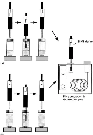
TheRbre SPME device consists of aRbre holder and Rbre assembly with built-inRbre inside the needle. In Rbre SPME, analytes are extracted directly from the sample onto a polymeric stationary phase coated on theRbre. When theRbre is inserted into the sample, the target analytes partition from the sample matrix into the stationary phase until equilibrium is reached. Two types ofRbre SPME techniques can be used to extract analytes: headspace SPME and immersion SPME. In headspace SPME, theRbre is exposed in the headspace of gaseous, liquid or solid samples. In immersion SPME, the Rbre is directly immersed in liquid samples. TheRbre with concentrated analytes is then transferred to an instrument for desorption, followed by separation and quantiRcation. Head-space and immersion SPME techniques can be used in combination with any GC, GC/MS, HPLC and LC/MS system. The process of the Rbre SPME/GC method is shown inFigure 1.
Figure 1 Schematic illustration of headspace and immersion SPME/GC methods. (A) Headspace SPME; (B) direct immersion SPME.
analytes. Fibre SPME techiques in combination with GC or GC/MS are unsuitable for the extraction of less volatile or thermally labile compounds. Thus derivat-ization approaches are frequently used to extract po-lar compounds from biological samples. Four types of derivatization techniques in combination with SPME are implemented. Direct derivatization in the sample matrix is similar to well-established approaches used in solvent extraction. Analytes are extracted by SPME after derivatization in the vial. For in-coating derivat-ization with the Rbre-doping method, simultaneous derivatization and extraction are directly performed in the Rbre coating by a two-step process: (1) dope Rbre with derivatization agent and (2) expose doped
Rbre to sample for extraction. This technique can be used for polar volatile compounds. Another in-coat-ing derivatization technique is performed by the fol-lowing two-step process: (1) dopeRbre to sample for extraction and (2) expose doped Rbre in the head-space of derivatizing agent. For derivatization in the injection port, the analyte extracted by SPME is de-sorbed in a GC injection port and then derivatized with additional reagent.
Figure 2 Schematic of the SPME-HPLC system. (a) Stainless steel (SS) 1/16 inch tee joint; (b) 1/16 inch o.d., 0.02 inch i.d., SS tubing; (c) 1/16 inch o.d. poly(ether ether ketone) (PEEK) tubing (0.02 inch i.d.); (d) two-piece finger-tight PEEK union; (e) PEEK tubing (0.005 inch i.d.) with a one-piece PEEK union. (Reproduced with permission from Pawliszyn J (1997)Solid Phase Microextrac-tion: Theory and Practice. Translated by permission of John Wiley & Sons, Inc. All rights reserved.)
injection port is dependent on the injection depth, injector temperature, and exposure time. A narrow-bore GC injector insert is required to ensure high linearSow and theRbre needs to be exposed immedi-ately after the needle is introduced into the insert. Needle exposure depth should be adjusted to place theRbre in the centre of the hot injector zone. Desorp-tion time is determined by the injector temperature and the linearSow rate around theRbre. The HPLC interface, on the other hand, consists of a six-port injection valve and a special desorption chamber, and requires solvent desorption of the analyte prior to HPLC or LC/MS analysis. A typical SPME/HPLC interface is shown inFigure 2. The desorption cham-ber is placed in the position of the injection loop. After sample extraction, theRbre is inserted into the desorption chamber at the ‘load’ position under am-bient pressure. When the injector is changed to the ‘inject’ position, mobile phase contacts theRbre, de-sorbs the analytes, and delivers them to the HPLC column for separation. Two desorption techniques can be used to remove the analytes from the Rbre: dynamic desorption and static desorption. In dy-namic desorption, the analytes can be removed by a moving stream of mobile phase. When the analytes
are more strongly adsorbed to theRbre, theRbre can be soaked in mobile phase or other strong solvent for a speciRed time by static desorption before injection onto the HPLC column. In each desorption tech-nique, rapid and complete desorption of analytes us-ing minimal solvent is important for optimizus-ing the SPME/HPLC or SPME/LC/MS methods.
In-tube SPME
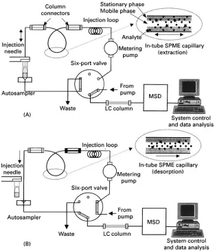
In-tube SPME using an open-tubular capillary col-umn as the SPME device was developed for coupling with HPLC or LC/MS. It is suitable for automation, and can continuously perform extraction, desorption and injection using a standard autosampler. With the in-tube SPME technique, organic compounds in aqueous samples are directly extracted from the sample into the internally coated stationary phase of a capillary column, and then desorbed by introducing a moving stream of mobile phase or static desorption solvent when the analytes are more strongly absorbed to the capillary coating. A schematic diagram of the automated in-tube SPME/LC/MS system is shown in
Figure 3. The capillaries selected have coatings
Figure 3 Schematic of the in-tube SPME/LC/MS system. (A) Load position (extraction phase); (B) injection position (desorption phase). (Reproduced with permission from Kataoka H, Narimatsu S, Lord HL and Pawliszyn J (1999)Analytical Chemistry 71: 4237. Copyright American Chemical Society.)
injection loop and the injection needle of the HPLC autosampler. While the injection syringe repeatedly draws and ejects sample from the vial under com-puter control, the analytes partition from the sample matrix into the stationary phase until equilibrium is reached. Subsequently, the extracted analytes are dir-ectly desorbed from the capillary coating by mobile phaseSow or by aspirating a desorption solvent. The desorbed analytes are transported to the HPLC col-umn for separation, and then detected with UV or a mass selective detector (MSD).
In in-tube SPME, the amount of analyte extracted by the stationary phase of the capillary column de-pends on the polarity of capillary coating, number and volume of draw/eject cycles and the sample pH. A capillary column 50}60 cm long is optimal for extraction. Below this level, extraction efRciency is reduced, and above this level, peak broadening is observed. In general, complete equilibrium extraction is not obtained for any of the analytes, because the analytes are partially desorbed into the mobile phase
during each eject step. The target analytes with higher
K-values need longer equilibration times. Although an increase in number and volume of draw/eject cycles can enhance the extraction efRciency, peak broadening is often observed in this case. The optimal Sow rate of draw/eject cycles is 50}100L min\1. Below this level, extraction requires an inconvenient-ly long time, and above this level, bubbles form on the inside of the capillary and extraction efRciency is reduced. The in-tube SPME technique does not need a special SPME/HPLC interface for desorption of analytes. The analytes extracted onto the capillary coating can be easily desorbed by a moving stream of mobile phase or desorption solvent when the analytes are more strongly adsorbed to the capillary coating. Carryover in the in-tube SPME method is lower or eliminated in comparison with the Rbre-SPME method.
is performed on the outer surface of the Rbre for Rbre-SPME and on the inner surface of the capillary column for in-tube SPME. Therefore, with the in-tube SPME method, it is necessary to prevent plugging of the capillary column andSow lines during extraction, and typically particles must be removed from samples by Rltration before extraction. On the other hand, with the Rbre-SPME method, it is not necessary to remove particles before extraction because they are removed by washing theRbre with water before inser-tion into the desorpinser-tion chamber of the SPME/HPLC interface. Another signiRcant difference between in-tube SPME and manualRbre-SPME/HPLC is the pos-sible decoupling of desorption and injection with the in-tube SPME method. In the Rbre-SPME method, analytes are desorbed during injection as the mobile phase passes over theRbre. On the other hand, in the in-tube SPME method, analytes are desorbed by mo-bile phase or by aspirating a desorption solvent from a second vial, and then transferred to the HPLC column by mobile-phase Sow. The Rbre-SPME/ HPLC method also has the advantage of eliminat-ing the solvent peak from the chromatogram, but peak broadening is sometimes observed because analytes can be slow to desorb from the Rbre. With the in-tube SPME method, peak broadening is not observed because analytes are completely desorbed before injection.
Biomedical Applications
:
Drug
Analysis
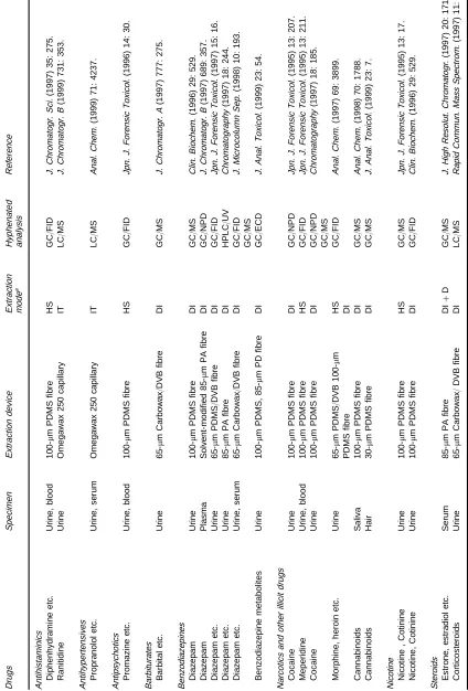
SPME methods applied to the analysis of various abused and therapeutic drugs in biological samples are listed in Table 1, according to the drug type, sample type, extraction device, extraction mode, and analytical technique. The SPME methods using m polydimethylsiloxane (PDMS)Rbres in combina-tion with GC or GC/MS are widely used for the analysis of various drugs. The SPME methods coupled with HPLC or LC/MS are used for the analy-sis of less volatile or thermally labile drugs. For recent reviews of some of these methods for drug analysis see Pawliszyn, Lord and Pawliszyn, Namera et al., Juntinget al., Kataokaet al.and Sporkert and Pragst in the Further Reading section.
Amphetamines and Related Compounds
Yashiki and co-workers developed a simple and rapid method for analysing amphetamine (AM) and meth-amphetamine (MA) in urine and blood samples by headspace SPME and GC/MS-selected ion monitor-ing (SIM). In order to move the analytes into the headspace, the sample was heated at 803C for 20 min under K2CO3 or NaOH alkaline conditions.
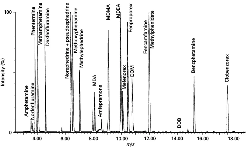
Sub-sequently, a 100-m PDMSRbre was exposed to the headspace for 5 min, and then inserted into the injec-tion port of GC/MS for desorption. The method was twenty times more sensitive than the conventional headspace method. Lord and Pawliszyn optimized several extraction parameters for the analysis of AM and MA in urine samples by headspace SPME/ GC-Same ionization detection (FID). Centini et al. and Battu et al. reported simultaneous analysis of amphetamines and their analogues, such as 3,4-methylenedioxyamphetamine (MDA), 3,4-methyl-enedioxymethamphetamine (MDMA) and 3,4-methy-lenedioxyethylamphetamine (MDEA), in urine sam-ples by headspace SPME using a 100-m PDMSRbre. As shown inFigure 4, a clean total-ion chromatogram is obtained from a urine sample spiked with 100 ng mL\1of each of the 21 central nervous system stimulants and extracted by the headspace SPME method. Koide et al. applied this technique to the analysis of amphetamines in hair samples.
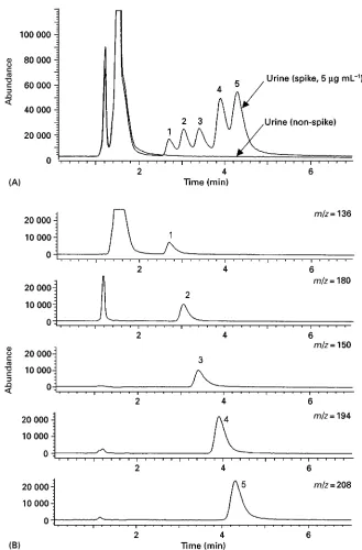
Degel, Penton, Ishii et al., Makino et al. and Myunget al. used the direct immersion technique in order to improve the extraction efRciency and sensi-tivity. The extraction recoveries of AM and MA by the immersion SPME method are several times higher than those by the headspace SPME method. Uglandet al. reported an SPME technique in combination with derivatization. After derivatization with alkylchloro-formate, amphetamines and their methylenedioxy analogues were analysed by immersion-Rbre SPME/ GC-nitrogen-phosphorus detection (NPD) or GC/MS. Kataokaet al. developed an in-tube SPME/LC/MS method for the analysis of amphetamines and their methylenedioxy analogues using Omegawax (Supelco, Bellefonte, PA, USA) capillary as the extraction device. As shown inFigure 5, these drugs spiked into urine samples were selectively analysed without inter-ference peaks by SIM-mode detection.
Anaesthetics
Figure 4 Total-ion chromatogram of a urine sample extract spiked with 21 central nervous system stimulants at 1000g L\1. SPME
conditions: fibre, 100m PDMS; extraction, at 803C headspace for 10 min with stirring; desorption, exposure for 10 min in GC injection port. GC/MS conditions: column, PTA-5 (30 m;0.32 mm i.d., 0.5m film thickness); injector, splitless mode at 2003C; split opening time, 2 min; oven temperature, programme from 60 to 1203C at 303C min\1, then to 2103C at 53C min\1, and finally to 2803C at
303C min\1and hold at 2803C for 5 min; transfer line and detector temperature, 2803C; helium flow-rate, 1.3 mL min\1, ionization,
70 eV. (Reproduced with permission from Battu C, Marquet P, Fauconnet AL, Lacassie E and Lacha(tre G (1998) Journal of Chromatographic Science 36: 1, by permission of Preston Publications, A Division of Preston Industries, Inc.)
Furthermore, Kumazawaet al. reported a method for analysis of phencyclidine in urine and whole blood by headspace-SPME and GC with a surface ionization detector (SID). Watanabeet al. developed a simple method for analysis ofRve local anaesthetics in blood samples by headspace SPME using a 100-m PDMSRbre and GC/MS-SIM. Kosteret al. reported direct immersion-SPME methods coupled with GC-FID and HPLC-UV for the determination of lidocaine in urine samples. Desorption of the PDMS Rbre in HPLC is more complicated than the desorption in GC, because it is dependent on the composition of the mobile phase or the desorption solvent.
Antidepressants
Kumazawa et al. developed a simple headspace-SPME method for the analysis of four tricyclic antide-pressants in urine and whole-blood samples. These drugs were extracted with a 100-m PDMSRbre after heating at 1003C in the presence of a NaOH solution. Namera et al. reported a headspace-SPME/GC-MS method for the analysis of three tetracyclic antide-pressants in whole-blood samples, and its application to a medicolegal case of setiptiline intoxication. Ulrich and Martens developed a direct immersion-SPME method for the simultaneous analysis of ten antidepressant drugs and metabolites in plasma
sam-ples, and applied the method to toxicological analysis after the accidental or suicidal intake of higher doses. The sample was extracted with a 100-m PDMSRbre for 10 min and the Rbre was exposed in the GC injection port at 2603C for 1 min after washing in 50% methanol and subsequent drying at room tem-perature. As shown inFigure 7, these drugs in plasma samples were selectively analysed by NPD without interference peaks. However, the recoveries of antide-pressants from plasma samples were very low due to the high protein binding of these drugs. The limits of quantiRcation for these drugs in plasma samples were 90}200 ng mL\1. The sensitivity can be considerably improved by increasing the extraction time and dilu-tion of plasma samples with water.
Benzodiazepines
Figure 5 Total ion and SIM chromatograms obtained from urine samples spiked with amphetamines by in-tube SPME/LC/MS. (A) Total ion chromatograms obtained from urine and spiked urine samples; (B) SIM chromatograms obtained from spiked urine sample. Urine sample (10L) was diluted ten times with water and used for analysis after filtration. Stimulants were spiked at a concentration of 5 mg mL\1urine. LC/MS conditions: column, Supelcosil LC-CN (3.3 cm;4.6 mm i.d., 3m particle size); column
temperature, 253C; mobile phase, acetonitrile/50 mMammonium acetate (15 : 85); flow-rate, 0.4 mL min\1; fragmentor voltage, 40 V;
ionization mode, positive ESI; SIM ion,m/z"136 (AM), 150 (MA), 180 (MDA), 194 (MDMA) and 208 (MDEA). In-tube SPME conditions: capillary, Omegawax 250 (60 cm;0.25 mm i.d., 0.25m film thickness); sample pH, 8.5; draw/eject cycles, 15; draw/eject volume, 35L; draw/eject flow-rate, 100L min\1, desorption solvent, mobile phase. Peaks: 1, AM; 2, MDA; 3, MA; 4, MDMA; and 5,
MDEA. (Reproduced with permission from Kataoka H, Lord HL and Pawliszyn J (2000)Journal of Analytical Toxicology 24: 263, by permission of Preston Publications, A Division of Preston Industries, Inc.)
solvent-modiRed SPME technique is limited by the incompatibility of the SPME coatings with most or-ganic solvents. Luoet al. developed a direct immer-sion-SPME method for the simultaneous analysis of
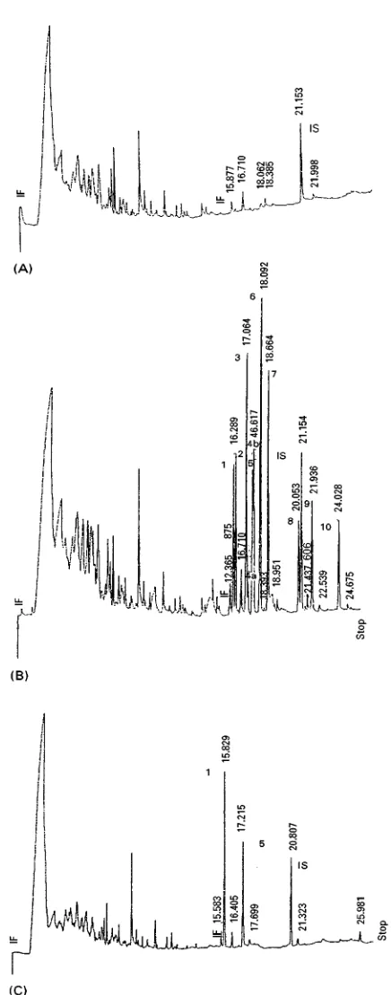
Figure 6 Capillary GC of ten local anaesthetics extracted from human whole blood by use of direct immersion-SPME. (A) The authentic drugs (50 ng each on column); (B) a drug extract at pH 7 without salt; (C) a drug extract at pH 7 in the presence of 0.5 g NaCl; (D) a blank extract at pH 7 in the presence of 0.5 g NaCl. The mixture of ten drugs (5g each) was added to 1 mL of human whole blood. SPME conditions: fibre, 100m PDMS; extraction, at room temperature for 40 min with stirring; desorption, 1 min exposure in GC injection port. GC conditions: column, DB-17 (30 m;0.25 mm i.d., 0.25m film thickness); column temperature, initially hold at 1003C for 1 min and increase to 2903C at 103C min\1; injector and detector temperatures, 2503C; He carrier gas flow-rate, 3 mL min\1;
injection, splitless; detector, FID. Peaks: 1, ethyl aminobenzoate; 2, prilocaine; 3, lidocaine; 4, procaine; 5, mepivacaine; 6, tetracaine; 7, bupivacaine; 8, p-(butylamino)benzoic acid-2-(diethylamino)ethyl ester; 9, benoximate; and 10, dibucaine. (Reproduced with permission from Kumazawa T, Sato K, Seno H, Ishii A and Suzuki O (1996)Chromatographia 43: 59.)
pH 7 and sampling at 453C with agitation, and ana-lysed by GC-MS.
Guan et al. analysed the metabolites of benzo-diazepines from acid-hydrolysed urine samples using a direct immersion-SPME method in combination with GC-electron capture detection (ECD). The de-tection limits were 2}20 ng mL\1for most drugs tes-ted. Jinno and Taniguchi, however, developed an SPME method coupled with HPLC for the analysis of six benzodiazepines in human urine samples.
Sensitiv-ity may be increased by the combination of saturated salt and weakly alkaline conditions in the extraction matrix. As shown inFigure 8, a 65-m PARbre was found to be more efRcient in the extraction of ben-zodiazepines than a 100-m PDMSRbre.
Narcotics and Other Illicit Drugs
Figure 7 Typical SPME-GLC-NPD chromatograms obtained from (A) blank plasma with internal standard, (B) plasma spiked with ten antidepressant drugs and metabolites, each 375 ng mL\1, and (C) a sample of a patient after suicidal intoxication with amitriptyline
(amitriptyline, 766 ng mL\1; nortriptyline, 489 ng mL\1. SPME conditions: fibre, 100-m PDMS; extraction, shaking at 700 rpm for
10 min at 223C; desorption, 1 min exposure in GC injection port. GC conditions: column, DB-1 (30 m;0.32 mm i.d., 0.25m film thickness); column temperature, programme from 1403C to 2203C at 203C min\1and from 2203C to 2703C at 23C min\1; injector and
detector temperatures, 2603C and 3003C, respectively; N2carrier gas flow-rate, 0.7 mL min\1; injection, splitless; detector, NPD.
Figure 8 Chromatograms of extracted drugs with (A) 100-m PDMS and (B) 85-m PA. SPME conditions: extraction, stirring at 840 rpm for 3 h at 603C; desorption, 30 min exposure in desorption chamber. HPLC conditions: column, Siperiorex ODS (250 mm;1.5 mm i.d.); mobile phase, acetonitrile/water; flow-rate, 100L min\1; detection, UV at 220 nm. Peaks: 1, nitrazepam; 2,
flunitrazepam; 3, fludiazepam; 4, diazepam; 5, clotiazepam; and 6, medazepam. (Reproduced with permission from Jinno K and Taniguchi M (1997)Chromatography 18: 244.)
samples. Recovery of cocaine by this technique using a 100-m PDMS Rbre was 20%, and the detection limit was about 12 ng mL\1. Lord and Pawliszyn applied the SPME/GC-FID method developed for amphetamines to the analysis of meperidine, codeine, methadone, morphine and her-oin in spiked urine samples. Furthermore, Hallet al. applied an immersion SPME technique to the analysis of four cannabinoids in human saliva. These drugs were extracted with a 100-m PDMSRbre and ana-lysed in the range from 5 to 500 ng mL\1by GC/MS. Using this method, 9-tetrahydrocannabinol (9 -THA) was detected in a saliva sample collected 30 min after the subject had smoked marijuana (Figure 9).
Strano-Rossi and Chiarotti reported an immers-ion SPME method using a 30-m PDMS Rbre in combination with GC/MS for the analysis of canna-binoids in alkaline hydrolysed hair samples. The method is also applied to the analysis of other drugs such as methadone, cocaine and cocaethylene in hair samples.
Other Drugs
Figure 9 Chromatograms after performing SPME on human saliva samples prior to and after marijuana smoking. (A) SIM chromatogram of saliva sample before marijuana smoking; (B) total ion chromatogram of saliva sample after marijuana smoking; (C) SIM chromatogram of saliva sample after marijuana smoking. SPME conditions: fibre, 100m PDMS; extraction, immersion for 10 min with stirring; desorption, exposure for 12 min in GC injection port. GC/MS conditions: column, DB-5ms (30 m;0.25 mm i.d., 0.5m film thickness); oven temperature, initially hold at 503C for 0.2 min and increase to 2803C at 153C min\1, and finally hold at
2803C for 2 min; transfer line temperature, 2803C; detection, ion trap (electron ionization mode); SIM ion,9-THC (m/z"231, 299,
314). (Reproduced with permission from Hall BJ, Satterfield-Doerr M, Parikh AR and Brodbelt JS (1998)Analytical Chemistry 70: 1788. Copyright American Chemical Society.)
urine samples. A 65-m Carbowax/DVB Rbre was suitable for the extraction of these drugs. The detec-tion limits reached 1 ng mL\1. Okeyoet al. developed a straightforward method for performing derivatizing reactions of Rve steroidsin situin SPMERbres. After extraction of drugs from serum samples by direct im-mersion SPME, the drugs extracted on 85-m PARbre were derivatized in the headspace of the silylating reagent bis(trimethylsilyl)triSuoro-acetamide, and then analysed by GC/MS. With derivatization, SPME and GC analysis can be easily extended to the analysis of semi- and non-volatile compounds.
Volmer and Hui developed a SPME/LC/MS method for isolating and analysing eleven cor-ticosteroids and two steroid conjugates from urine samples. After extraction in the vial by direct
immersion SPME using 65-m Carbowax/DVB Rbre, the drugs extracted in theRbre were desorbed in the desorption chamber of the SPME/HPLC interface, and then analysed by electrospray LC/MS. As shown in Figure 10, several corticosteroids and steroid sulfates spiked in urine samples were selectively analysed, although a minor peak was observed in the blank control urine in the SIM trace for cortisone.
Figure 10 SPME/LC/MS analysis of several corticosteroids and steroid conjugates by time-scheduled SIM. The original urine sample was spiked at the 20 mg mL\1level. (A) Blank control urine; (B) spiked urine. LC/MS conditions: column, YMC ODS-AQ (50
mm;4.0 mm i.d., 3m particle size); column temperature, 253C; mobile phase, A"100 mMammonium acetate and B" acetonit-rile/methanol (50 : 50:#100 mM ammonium acetate), A : B was gradient programmed from 60 : 40 to 20 : 80 in 10 min; flow-rate, 1 mL min\1; fragmentor voltage, 40 V; ionization mode, negative ESI. SPME conditions: fibre, 65m carbowax/DVB; sample pH, 8.5;
extraction, immersion for 15 min with stirring; desorption, methanol/water (50 : 50) for 5 min. Peaks: 1, estriol-3-sulfate; 2, cortisone; 3, fludrocortisone; 4, estrone-3-sulfate; 5,6-methylprednisolone; 6, budesonide (epimer B); 7, budesonide (epimer A); IS"internal standard (niflumic acid) at 20g mL\1. (Reproduced with permission from Volmer DA and Hui JPM (1997)Rapid Communications in
Mass Spectrometry 11: 1926. Copyright John Wiley & Sons Limited.)
methanol, and then analysed by electrospray LC/MS. Using this technique, nine beta-blockers and metab-olites in urine and serum samples were also analysed. These methods were simple, rapid, selective and sen-sitive, and directly applied to urine samples and serum samples after ultraRltration. Propranolol (PL) and its metabolites were successfully detected in the serum sample of a patient administrated PL (see
Figure 11).
Prospective of SPME in Biomedical
Analysis
Figure 11 Total ion and SIM chromatograms obtained from standard propranolol and its metabolites, and a clinical serum sample by in-tube SPME/LC/MS. (A) Standard solution containing 200 ng mL\1propranolol (PL), 50 ng mL\14-hydroxypropranolol (4-OH-PL)
and 7-hydroxypropranolol (7-OH-PL), 20 ng mL\15-hydroxypropranolol (5-OH-PL) andN-desisopropylpropranolol (NDP). (B) Clinical
serum sample (100L). Serum sample was diluted five times with 1%acetic acid and used for analysis after ultrafiltration. LC/MS conditions: column, Hypersil BDS C18(5.0 cm;2.1 mm i.d., 3m particle size); column temperature, 253C; mobile phase,
acetonit-rile/methanol/water/acetic acid (15 : 15 : 70 : 1); flow-rate, programme from 0.25 to 0.45 mL min\1for 20 min run; fragmentor voltage,
70 V; ionization mode, positive ESI; SIM ion,m/z"218 (NDP), 276 (hydroxypropranolols) and 260 (PL). In-tube SPME conditions: capillary, Omegawax 250 (60 cm;0.25 mm i.d., 0.25m film thickness); sample pH, 8.5; draw/eject cycles, 15; draw/eject volume, 35L; draw/eject flow-rate, 100L min\1, desorption solvent, mobile phase. Peaks: 1, 5-OH-PL; 2, 4-OH-PL; 3, 7-OH-PL; 4, NDP; and
5, PL. (Reproduced with permission from Kataoka H, Narimatsu S, Lord HL and Pawliszyn J (1999)Journal of Analytical Chemistry 71: 4237. Copyright American Chemical Society.)
and hair. The afRnity of the Rbre coating for an analyte is the most important factor in SPME. As shown in Table 1,Rbre coatings of different polarity and thickness were selected for each drug. Most drugs
The extraction of analytes is performed on the outer surface of the Rbre for Rbre SPME and in the inner surface of the capillary for in-tube SPME. Commer-cially available SPME Rbres for drug analysis are limited, by GC capillary columns with a vast array of stationary phases are commercially available for in-tube SPME. HeadspaceRbre SPME is suitable for the extraction of drugs in gaseous, liquid and solid sam-ples, because of the avoidance of contact with an aggressive matrix incompatible with theRbre. Direct immersion Rbre SPME can be used to extract drugs from clear and cloudy liquid samples, however, in-tube SPME is limited to the extraction of clear liquid samples. The headspace SPME technique, therefore, is suitable for direct extraction from whole blood samples, while immersion Rbre SPME or in-tube SPME methods require deproteinization or ultraR l-tration of these samples prior to extraction. As men-tioned above, the extraction efRciency ofRbre SPME depends on extraction time, agitation, heating, sample pH and salt concentration. For in-tube SPME, number, volume and speed of draw/eject cycles, and sample pH are important factors for efRcient extrac-tion. On the other hand, the desorption of analyte from a Rbre or capillary coating depends on the temperature of the injection port and exposure time in combination with GC or GC/MS, or component and volume of solvent when used in combination with HPLC or LC/MS. Therefore, these SPME para-meters should be optimized when developing a new SPME method for drug analysis.
With further development of new coating mater-ials, such as afRnity coatings for target drugs and chiral coatings for optically active drugs, the further development of derivatization methods, further coup-ling with different analytical instruments, such as capillary electrophoresis, and improvement of the extraction and desorption conditions, the SPME tech-nique is expected to be widely applied in the future for highly efRcient extraction of drugs from various biological samples.
See also: II/Chromatography: Gas: Derivatization. Extraction: Solid-Phase Microextraction.
Further Reading
Battu C, Marquet P, Fanconnet AL, Lacassie E and Lachatre G (1998) Screening procedure of 21
ampheta-metry.Journal of Chromatographic Science36: 1}7. Degal F (1996) Comparison of new solid-phase extraction
methods for chromatographic identiRcation of drugs in clinical toxicological analysis.Clinical Biochemistry29: 529}540.
Eisert R and Levsen K (1996) Solid-phase microextraction coupled to gas chromatography: a new method for the analysis of organics in water. Journal of Chromato-graphy A733: 143}157.
Junting L, Peng C and Suzuki O (1998) Solid-phase micro-extraction (SPME) of drugs and poisons from biological samples.Forensic Science International97: 93}100. Kataoka H, Narimatsu S, Lord HL and Pawliszyn J (1999)
Development of on-line in-tube solid-phase microex-traction/LC/MS system.Chromatography20: 237}246. Kroll C and Borchert HH (1998) Solid phase microextrac-tion (SPME) for sample preparamicroextrac-tion during drug meta-bolism studies.Pharmazie53: 172}177.
Lord HL and Pawliszyn J (1998) Recent advances in solid-phase microextraction.LC-GCS41}S46.
Namera A, Yashiki M, Kojima T and Fukunaga N (1998) Solid phase microextraction in forensic toxicology. Japanese Journal of Forensic Toxicology16: 1}15. Pawliszyn J (1995) New directions in sample preparation
for analysis of organic compounds.Trends in Analytical Chemistry14: 113}122.
Pawliszyn J (1997)Solid Phase Microextraction: Theory and Practice. New York: John Wiley.
Pawliszyn J (1999)Applications of Solid Phase Microex-traction. Cambridge, UK: The Royal Society of Chemistry.
Penton ZE (1997) Sample preparation for gas chromato-graphy with solid-phase extraction and solid-phase microextraction. Advances in Chromatography 37: 205}236.
Sporkert F and Pragst F (2000) Use of headspace solid-phase micro-extraction (HS-SPME) in hair analysis for organic compounds.Forensic Science International107: 129}148.
Ulrich S and Martens J (1997) Solid-phase microextraction with capillary gas}liquid chromatography and nitro-gen}phosphorus selective detection for the assay of anti-depressant drugs in human plasma. Journal of Chromatography B: Biomedical Applications 69: 217}234.
Volmer DA and Hui JPM (1997) Rapid determination of corticosteroids in urine by combined solid-phase micro-extraction/liquid chromatography/mass spectrometry. Rapid Communications in Mass Spectrometry 11: 1926}1934.