The effect of a degraded core on the mechanical behaviour of
tissue-engineered cartilage constructs: a poroelastic finite element analysis
D.J. Kelly & P.J. Prendergast
Centre for Bioengineering, Department of Mechanical Engineering, Trinity College, Dublin, Ireland.
Address correspondence to: Prof. Patrick J Prendergast Centre for Bioengineering
Department of Mechanical Engineering Parsons Building
Trinity College Dublin 2 Ireland
Tel: +353.1.6081383
Fax: +353.1.6795554
Email: [email protected]
Submitted to: Medical and Biological Engineering and Computing
First submitted: December 2002
Abstract
The structure and functionality of tissue-engineered cartilage is determined by the tissue culture conditions and mechanical conditioning during growth. The quality of tissue-engineered cartilage may be evaluated using tests such as the confined compression test. Tissue-engineered cartilage constructs usually consist of an outer layer of cartilage and an inner core of either undeveloped cartilage or degrading scaffold material. In this paper, a biphasic poroelastic finite element model is used to demonstrate how such a core influences the reaction force vs. time curve obtained from a confined compression test. The finite element model predicts that higher volumes of degraded scaffold in the inner reduces the aggregate modulus calculated from the confined compression test and raises the estimate of tissue’s permeability. The predicted aggregate modulus reduces from 0.135 MPa for a homogenous construct, to 0.068 MPa for a construct that is only 70% cartilaginous. We find that biphasic poroelastic finite modelling should be used in preference to a one-dimensional model which assumes homogeniety in estimating the properties of tissue-engineered cartilage.
1. Introduction
Cartilage tissue has limited reparative capabilities, and this has made it a candidate for tissue-engineered solutions. An important step in the development of engineered tissues is to quantify their biomechanical functionality. The problem with testing tissue-engineered cartilage is that in vitro culture conditions can lead to cartilage formation only in an outer layer of the scaffold (Vunjak-Novakovic et al, 1999; Pei et al., 2002), with the center of the scaffold presumably consisting of either undeveloped
tissue, or degrading polymer and dead cells, see Fig. 1.
Normal articular cartilage has been tested in many loading modes: uni-axial tension (Akizuki et al., 1986; Woo et al., 1976), confined compression (Mow et al., 1980; Korhonen et al., 2002; Bursac et al., 1999), unconfined compression (Korhonen et al., 2002; Bursac et al., 1999), indentation (Korhonen et al., 2002; Elmore et al.,
1963; Suh and Bai, 1997) and torsion (Hayes et al., 1971). The tissue has been found to be visceoelastic (Hayes et al., 1971; Woo et al., 1980) anisotropic and inhomogeneous (Woo et al., 1976). The most common mechanical test of tissue-engineered cartilage has been the confined compression test (Vunjak-Novakovic et al., 1999; Pei et al., 2002; Ma et al., 1995, Ma and Langer, 1999; Davisson et al.,
2002, Mauck et al., 2002). This test typically consists of applying a ramp displacement to a radially confined plug of cartilage and holding the displacement for a period of time. Under this loading condition the reaction force increases to a maximum and then relaxes to an equilibrium value. The rise and relaxation of the reaction force is measured during the test. When the force equilibrates, the aggregate modulus (denoted Ha) of the tissue can be determined as the equilibrium reaction
equilibrium reaction force after each loading increment to obtain an equilibrium stress-strain curve – the slope of this curve gives Ha. When the aggregate modulus of the tissue is known an estimate of the permeability (ko) can be found by varying ko to fit the analytical prediction of a biphasic model to the experimentally obtained reaction force vs. time curve – this is the procedure followed by Ma et al (1995). Testing of the tissue in unconfined compression (a test between two platens) yields a value for the Young’s Modulus (Es) of the tissue. Knowing both the aggregate
modulus (confined compression test) and the Young’s modulus (unconfined compression) allows for a calculation of the Poisson’s ratio (vs) using the relationship:
) 2 1 )( 1 ( ) 1 ( s s s s a v v v E H − + − =
This method of determining Poisson’s Ratio (vs) has been confirmed by comparing
with values obtained using optical techniques (Korhonen et al., 2002). However, using this method to determine the mechanical properties of tissue-engineered cartilage implicitly assumes the tissue construct to be homogenous, which is commonly not the case (Fig. 1).
In this paper, finite element modeling of the mechanical behavior of tissue-engineered cartilage constructs with degraded non-cartilaginous cores is carried out. The extent of the error in the determination of cartilage tissue properties that would be obtained if the tissue-engineered construct assumed to be homogeneous, as is often done, is then quantified.
2. Methods
2.1 Finite element model
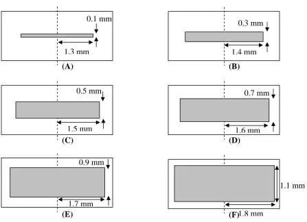
piece of tissue-engineered cartilage, while another six consisted of tissue-engineered cartilage with a degraded scaffold centre, see Fig. 2 (a)-(f). Boundary conditions to simulate the confined compression test were applied, see Fig. 3. The construct is 4 mm in diameter and 1.5 mm high. The lower surface was restrained axially and the outer periphery was restrained radially. The pore pressure on the upper surface was set to zero. A ramp displacement of 0.15 mm (εo = 10% strain) was applied to the specimen at a strain rate of 0.03 µm/s for a ramp time (to) of 5000 s. This
displacement is then held for a further 5000 s. The cartilage is modeled as a biphasic poroelastic material with strain dependent permeability:
k = koexp[Mε]
where ε is the dilation of the solid phase, ko the intrinsic permeability and M is a
material parameter which describes the degree to which the permeability decreases with increasing strain. All elements were modelled as biphasic using the poroelastic theory, implemented in DIANA (TNO, Delft, The Netherlands), see DIANA online user’s manual – release 7.2. Strain dependent permeability was implemented using the porosity dependent permeability option in DIANA.
To assess the effect of the constitutive model for the degraded scaffold core, it is modelled in two ways:
(i) as a biphasic poroelastic material with strain-independent permeability, (ii) as a solid linear elastic material.
The Young’s modulus (Es), Poisson’s ratio (vs), permeability (ko) and porosity (n) for
2.2 Calculation of aggregate properties from finite element results
Using the predictions of the finite element model, the aggregate modulus (Ha) of the
tissue-engineered construct is determined by dividing the predicted stress at the end of the relaxation period by the applied strain.
A common technique for estimating the permeability of tissue-engineered cartilage is to fit the force vs. time curve to the biphasic equations for soft tissues in confined compression, as written by Mow et al (1980). For a slow compression rate
problem, defined by the inequality
k H h t a 2
0 >> , the stress rise (0 < t < to) due to the
application of a ramp displacement is given by the expression (Mow et al., 1984, 1990): ⎟ ⎟ ⎠ ⎞ ⎜ ⎜ ⎝ ⎛ + + = − ... 3 1 ) ( 0 0 0 0 2 0 o c t t M a a e t k H h t t H
t ε ε
σ , (1)
while the stress response during the initial period of relaxation (t >to) is given by:
... 1 ) exp( 2 ) ( 0 0 0 2 0 + ⎟⎟ ⎠ ⎞ ⎜⎜ ⎝ ⎛ − − = = t t M t k H h H t o a o a t t c c ε π ε σ
σ , (2)
where the intrinsic permeability k is defined as
[
o]
o M
k
t being the time and h the thickness of the sample. This model incorporates
strain-dependent permeability (equation 3). All the parameters, except the permeability ko
and the material constant M, can be determined from the confined compression test. The unknown parameters can be determined by systematically varying their values until the analytical reaction force vs. time curve best fits the data from the finite element model. In this case the simulated experimental data from 3000 to 5000 s is fitted to the compressive stress prediction of equation (1) by varying the value of the permeability k0 and material constant M to minimize the sum of the squares of the
differences between the simulated experimental data and the biphasic prediction.
3. Results
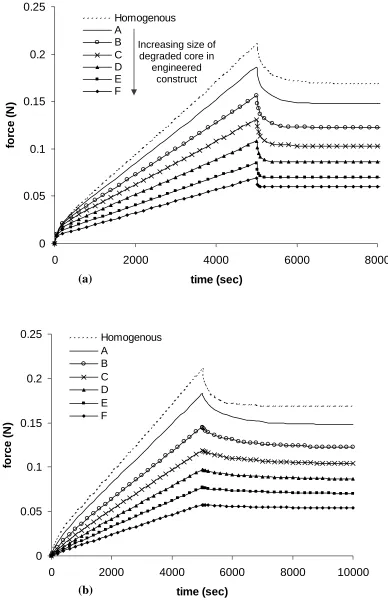
The force vs. time curves computed from the finite element models show that increasing the size of the non-cartilage core region in the tissue-engineered construct reduces the peak reaction force and equilibrium force. This is true when the non-cartilage core region is modeled as biphasic [Fig. 4 (a)] and when it is modeled as linear elastic [Fig. 4(b)]. The predicted peak force is slightly lower when the core is modelled as a linear elastic material compared to a biphasic material; however the differences in the predicted equilibrium forces are quite small, as would be expected.
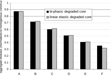
entirely of cartilage would be 50% lower than the modulus of the actual cartilage tissue within the construct.
To estimate the permeability that would be obtained if the constructs were assumed to be homogenous, equation (1) is fitted to the curves of Fig. 4 (a) and Fig. 4 (b) using the method detailed in Section 2.2. Different results are obtained depending on the constitutive model used for the non-cartilage core. When the non-cartilage core is modelled as biphasic, it is predicted that the permeability of constructs A, B and C is not very different from the permeability of the homogenous construct, but that further increasing the size of the non-cartilage region within the construct (constructs D, E and F) results in an increase in the predicted permeability (Fig. 6a). The predicted value for the material constant M is seen to decrease as the size of the degraded non-cartilage core is increased (Fig. 6b).
When the non-cartilage core is modeled as linear elastic, the predicted permeability increases as the size of the degraded non-cartilage core is increases (Fig. 6a). However no significant differences are observed in the predicted value of the material constant M as the size of the degraded non-cartilage core is changed (Fig. 6b).
Discussion
lead to an underestimate of the elastic modulus of cartilage component is within the construct (Fig. 5). Such a result may lead to the conclusion that the engineered tissue is of poorer quality than is actually the case. For example, treatment of engineered cartilage constructs with IGF-I during the culture period has been shown to promote the synthesis of collagen type II rather than type I (Pei et al., 2002); however the aggregate moduli reported for these constructs was low. The results presented in this paper indicate that this is not an intrinsic property of the cartilage but rather results from the structural inhomogeneity of the tissue-engineered construct as a whole.
Similarly the permeability of tissue-engineered cartilage is usually quantified by fitting the force vs. time curve to a solution of a biphasic constitutive model that assumes the tissue to be homogenous. The results presented in this paper (Fig. 6) show that such a test may overestimate the permeability of engineered cartilage component of the tissue-engineered cartilage constructs. We propose that a more appropriate method to determine the permeability of the tissue is to use a finite element based technique that takes account of any inhomogeneity in the construct. Such homogeneity can be readily determined using histological techniques (Fig. 1). The biphasic model used here to estimate the permeability of the constructs was also limited to slow rate of compression experiments. Experiments with high strain rates should implement finite deformation biphasic theory to estimate the permeability of tissue-engineered cartilage (Kwan et al., 1990; Holmes and Mow, 1990), or use a hyperelastic constitutive model for the solid phase (Almedia and Spilker, 1998).
Acknowledgements
References
Akizuki, S., Mow, V.C., Muller, F., Pita, C.J., Howell, D.S., Manicourt, D.H., 1986. Tensile properties of human knee joint cartilage: I. Infleunce of ionic conditions, weight bearing, and fibrillation on the tensile modulus. Journal of Orthopaedic Research 4, 379-392.
Almedia, E. S., Spilker, R. L., 1998. Finite element formulations for hyperelastic transversely isotropic biphasic soft tissues. Comput. Methods Appl. Mech. Engrg. 151, 513-538.
Bursac, P.M., Obitz, T.B., Eisenberg, S.R., Stamenovic, D., 1999. Confined and unconfined stress relexation of cartilage: appropriateness of a transversely isotropic analysis. Journal of Biomechanics 32, 1125-1130.
Davisson, T., Kunig, S. Chen, A., Sah, R., Ratcliffe, A., 2002. The effects of
perfusion and compression on modulation of tissue-engineered cartilage. Procs 48th Ortho Res Soc, Dallas, Texas, poster no. 0488.
Elmore, S.M., Sokoloff, L. Norris, G., Carmeci, P., 1963. Nature of ‘imperfect’ elasticity of articular cartilage. Journal of Applied Physiology 18, 393-396. Hayes, W.C., Mockros, L.F., 1971. Visceoelastic properties of human articular
cartilage. Journal of Applied Physiology 31, 562-568.
Holmes, M. H., Mow V. C., 1990. The nonlinear characteristics of soft gels and hydrated connective tissues in ultrafiltration. Journal of Biomechanics 23, 1145-1156.
Korhonen, R.K., Laasanen, M.S., Toyras, J., Rieppo, J., Hirvonen, J., Helminen, H.J., Jurvelin, J.S., 2002. Comparison of the equilibrium response of articular cartilage in unconfined compression, confined compression and indentation. Journal of Biomechanics 35, 903-909.
Kwan, M. K., Lai, W. M., Mow, V. C., 1990. A finite deformation theory for cartilage and other soft hydrated connective tissues – I. Equilibruim results. Journal of Biomechanics 23, 145-155.
Ma, P.X., Schloo, B., Mooney, D., Langer R., 1995. Development of biomechanical properties and morphogenesis of in vitro tissue-engineered cartilage. Journal of Biomedical Materials Research 29, 1587-1595.
Mauck, R.L., Seyhan, S.L., Jamieson, K.V., Nicoll, S.B., Ateshian, G.A., Hung, C.T., 2002. Synergistic effects of growth factors and dynamic loading for cartilage tissue engineering. Procs 48th Ortho Res Soc, Dallas, Texas, paper no. 0213.
Mow, V.C., Holmes, M. H., Lai, W. M., 1984. Fluid transport and mechanical properties of articular cartilage: a review. Journal of Biomechanics 17, 377-394. Mow, V. C., Hou, J. S., Owens, J. M., Ratcliffe, A., 1990. Biphasic and quasilinear
viscoelastic theories for hydrated soft tissues. In: Biomechanics of diarthrodial joints, Vol. 1, 215-260. Springer-Verlag, New York.
Mow, V.C., Kuei, S.C., Lai, W.M., Armstrong, C.G., 1980. Biphasic creep and stress relexation of articular cartilage in compression: Theory and experiments. Journal of Biomechanical Engineering 102, 73-84.
Pei, M., Seidel, J., Vunjak-Novakovic, G., Freed, L.E., 2002. Differential effects of growth factors (TGF Beta-1, FGF-2, IGF-I) on engineered cartilage cellularity, structure and function. Procs 48th Ortho Res Soc, Dallas, Texas, poster no. 0484. Suh, J.-K., Bai, S., 1997. Biphasic poroviscoelastic behaviour of articular cartilage in
creep indentation test. Transanctions 43rd Annual Meeting of the Orthopaedic Research Society, San Francisco, CA, 22, p.823.
Vunjak-Novakovic, G., Martin, I., Obradovic, B., Treppo, S., Grodzinsky, A.J., Langer, R., Freed, L.E., 1999. Bioreactor cultivation conditions modulate the composition and mechanical properties of tissue-engineered cartilage. Journal of Orthopaedic Research 17, 130-138.
Woo, S.L-Y., Akeson, W.H., Jemmott, G.F., 1976. Measurements of
nonhomogeneous, directional mechanical properties of articular cartilage in tension. Journal of Biomechanics 9, 785-791.
Tables
Cartilage Degraded Scaffold
(biphasic)
Degraded Scaffold (solid) Young’s modulus
(MPa) 0.1 0.001 0.001
Permeability
(mm4/Ns) 1e-2 10 -
Poisson’s ratio 0.3 0.49 0.49
Porosity 0.8 0.99 -
List of figures
Fig. 1. Cross-section of an inhomogenous tissue-engineered cartilage construct, adapted from Pei et al [16].
Fig. 2. Illustrations of the inhomogenous constructs showing the sizes of degraded polymer in each.
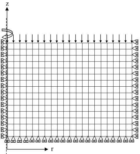
Fig. 3. Axi-symmetric finite element mesh of the construct, illustrating the loads and boundary conditions applied to the model.
Fig. 4. Predicted force against time curves for the confined compression of homogeneous and inhomogeneous cartilage constructs. (a) Non-cartilage region modelled as a biphasic material. (b) Non-cartilage modelled as a solid material. (For keys see Fig. 2)
Figures
1.1 mm 1.5 mm
0.9 mm
0.7 mm 0.5 mm
0.3 mm
1.4 mm 0.1 mm
1.3 mm
(F) 1.8 mm (E)
1.7 mm
(C) (D)
1.6 mm
[image:16.595.97.544.95.419.2](A) (B)
z
[image:17.595.178.419.69.333.2]r
0 0.05 0.1 0.15 0.2 0.25
0 2000 4000 6000 8000
time (sec) fo rc e ( N ) Homogenous A B C D E F
Increasing size of degraded core in
engineered construct (a) 0 0.05 0.1 0.15 0.2 0.25
0 2000 4000 6000 8000 10000
[image:18.595.100.488.85.693.2]time (sec) fo rce ( N ) Homogenous A B C D E F (b)
Fig. 4. Predicted force against time curves for the confined compression of homogeneous and inhomogeneous cartilage constructs. (a) Non-cartilage region modelled as a biphasic material. (b) Non-cartilage modelled as a solid material. (For
0 0.1 0.2 0.3 0.4 0.5 0.6 0.7 0.8 0.9 1
A B C D E F
A g gr e g a te Mod u lu s ( N or ma lis e d t o ho m o ge no us c o n s tr uc t)
bi-phasic degraded core
[image:19.595.112.495.84.355.2]linear elastic degraded core
0 1 2 3 4 5 6 7 8 9 10
A B C D E F
P e rm e a b il it y ( N or m a li s e d t o ho m o ge no us c o n s tr uc t)
bi-phasic degraded core
linear elastic degraded core
(a) 0 1 2 3 4 5 6 7 8 9 10
A B C D E F
M a te ri a l c ons ta nt M
bi-phasic degraded core
linear elastic degraded core
(b)
Fig. 6. (a) The predicted change in permeability (due to curve fitting to biphasic model with strain-dependent permeability) of inhomogeneous constructs with
increasing size of degraded scaffold centre. The values are normalised to the permeability of a homogenous cartilage construct, which is 1e-2 mm4/Ns. (b) Changes
[image:20.595.136.459.84.634.2]