Mullin JW (1993)Crystallization, 3rd edn. Oxford: Butter-worth-Heineman Ltd.
Myerson AS, ed. (1999)Molecular Modeling Applications in Crystallization. Cambridge: Cambridge University Press.
Sherwood J (1969) Defects in organic crystals.Molecular Crystals and Liquids9: 37.
Sloan ED Jr (1990)Clathrate Hydrates of Natural Gases. New York: Marcel Dekker Inc.
Biomineralization
D. Volkmer, University of Bielefeld, Bielefeld, Germany
Copyright^ 2000 Academic Press
Many organisms have developed sophisticated strategies to direct the growth of the inorganic con-stituents of their mineralized tissues. Active control mechanisms are effective at almost all levels of struc-tural hierarchy, ranging from the nanoscopic regime
}the nucleation of a crystallite at a speciRc site}up to the macroscopic regime, where the biophysical prop-erties of the mineralized tissue have to be matched to a certain function.
Among the many open questions, one of the most challenging scientiRc problems is to gain insights into the molecular interactions that occur at the interface between the inorganic mineral and the macromolecu-lar organic matrix. Biogenic crystals often express exceptional habits that are seemingly unrelated to the morphology of the same type of crystals when grown under equilibrium conditions. For the most wide-spread calciRed tissues it is frequently assumed that a structurally rigid composite matrix consisting of
Rbrous proteins and thereon adsorbed acidic macro-molecules acts as a supramolecular blueprint that templates nucleation of the inorganic phase. Sub-sequent crystal growth proceeds within a specialized compartment which encloses a suitable aqueous microenvironment. The particular composition of solutes, which often comprises a complex mixture of dissolved electrolytes and macromolecules, has a strong inSuence on the morphology of the crystals. In the course of mineral deposition, growth modiRers may interact with the maturing crystal in different ways: dissolved macromolecules may be adsorbed onto speciRc crystal faces, thus slowing down or inhibiting deposition rates along certain crystallo-graphic directions. Adsorbed macromolecules may be completely overgrown by the mineral to produce lat-tice defects or to introduce discontinuities in the crys-tal texture.
Efforts in trying to separate and mimic aspects of these complex interactions with simple model sys-tems will help to improve our understanding of crys-tallization processes that are under biological control.
A proRtable knowledge transfer in the direction of biologically inspired design strategies for building new and improved composite materials can be pre-dicted for the near future.
The following account of biomineralization fo-cuses on two special topics of this wide research
Reld, namely ferritin and mollusc shell mineraliz-ation, which are considered here as illustrative examples. For a more comprehensive survey which includes further important types of biominerals (e.g. bone or biogenic structures made of amorphous silica) the reader should consider one of the many excellent monographs and review articles on the sub-ject.
Ferritin
:
From Iron Storage
to Nanoparticle Synthesis
Mineral deposition in the iron storage protein (fer-ritin) may be regarded as an archetypal biological model for the formation of a nanocrystalline mineral phase within a conRned space. The structure and function of ferritin have been reviewed in great detail. Ferritin consists of an oligomeric protein shell (apoferritin) and a core of poorly crystalline Fe(III) oxyhydroxide (presumably ferrihydrite, 5 Fe2O3)
9 H2O). Iron is temporarily stored within and
re-leased from the central cavity of the encapsulating protein shell. The availability of several high resolu-tion three-dimensional structures of apoferritins ori-ginating from different organisms provides a reliable basis to discuss possible pathways of iron biomineral-ization. Current biomimetic strategies to achieve similar properties include mineralization in oil} water microemulsions, block copolymer micelles, or biotechnologically produced capsule-forming proteins.
Apoferritin Structure and Biological
Function
Table 1 Characteristics of ferritins
Iron storage protein Source Composition Physiological functions
Ferritins Vertebrates 24-mer, predominantly heteropolymeric Mobile iron storage Invertebrates composed of H-, L- and M-chains Iron detoxification
Core of crystalline ferrihydrite (polydisperse) Prevention from hydroxyl Fe/P ratio510 : 1, 1000}3000 Fe(III)/core radical formation
Bacterioferritins Eubacteria 24-mer, predominantly homopolymeric Precursor to magnetite in Fungi Up to 12 haem (cytochomeb557) groups magnetotactic bacteria
Core of amorphous hydrous ferric phosphate Fe/P ratio 1.1 : 1}1.9 : 1, 600}2300 Fe(III)/core
Adapted with permission from Le Brun N, Thomson AJ and Moore GR (1997) Metal centres of bacterioferritins or non haem-iron-containing cytochromesb557.Structure and Bonding 88: 103}138
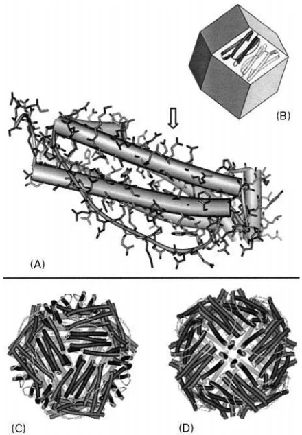
Figure 1 (A) Single apoferritin subunit;-helix regions of the secondary protein structure are represented as cylinders. The arrow marks a putative mineral nucleation site (here: Glu 57, 60, 61 and 64 of L-chain horse apoferritin, PDB code: 1AEW). (B) Supramolecular architecture of the apoferritin protein shell: 12 subunit dimers form the faces of an imaginary rhombic dodecahedron. (C, D) View along the three-fold channels (the four-fold channels, respectively) of the apoferritin structure. Adapted from Harrison and Arosio (1996).
prokaryots (Table 1). Sequence similarities of haem-free ferritins and haem-containing bacterioferritins may fall below 20%. However, their quaternary pro-tein structures are almost identical, suggesting that a convergent molecular evolution within different groups of organisms has independently led to an optimal solution for the availability of a mobile tem-porary iron storage.
While the general physiological effect of ferritin originating from different organisms may differ, it clearly functions as an iron deposit on the molecular scale, owing to several remarkable features:
E the apoferritin creates a conRned space which ulti-mately restricts the maximum size of the inwardly growing mineral phase;
E several anionic residues induce a net negative charge on the inner protein surface which compen-sates for the positive surface charges of initially formed polycationic Fe(III) oxyhydroxy species;
E the H-chain ferritin subunit contains a ferroxidase centre that catalyses the oxidation of Fe(II) by molecular oxygen to yield Fe(III);
E the L-chain ferritin subunit bears glutamic acid residues in close proximity which point towards the central cavity, thus possibly acting as an active site for crystal nucleation;
E the apoferritin supports long range electron trans-fer across the protein coat, enabling fast reductive release of Fe(II) ions from the highly insoluble Fe(III) oxyhydroxide mineral.
Apoferritin is built up by 24 structurally com-plementary subunits that self-assemble to form a hol-low shell of an approximate outer diameter of 11 nm. An individual subunit consists of a long 4--helix bundle with an additional short -helix lying at an angle of about 603 to the bundle axis at the C-terminal side of the amino acid chain (Figure 1A). At the beginning of apoferritin self-assembly dimers form mainly through a multitude of hydrophobic
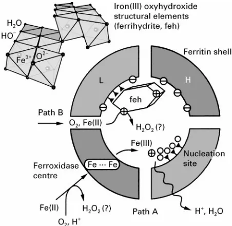
[image:2.568.294.514.301.618.2]Figure 2 Iron mineral formation in ferritin. A self-assembled heteropolymeric shell forms a spatially confined mineralization compartment composed of H- and L-chain subunits that possess different roles in mineralization. The H-chain contains a pre-organized arrangement of coordinating amino acid residues (fer-roxidase centre) which is able to take up two Fe(II) ions and to catalyse their oxidation to Fe(III) ions. The L-chain possesses four highly conserved glutamic acid residues, that are assumed to play a decisive role in Fe(III) mineral nucleation.
a nearly spherical cavity of an approximate diameter of 8 nm. The central cavity is accessible through channels of three-fold and four-fold symmetry which are situated at the vertices of the rhombic dodecahed-ron (Figure 1C, D). While the three-fold channels possess a hydrophilic surface, the four-fold channels are more hydrophobic in nature. The transport char-acteristics of the ferritin channels are still a matter of controversy. The three-fold channels are likely to be involved in the uptake and release of iron ions as well as in the regulation of the ferritin water content. For the four-fold channels an active role in the uptake of dioxygen has been proposed.
Ferritin Biomineralization
Since ferritin subunits spontaneously self-assemble
in vivo as well as in vitro to yield the complete apoferritin shell, it has been possible to study mineral deposition within the ferritin cavity under various experimental conditions.
Mineral formation within the ferritin cavity pro-ceeds via two different pathways (Figure 2): At low iron content, hydrated Fe(II) ions are taken up from the external medium by the H-chain ferroxidase centre where rapid oxidation takes place. Hydrated Fe(III) ions are released from the ferroxidase centre to
enter the ferritin cavity (path A). The inner apofer-ritin surface bears a multitude of primarily hy-drophilic and anionic amino acid residues which point towards the central cavity. In particular, each L-chain subunit contains a distinct array of four potentially coordinating glutamate residues (Fig-ure 1A) that could accumulate a small number of hydrated Fe(III) ions. The immobilized Fe(III) ions start to polymerize, leading to an OH\-bridged multinuclear Fe(III) oxyhydroxide cluster. Further cluster growth proceeds via addition of Fe(III) hexa-qua cations to the cluster surface with concomitant loss of an H2O ligand for each established
coor-dinative bond.
On reaching a critical crystallite size, autocatalytic Fe(II) oxidation at the surface of the mineral nucleus outweighs ferroxidase-induced oxidation, and min-eral growth continues until most of the cavity isRlled by one (or several) Fe(III) oxyhydroxide nanocrys-tal(s) (path B). The pathways of iron transport into the cavity, as well as the primary product(s) of iron(II) oxidation, are not yet fully established.
Since hydrated Fe(III) ions form polycationic oligomers at the initial stages of polymerization, the general consensus is that L-chain glutamate residues contribute to a negative surface potential on the in-side of the apoferritin shell in order to create a ther-modynamic sink for Fe(III) oxyhydroxide deposition. In addition to this general effect, a more speciRc template effect on iron mineral formation has been proposed which takes into account the special ar-rangement of glutamate residues and the fact that the sequence motif is highly conserved among ferritins from different classes of organisms. The poor crystallinity of the ferritin mineral on the other hand, as well as the fact that nanosized Fe(III) oxyhydroxide particles spontaneously grow from Fe(III)-containing aqueous solutions, challenges this interpretation.
Ferritin Core Structure
The mineral phase of ferritin displays properties simi-lar to the iron oxyhydroxide ferryhydrite which is widespread in nature. This mineral has been tradi-tionally described as amorphous or colloidal ferric hydroxide}Fe(OH3)}due to its poor X-ray
diffract-ing properties. The actual composition however, is Fe5HO8)4 H2O and recent X-ray absorption studies
Rrst oligomerize to yield chains of water-soluble poly-cations in which octahedrally coordinated Fe(III) ions are bridged by hydroxide anions. Further deprotona-tion and polymerizadeprotona-tion yield ferrihydrite, which, at close to its zero point of charge at pH 7}8, slowly and irreversibly dehydrates and rearranges to the thermo-dynamically more stable iron oxide hematite ( -Fe2O3). Although the details of the crystal structure
and surface properties of ferrihydrite are still contro-versial, the current data support a structural model in which double chains of edge-sharing iron(III) octa-hedra are cross-linked via corners (Figure 2, top), similar to the essential structural elements of the mineral goethite (-FeOOH). The dimensions of coherent X-ray scattering domains for synthetic ferrihydrite are within the range of 1}6 nm, which accounts for the characteristic broadening of X-ray diffraction lines. Temperature-dependent57Fe MoK
ss-bauer spectra of synthetic ferrihydrite and ferritin cores display identical features, indicating a super-paramagnetic behaviour of the crystalline phase at room temperature and below. Therefore synthetic ferrihydrite precipitates, as well as ferritin cores, may be better described as nanocrystals rather than nanocolloids.
Biologically Inspired Nanoparticle
Synthesis
The intriguing supramolecular architecture of fer-ritin, as well as its unique functional properties, might be taken as a model for the construction of artiRcial nanocompartments where crystal growth takes place within a spatially conRned microenvironment under controlled conditions. Classical approaches that make use of the entrapped water content of oil}water microemulsions are likewise simple to carry out, but as a rule suffer from relatively broad size distributions of the precipitated nanoparticulate materials. For ad-vanced applications, novel synthetic routes will there-fore be required to control the dimensions of the desired inorganic nanoparticles. Examples of techno-logically important compounds which exhibit strong-ly size-dependent physical and chemical properties range from catalytically active, highly dispersed metal nanocolloids (e.g. Pt, Pd, Rh), quantum-conRned semiconductor nanoparticles (CdS, CdSe) to nano-scale ferrimagnetic particles (e.g. -Fe2O3) and
nanocomposite magnetic alloys. Recent examples of biologically inspired synthetic approaches to synthe-size nanoscale inorganic materials include the use of biotechnologically engineered apoferritin shells and virus protein cages (capsids), or the use of monodis-perse block copolymer micelles as nanoscale reaction compartments (see Further Reading).
From Calci
\
ed Tissues to Engineered
Crystals
While ferritin represents an example of a nanosized crystalline biomineral, the architecture for example of a vertebrate bone or a mollusc shell spans several length scales. The morphology of the calciRed tissue is ultimately encoded in the genome that governs the biosynthesis of required materials at the cellular level of structural hierarchy. Biomineralization therefore, as a highly complex phenomenon of living organisms, cannot be reduced to a single mechanistic aspect. The following representation of CaCO3 mineralization
in molluscs is admittedly a crude simpliRcation which mainly concentrates on structural aspects, while at the same time ignoring the dynamic charac-ter of the entire process. Special emphasis here is put on induced CaCO3 crystal nucleation, i.e. the early
stages of crystal growth where the system properties can be described by supramolecular recognition events occurring at the mineral}matrix interface. At this level, common features of ferritin, mollusc shell or bone mineralization do exist: the inter-action of highly specialized acidic macromolecules with different surfaces of the growing single crystal. Current research efforts focus on the isolation and characterization of macromolecules from calciRed tissues. Functional properties of isolated macromolecules or fractions of macromolecules are systematically investigated for their ability to inSuence CaCO3 nucleation, growth and
poly-morphism. Biologically inspired synthetic strategies try to assemble artiRcial matrices in order to mimic structural and functional properties of mineralizing tissues.
Crystallochemical Aspects of CaCO
3Biomineralization
CaCO3, together with amorphous silica, is the most
abundant biomineral. There exist three CaCO3
poly-morphs}calcite, aragonite and vaterite}all of which occur in calciRed tissues. A monohydrate (mono-hydrocalcite) and a hexahydrate form (ikaite) of CaCO3 have been characterized as metastable
precursor phases during the incipient stages of crystal formation (Table 2).
At ambient conditions, calcite is the thermody-namically most stable CaCO3polymorph. However,
from oversaturated aqueous solutions containing Mg2# at a molar ratio Mg/Ca'4 (comparable to
Table 2 Characteristics of the most important CaCO3mineral phases
Mineral Crystal system (space group)
Specific density (g cm\3)
Solubility (!log Ksp)
Biological occurrence
Calcite (CaCO3) Trigonal (R3c) 2.71 8.48 Very common
Aragonite (CaCO3) Orthorhombic (Pmcn) 2.93 8.34 Very common
Vaterite (CaCO3) Orthorhombic (Pbnm) 2.54 7.91 Rare
Monohydrocalcite Trigonal (P3121) 2.43 7.60 Very rare
(CaCO3)H2O)
Ikaite (CaCO3)6H2O) Monoclinic (C2/c) 1.77 7.12 Unknown
Adapted with permission from Morse JW and Mackenzie FD (1990)Geochemistry of Sedimentary Carbonate, p. 41. Amsterdam: Elsevier.
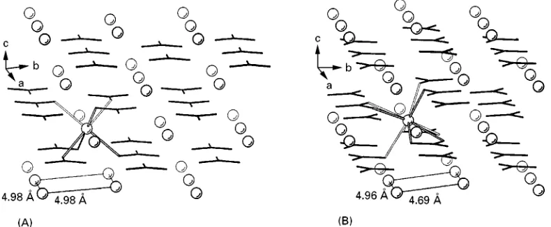
Figure 3 Ion-packing arrangement in the crystal structures of (A) calcite and (B) aragonite. The coordinative bonds between CO2\ 3 anions (black sticks) and one of the Ca2#ions (open circles) are emphasized with open lines. The minimum distances of Ca2#ions in
theab planes for both crystal lattices are indicated at the bottom.
The arrangement of the ions in crystalline CaCO3
may be described in terms of separate layers of cations and anions. Coordination environments for Ca2# ions (CO2\
3 , respectively) in the polymorphs
differ from each other, as a result of different successions of layers, as well as different crystallo-graphic orientations of the planar carboxylate groups in the crystal lattices (Figure 3). In calcite, each single densely packed Ca2#layer parallel to theabplane is
situated between single layers of CO2\
3 with each
layer containing anions oriented in opposite directions. Each Ca2# ion is situated in a distorted octahedral
coordination environment of six different CO2\
3
an-ions. In aragonite, the positions of Ca2#ions in theab
plane are nearly identical to those of the calcite struc-ture. In contrast, the CO2\
3 anions below and above
the Ca2#layer are separated into two layers, which are
lifted by 0.98 A> along the c direction, leading alto-gether to a ninefold coordination of Ca2#ions.
Shell Formation in Molluscs
It has long been recognized that calcifying organisms have developed active mechanisms to select the
poly-morph, and to control the distribution, shapes and orientations of crystals in their mineralized tissues. Molluscs are among the most thoroughly investigated organisms in this regard; they build concrete shells from CaCO3. The mollusc shell may be regarded as
a microlaminate composite consisting of layers of highly oriented CaCO3crystals which are
intersper-sed with thin sheets of an organic matrix. Crystals within separate shell layers usually consist of either pure aragonite or pure calcite. Vaterite, when present, is usually associated with shell repair. The succession of shell layers, as well as their pronounced ultrastruc-tural features (seeTable 3) are important characters in mollusc taxonomy. The main protective functions of the shell are to prevent desiccation, predation and abrasion. The shell also provides support for the body and a site for muscle attachment.
Shell formation occurs in two principal phases. The
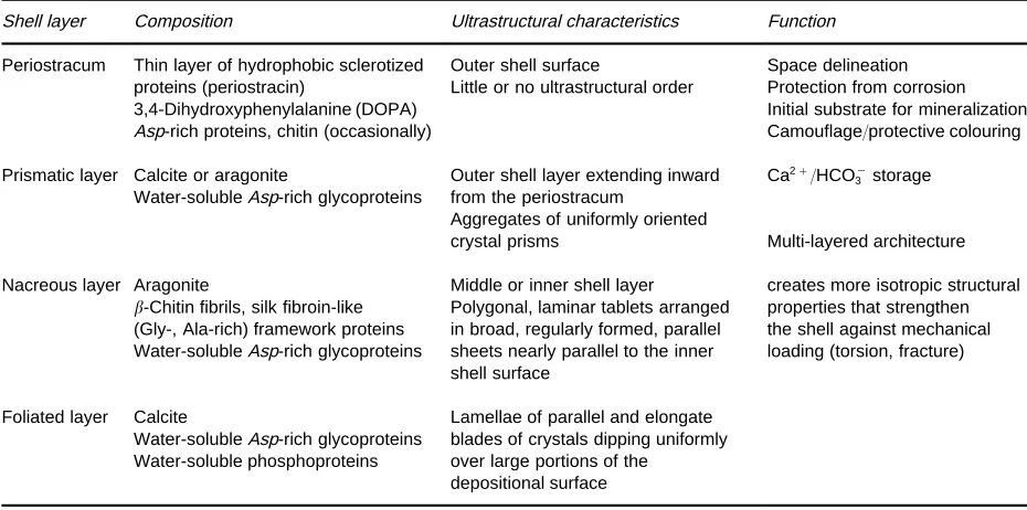
[image:5.568.90.485.512.675.2]Table 3 Classification of mollusc shell layers
Shell layer Composition Ultrastructural characteristics Function
Periostracum Thin layer of hydrophobic sclerotized Outer shell surface Space delineation proteins (periostracin) Little or no ultrastructural order Protection from corrosion 3,4-Dihydroxyphenylalanine (DOPA) Initial substrate for mineralization Asp-rich proteins, chitin (occasionally) Camouflage/protective colouring
Prismatic layer Calcite or aragonite
Water-solubleAsp-rich glycoproteins
Outer shell layer extending inward from the periostracum
Ca2#/HCO\
3 storage
Aggregates of uniformly oriented
crystal prisms Multi-layered architecture
Nacreous layer Aragonite Middle or inner shell layer creates more isotropic structural -Chitin fibrils, silk fibroin-like Polygonal, laminar tablets arranged properties that strengthen (Gly-, Ala-rich) framework proteins in broad, regularly formed, parallel the shell against mechanical Water-solubleAsp-rich glycoproteins sheets nearly parallel to the inner
shell surface
loading (torsion, fracture)
Foliated layer Calcite Lamellae of parallel and elongate Water-solubleAsp-rich glycoproteins blades of crystals dipping uniformly Water-soluble phosphoproteins over large portions of the
depositional surface
Adapted with permission from Wilbur KM and Saleuddin ASM (eds) (1983) Shell formation. In:The Mollusca, vol. 4, pp. 236}287. San Diego: Academic Press.
Figure 4 (A) Transverse section of the mantle edge of a bivalve showing the system of compartments. (B) Scheme of ion fluxes at the interface between the outer epithelial cells of the mollusc tissues and the incipient layer of nacre. (Simplified representations, not to scale.)
the so-called extrapallial space (Figure 4B). Crystals grow in intimate association with a secreted, highly specialized organic matrix. In order for crystals to form, the extrapallialSuid must become supersaturated with CaCO3, which imposes active accumulation
strategies upon molluscs that inhibit a freshwater environment, where the external medium is depleted of Ca2# ions. The concentration of Ca2# ions is
actively regulated by a Ca membrane transport sys-tem that is located in the body and the mantle epi-thelium. Active carbonate transport has also been postulated for the regulation of HCO\3 ion
concen-tration. However, as an additional source of hydro-gen carbonate, the mollusc may utilize metabolic carbon dioxide from its respiratory system. Any sup-ply of HCO\3 ions is tightly associated with carbonic
anhydrase (CA) activity, an enzyme that catalyses the interconversion of carbon dioxide (CO2) and
carbon-ic acid (H2CO3). High carbonic anhydrase activity is
[image:6.568.70.505.513.676.2]Figure 5 Fractured surface of the nacreous layer of the bivalve molluscAtrina rigida. The inset shows the inner nacreous layer of tabular aragonite crystals (top) and the outer prismatic layer of columnar calcite crystals (bottom). SEM micrographs, scale bar denotes 1m. Courtesy of Y. Levi, Department of Structural Biology, Weizmann Institute of Science, Israel.
The extrapallialSuid also contains a complex mix-ture of inorganic and organic substances. Analysis of several species of marine and freshwater species showed that the major cations present are Na#, K#, Ca2#and Mg2#while the major anions are HCO\
3,
Cl\ and SO2\
4 . The organic components of the
ex-trapallial Suid include amino acids, proteins, muco-polysaccharides and low molecular organic acids (e.g. succinic acid). The organic compounds are secreted by mantle epithelial cells, but their metabolic origins in mollusc tissue have not yet been located.
The Structure of Nacre
Biogenic crystals may use the inner surface of the periostracum, the surface of other, already formed crystals, or the organic matrix as a substrate. Special attention has been drawn to the microstructure of nacre which exhibits an exceptionally regular ar-rangement of tabular aragonite crystals. Thin inter-digitated aragonite plates develop in close association with thin horizontally aligned organic layers (inter-lamellar sheets) upon which aragonite crystals are nucleated (Figure 4B, Figure 5). In order to grow crystals into a highly regular brickwork-like pattern, numerous nucleation events would have to be syn-chronized with each other at distant locations. An alternative growth mechanism was proposed to ex-plain the precision by which aragonite platelets are uniformly co-aligned within the same and consecu-tive layers. According to this model, nacre may be constituted of extended, continuous single crystalline domains of aragonite platelets that are interconnect-ing by mineral bridges through the perforated inter-lamellar sheets.
Biologically Induced Crystal
Nucleation
Putative Structure of Acidic Nucleation Sites in Calci\ed Tissues
One of the critical problems in understanding the mechanisms of matrix-associated mineralization is the lack of information on the three-dimensional structures of biological macromolecules that interface with the mineral. A literature search up to the middle of 1999 yielded very few examples where complete or partial information about the primary structure of macromolecules directly involved in mineralization have been determined (Table 4). Macromolecules isolated from mollusc tissues have traditionally been distinguished into two different classes, based on solubility properties. Chemical analysis showed that the insoluble fraction consists
mainly of Rbrous proteins (collagen, chitin) and/or polysaccharides. These macromolecules together build a rigid framework upon which speciRc macro-molecules from the soluble fraction may become ad-sorbed. The primary function of the insoluble organic matrix is to subdivide the mineralization compart-ment into an organized network of microcompart-ments and thus to delimit the available space for crystal growth and/or to constrain the crystal packing arrangement to a certain extent. The surface of this macromolecular assembly may serve as a supramolecular template for oriented nucleation of single crystals, although this structure}function relation is difRcult to prove for biological systems
in vivo.
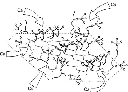
Figure 6 Schematic representation of a putative nucleation site in molluscan tissues. An acidic glycoprotein is anchored to a rigid substrate (as schematized by the broken lines) through hydro-phobic or electrostatic interactions. The sulfate groups, linked to flexible oligosaccharide side chains, concentrate Ca2#ions on an
Asp-rich oligopeptide domain that is assumed to adapt a highly regular-sheet conformation. A first layer of Ca ions may thus be fixed and oriented in space, upon which further mineral growth ensues. Reproduced with permission from Addadi and Weiner (1989).
chemical nature, and their association with different mineral phases clearly rule out a uniform function. They may roughly be divided intoRve different func-tional classes:
E concentration regulators: macromolecules that are linked to Ca2#and/or CO2\
3 transport and
meta-bolism (e.g. carbonic anhydrase)
E growth inhibitors: acidic macromolecules that strongly bind Ca2#ions and become
nonselective-ly adsorbed on to any arbitrary crystal face which is exposed to the mother liquor
E growth modiRers: acidic macromolecules that in-teract stereoselectivity with distinct faces of a nascent crystal
E texture modiRers: acidic macromolecules that be-come occluded and modify texture and mechanic properties of crystals
E nucleators: immobilized acidic macromolecules that form a highly regular template for induced crystal nucleation
Due to the complex nature of interactions in biolo-gical matrices, the same acidic macromolecule may belong to more than one of the above mentioned categories and its functional properties may change within different organisms and microenvironments.
To demonstrate a possible mode of molecular in-teraction between acidic macromolecules and crystal surfaces, this survey of mollusc mineralization will conclude with a brief section about induced CaCO3
nucleation in biological systems. The bodySuids of mineralizing organisms contain crystallization inhibi-tors that prevent spontaneously formed crystal nuclei from growing into larger crystals. To direct mineral deposition to the appropriate location, active nuclea-tion sites have to exist in mineralizing compartments. The opening section about iron storage has already indicated how the molecular architecture of ferritins may be associated with nucleation of iron minerals. For induced calcite and aragonite nucleation, system-atic investigations on biological and suitably assem-bled artiRcial systems have shed some light on the structural requirements of a putative nucleation site, especially in mollusc shells. The model of Addadi and Weiner proposes structurally pre-organized domains of acidic residues, that could serve as a supramolecu-lar template for oriented crystal nucleation. Such highly ordered domains could result from acidic mac-romolecules being adsorbed on a rigid scaffold of insoluble matrix proteins (Figure 6). As an example, the interlamellar organic sheets of mollusc shell nacre consist of thin sheets of -chitin (a water-insoluble (1P4)-linked 2-acetamido-2-deoxy-D-glucan)
sand-wiched between thicker sheets of silk Rbroin-like proteins. SilkRbroin itself possesses microcrystalline
domains of repeating [Gly-Ala-Gly-Ala-Gly-Ser]n
units that adopt an antiparallel-pleated sheet con-formation. These domains have a highly regular and hydrophobic surface upon which acidic macro-molecules are adsorbed from solution. In the course of adsorption, the acidic macromolecules has to fold into the appropriate conformation, in order to maxi-mize its hydrophobic interactions with the silkRbroin surface. Possible candidates for acidic macro-molecules interacting with silk Rbroin in the de-scribed way are oligopeptides that include sequence motifs of [Asp-X]n, (X"Gly, Ser), which have
a strong tendency to fold into a-sheet conformation in the presence of Ca2# ions. As a consequence, the
aspartic acid residues of [Asp-X]nsequences would be
positioned at only one side of the -pleated sheet, resulting in an organized two-dimensional array of carboxylate ligands.
It is tempting to assume that the carboxylate resi-dues coordinate a Rrst layer of Ca2# ions which
would in turn become theRrst layer of an epitaxially growing CaCO3crystal. However, a more profound
analysis has so far failed to provide evidence for an epitaxial growth mechanism or a close stereochem-ical complementarity between the nucleating macro-molecules and the incipient CaCO3 crystal surface.
nucleate from the ab planes. The arrangement of Ca2#ions in this plane (the shortest distance between
Ca2# ions is 4.99 A> in calcite, and 4.69 A> in
arago-nite, respectively; Figure 3) is geometrically not com-mensurate with the period of amino acid residues in a protein -strand (approx. 6.9 A> ). Moreover, on pointing more or less perpendicular towards the Ca2#ions in the crystal (0 0 1) face, the carboxylate
residues of the -pleated sheet cannot continue the parallel arrangement of planar carbonate anions in the underlying layer(s). The current nucleation model thus does not support the picture of a calcite or aragonite single crystal being nucleated from (0 0 1) crystal faces by virtue of stereochemical selection principles.
Despite the similar positioning of Ca2#ions in the abplane of calcite and aragonite, mollusc shells dis-criminate between the two polymorphs by secreting them separately in different layers (e.g. prismatic layer and nacre). This suggests that additional factors participate in nucleation. One possibility is that dif-ferent Mg2#concentrations in theSuids of aragonitic
and calcitic layers may be present that could help to shift the balance between the two polymorphs. An-other possibility is the presence of polymorph-speciRc macromolecules that interact with more than one face of the nascent crystal. For a valid explanation of selective nucleation of either polymorph, the current, essentially geometric model will have to be reRned. A lot is expected from the Rrst three-dimensional structure of a nucleating macromolecule, although its active conformation may depend on the accompany-ing insoluble organic matrix in the biological tissues. Finally, novel theoretical approaches are currently being investigated to explore realistic surface proper-ties of the CaCO3polymorphs which consider surface
relaxation as well as hydration of the outermost ionic layers.
Biologically Inspired Engineering
of CaCO
3Crystals
Several studies have been directed to the phenomenon of heteroepitaxy in CaCO3 biomineralization.
Semi-nal contributions have come from the group of Mann and co-workers who studied the inSuence of nega-tively charged surfactants on crystal nucleation. The group made use of a Langmuir Rlm balance that allows for spreading surfactant molecules as a mono-layer at the air}water interface with the charged head groups pointing toward the aqueous subphase. Nucleation of calcite single crystals was observed from monolayers of aliphatic monocarboxylic acids, sulfates or phosphonates. The crystals that grew
underneath the monolayers in general showed a sig-niRcantly narrower size distribution and reduced nucleation time, as compared to calcite crystals precipitated spontaneously from supersaturated solu-tions. Moreover, the crystals grew crystallographi-cally oriented relative to the monolayer. Calcite single crystals nucleated preferentially from the
10.0 face underneath compressed monolayers of amphiphilic carboxylic acids, while monolayers of alkylsulfates and -phosphonates led to calcite single crystals that nucleated from the (0 0 1) face. Detailed schemes were proposed to rationalize the different modes of interaction between the different head groups of amphiphiles in the monolayers and the corresponding faces from which the crystals were nucleated.
To gain more precise controls over the relative positions of coordinating residues, patterned self-assembled monolayers of bifunctional-terminated alkanethiols (HS(CH2)nX, X"CO2\, SO\3, PO23\and
OH) have been produced very recently on different supporting metals (Au, Ag) by means of microcontact printing. Depending on the appropriate combination of functional groups, length of alkyl chains, metal substrate and nucleating area, the selective nucleation of calcite single crystals has been achieved for a huge variety of crystallographic orientations. As a unique feature, the self-assembled monolayers allow one to grow isolated and oriented single crystals with a
de-Rned separation which is encoded in the tiling of the nucleating areas. The technique has been further em-ployed to grow crystalline CaCO3layers that consist
of alternating domains of differently oriented calcite single crystals.
The few cited examples demonstrate how model studies may focus on aspects of induced nucleation at organized organic surfaces. Single structural para-meters of the organic matrix can be varied systemati-cally and analysed for their inSuence on crystal nucleation and growth. Results gleaned from these experiments provide important informations that could help to interpret the observed, often highly complex, structures of biominerals. The models stud-ies, furthermore, indicate as to how biologically inspired design and novel technical approaches com-bine into innovative synthetic strategies for engineer-ing artiRcial crystalline architectures.
Acknowlegements
The author would like to thank Cia Addadi and Stephen Weiner for valuable discussions. Generous
See also: II / Crystallization: Polymorphism. III / Biolo-gical Systems: Ion Exchange BioloBiolo-gically Active Compounds and Xenobiotics: Magnetic Affinity; Supercritical Fluid Crystallization.
Further Reading
Addadi L and Weiner S (1989) Stereochemical and struc-tural relations between macromolecules and crystals. In: Mann S, Webb J and Williams RJP (eds) Biomineraliz-ation, pp. 133}156. Weinheim: VCH.
Aizenberg J, Black AJ and Whitesides GM (1999) Oriented growth of calcite controlled by self-assembled mono-layers of functionalized alkanethiols supported on gold and silver. Journal of the American Chemistry Society
121: 4500}4509.
Briat J-F and LobreHaux S (1998) Iron storage and ferritin in plants. In: Sigel A and Sigel H (eds) Metal Ions in Biological Systems, vol. 35, pp. 563}584.
Chasteen ND (1998) Ferritin. Uptake, storage, and release of iron. In: Sigel A and Sigel H (eds) Metal Ions in Biological Systems, vol. 35, pp. 479}514.
de Leeuw NH and Parker CS (1998) Surface structure and morphology of calcium carbonate polymorphs calcite, aragonite, and vaterite: an atomistic approach.Journal of Physical Chemistry B102: 2914}2922.
Douglas T and Young M (1998) Host}guest encapsulation of materials by assembled virus protein cages. Nature
393: 152}155.
Fendler JH (ed.) (1998)Nanoparticles and Nanostructured Films:Preparation,Characterization and Applications. Weinheim: VCH.
Gider S, Awschalom DD, Douglas Tet al. (1995) Classical and quantum magnetic phenomena in natural and artiR -cial ferritin proteins.Science268: 77}80.
Harrison PM and Arosio P (1996) The ferritins: molecular properties, iron storage function and cellular regulation.
Biochimica et Biophysica Acta } Bioenergetics 1275: 161}203.
Harrison PM, Hempstead PC, Artymiuk PJ and Andrews SC (1998) Structure}function relationship in the fer-ritins. In: Sigel A and Sigel H (eds)Metal Ions in Biolo-gical Systems, vol. 35, pp. 435}477.
Heywood B (1996) Template-directed nucleation and growth of inorganic materials. In: Mann S (ed.)
Biomimetic Materials Chemistry, pp. 143}173. Weinheim: VCH.
Jambor JL and Dutrizac JE (1998) Occurrence and consti-tution of natural and synthetic ferrihydrite, a wide-spread iron oxyhydroxide. Chemical Reviews 98: 2549}2585.
Lippmann F (1973)Sedimentary Carbonate Minerals. Min-erals, Rocks and Inorganic Materials, vol. 6. Berlin: Springer-Verlag.
Lowenstam HA and Weiner S (1989)On Biomineralization
(Eds Mann, Webb and R.J.P. Williams) New York: Oxford University Press, 7}49.
MoKller M and Spatz JP (1997) Mineralization of nanopar-ticles in block copolymer micelles.Current Opinion in Colloid and Interface Science2: 177}187.
Powell AK (1998) Ferritin. Its mineralization. In: Sigel A and Sigel H (eds)Metal Ions in Biological Systems, vol. 35, pp. 515}561.
SchaKffer TE, Ionescu-Zanetti C, Proksch R et al. (1997) Does abalone nacre form by heteroepitaxial nucleation or by growth through mineral bridges?Chemistry of Materials9: 1731}1740.
Simkiss K and Wilbur KM (1989) Molluscs } Epithelial control of matrix and minerals. In:Biomineralization.
Cell Biology and Mineral Deposition, pp. 230}260. San Diego: Academic Press.
Weiner S and Addadi L (1997) Design strategies in min-eralized biological materials. Journal of Materials Chemistry7: 689}702.
Control of Crystallizers and Dynamic Behaviour
H. J. M. Kramer, Delft University of Technology, Delft, The Netherlands
Copyright^ 2000 Academic Press
Introduction
Ideally industrial crystallizers are operated in such a way that the product speciRcations are met under conditions that permit proRtable, trouble-free pro-duction of the desired crystalline material. In indus-trial practice, however, many operational problems can be encountered that reduce the performance of the crystallizer. The most commonly encountered problems are listed here.
E Deposition of crystal solids on the crystallizer in-ternals, often called scaling or fouling. This results in a reduction of the heat transfer in the heat exchanger or leads to plugging of the process lines and can even hamper theSow pattern and thus the mixing in the crystallizer.
E Alternate feed composition. The resulting changes in the level of supersaturation in the crystallizer can lead to nucleation bursts or depletion of secondary nuclei, having a severe effect on the dynamics of the process.